. 2014 Feb 21;289(8):5320-9.
doi: 10.1074/jbc.M113.505636. Epub 2014 Jan 7.

Localization and functionality of the inflammasome in neutrophils

Affiliations

Localization and functionality of the inflammasome in neutrophils

Martina Bakele et al. J Biol Chem. .

Abstract

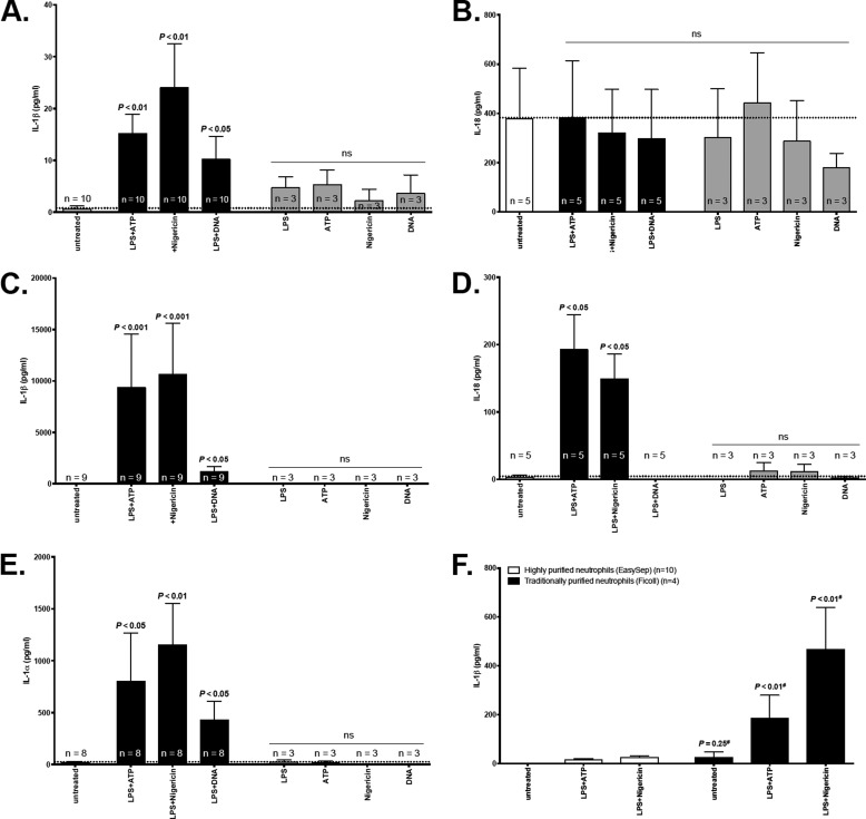
Neutrophils represent the major fraction of circulating immune cells and are rapidly recruited to sites of infection and inflammation. The inflammasome is a multiprotein complex that regulates the generation of IL-1 family proteins. The precise subcellular localization and functionality of the inflammasome in human neutrophils are poorly defined. Here we demonstrate that highly purified human neutrophils express key components of the NOD-like receptor family, pyrin domain containing 3 (NLRP3), and absent in melanoma 2 (AIM2) inflammasomes, particularly apoptosis-associated speck-like protein containing a CARD (ASC), AIM2, and caspase-1. Subcellular fractionation and microscopic analyses further showed that inflammasome components were localized in the cytoplasm and also noncanonically in secretory vesicle and tertiary granule compartments. Whereas IL-1β and IL-18 were expressed at the mRNA level and released as protein, highly purified neutrophils neither expressed nor released IL-1α at baseline or upon stimulation. Upon inflammasome activation, highly purified neutrophils released substantially lower levels of IL-1β protein compared with partially purified neutrophils. Serine proteases and caspases were differentially involved in IL-1β release, depending on the stimulus. Spontaneous activation of the NLRP3 inflammasome in neutrophils in vivo affected IL-1β, but not IL-18 release. In summary, these studies show that human neutrophils express key components of the inflammasome machinery in distinct intracellular compartments and release IL-1β and IL-18, but not IL-1α or IL-33 protein. Targeting the neutrophil inflammasome may represent a future therapeutic strategy to modulate neutrophilic inflammatory diseases, such as cystic fibrosis, rheumatoid arthritis, or sepsis.

Keywords: Cytokine; Immunity; Inflammation; Innate Immunity; Neutrophils; Pathogen-associated Molecular Pattern (PAMP); Toll-like Receptor (TLR).


Figures

FIGURE 1.
FIGURE 1.
mRNA expression profiling of inflammasome components in highly purified neutrophils mRNA expression levels for ASC, caspase-1, AIM2, NLRC4, NAIP, NLRP3, IL1B, IL18, IL1A, and IL33 were measured in highly purified neutrophils and were compared with peripheral blood monocytes. Target mRNA levels were normalized to the housekeeping gene β-actin. Monocytic mRNA levels were set to 1.0, and neutrophilic mRNA levels are shown in relation. Means ± S.E. (error bars) are shown. Priming of neutrophils was done using ultrapure LPS (10 ng/ml) in cRPMI at 37 °C, 5% CO2. *, p < 0.05 LPS versus medium-treated neutrophils.
FIGURE 2.
FIGURE 2.
Subcellular localization of inflammasome components in neutrophils. A, Western blot analysis of ASC (left panel), caspase-1 (middle panel), and AIM2 (right panel) in neutrophil subcellular fractions in human neutrophils. SV, secretory vesicles; PM, plasma membranes; 3°, tertiary granules; 2°, tertiary granules; 1°, primary granules; CY, cytosol. Target protein expression was normalized to total protein in the respective subcellular fractions. B, subcellular localization of ASC and AIM2 in neutrophils by immuno-transmission electron microscopy, indicating ASC and AIM2 protein in vesicular intracellular compartments. The red arrows mark AIM2 or ASC associated to a vesicular structure. Scale bars, 0.5 μm. C, subcellular localization of AIM2 in neutrophils. Co-localization of AIM2 (red) with markers for tertiary granules (matrix metalloproteinase 9), secondary granules (CD66b), primary granules (CD63), concanavalin A (cytosolic), and secretory vesicles (CD35/CR1) (all green) was studied by confocal laser scanning microscopy. DNA was stained with DAPI (blue).
FIGURE 3.
FIGURE 3.
Extrinsic inflammasome activation drives IL-1β, but not IL-1α, IL-18, or IL-33 secretion by highly purified neutrophils. Highly purified neutrophils (A, B, and F), PBMCs (C, D, and E), or partially purified neutrophils (F) were treated with LPS, ATP, nigericin, or DNA alone or in combination as indicated in the respective panels and described in detail in the methods section. Protein levels of IL-1β, IL-1α, IL-18, or IL-33 were quantified in cell culture supernatant by ELISA. IL-1α protein was undetectable in supernatants from neutrophils. IL-33 protein was undetectable in supernatants from neutrophils or PBMCs. F, IL-1β levels were measured in cell culture supernatants of highly purified neutrophils (white columns) and partially purified (Ficoll density gradient) isolated neutrophils (black columns). Two groups were compared using the Mann-Whitney U test. Identical cell input numbers (20 × 106) were used. Dashed lines indicate the cytokine concentration of the untreated control. Concentrations are given as pg/ml (mean ± S.E. (error bars)).
FIGURE 4.
FIGURE 4.
Intrinsic NLRP3 inflammasome activation in highly purified neutrophils. IL-1β, IL-1α, and IL-18 protein secretion of highly purified neutrophils from healthy subjects (white columns) and patients suffering from MWS (black columns) with or without inflammasome induction via LPS/ATP or LPS/nigericin is shown. Baseline IL-1β levels (untreated) were higher in MWS patients compared with healthy controls (left panel). Following stimulation with LPS + ATP or nigericin cells from healthy individuals secreted higher levels of IL-1β compared with MWS patients. In accordance to our observation in Fig. 3, IL-18 secretion by neutrophils from both healthy controls and MWS patients was inflammasome-independent (right panel). Concentrations are given as pg/ml (mean ± S.E. (error bars)).
FIGURE 5.
FIGURE 5.
Caspases and proteases are involved in IL-1β production by neutrophils. Highly purified neutrophils (A) and PBMCs (B) were stimulated with LPS/ATP or LPS/nigericin (black columns) with or without pretreatment with a serine protease inhibitor (PMSF, 1 mm, light gray columns) or a caspase inhibitor (Z-VAD-fluoromethyl ketone, 20 μm, dark gray columns). In neutrophils, inflammasome activation with LPS/ATP was reduced to a greater degree by inhibition of serine protease, whereas activation with LPS/nigericin was mainly suppressed by caspase inhibition (left panel). In PBMCs, serine protease inhibition had no effect on IL-1β secretion, whereas the caspase inhibitor fully abrogated IL-1β secretion after stimulation with LPS/ATP or LPS/nigericin (right panel). Concentrations are given as pg/ml (mean ± S.E. (error bars)).

References

    1. Nathan C. (2006) Neutrophils and immunity: challenges and opportunities. Nat. Rev. Immunol. 6, 173–182 - PubMed
    1. Kolaczkowska E., Kubes P. (2013) Neutrophil recruitment and function in health and inflammation. Nat. Rev. Immunol. 13, 159–175 - PubMed
    1. Häger M., Cowland J. B., Borregaard N. (2010) Neutrophil granules in health and disease. J. Intern. Med. 68, 25–34 - PubMed
    1. Cassatella M. A. (1995) The production of cytokines by polymorphonuclear neutrophils. Immunol. Today 16, 21–26 - PubMed
    1. Gross O., Thomas C. J., Guarda G., Tschopp J. (2011) The inflammasome: an integrated view. Immunol. Rev. 243, 136–151 - PubMed

Publication types