. 2014 Feb 21;289(8):4919-27.
doi: 10.1074/jbc.M113.511972. Epub 2014 Jan 7.

Proteomic analysis of tendon extracellular matrix reveals disease stage-specific fragmentation and differential cleavage of COMP (cartilage oligomeric matrix protein)

Affiliations

Proteomic analysis of tendon extracellular matrix reveals disease stage-specific fragmentation and differential cleavage of COMP (cartilage oligomeric matrix protein)

Stephanie Georgina Dakin et al. J Biol Chem. .

Abstract

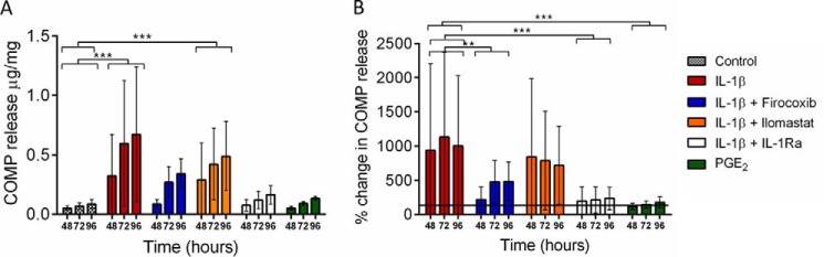
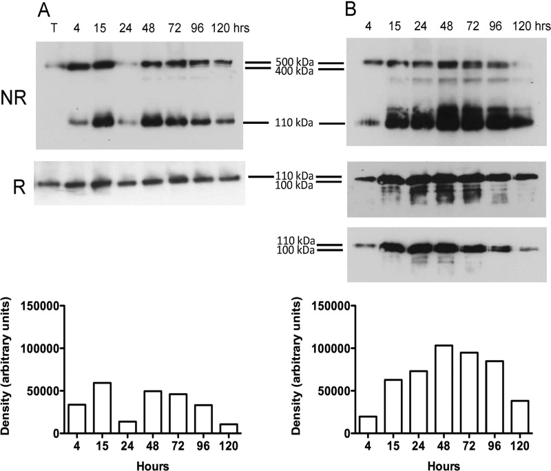
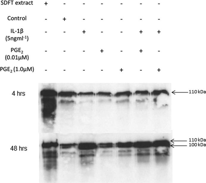
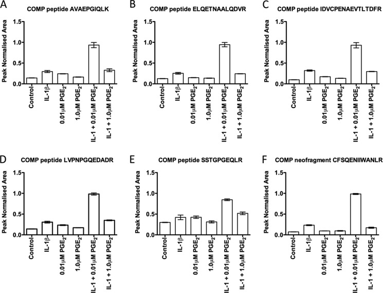
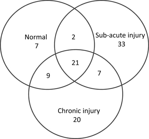
During inflammatory processes the extracellular matrix (ECM) is extensively remodeled, and many of the constituent components are released as proteolytically cleaved fragments. These degradative processes are better documented for inflammatory joint diseases than tendinopathy even though the pathogenesis has many similarities. The aims of this study were to investigate the proteomic composition of injured tendons during early and late disease stages to identify disease-specific cleavage patterns of the ECM protein cartilage oligomeric matrix protein (COMP). In addition to characterizing fragments released in naturally occurring disease, we hypothesized that stimulation of tendon explants with proinflammatory mediators in vitro would induce fragments of COMP analogous to natural disease. Therefore, normal tendon explants were stimulated with IL-1β and prostaglandin E2, and their effects on the release of COMP and its cleavage patterns were characterized. Analyses of injured tendons identified an altered proteomic composition of the ECM at all stages post injury, showing protein fragments that were specific to disease stage. IL-1β enhanced the proteolytic cleavage and release of COMP from tendon explants, whereas PGE2 had no catabolic effect. Of the cleavage fragments identified in early stage tendon disease, two fragments were generated by an IL-1-mediated mechanism. These fragments provide a platform for the development of neo-epitope assays specific to injury stage for tendon disease.

Keywords: Extracellular Matrix; Inflammation; Mass Spectrometry (MS); Peptides; Proteomics; Tendon.


Figures

FIGURE 1.
FIGURE 1.
Viability of tendon fibroblasts in explant culture. Confocal fluorescence micrographs of tendon sections treated with calcein AM to denote cytoplasmic staining of live cells (green) and ethidium bromide for nuclear staining (red) of live and dead cells. Explants after 24 h (A) and 120 h (B) are shown. Cell viability was 90 ± 5% after 24 h in culture and 60 ± 5% after 120 h. C, azide-treated explant after 24 h was used as a negative control. All experiments were performed within the 120-h culture period. Scale bar = 20 μm.
FIGURE 2.
FIGURE 2.
Release of COMP into media from SDFT explants in culture. Normal SDFT explants were derived from 3 horses ages between 9 and 13 years (mean 10 ± 2 years). Explants were rested for 24 h after cutting before media replacement and stimulation commencing. Media were harvested at 48, 72, and 96 h with complete replacement at each time point. A, mean cumulative COMP release showing significantly increased release with IL-1β stimulation alone and in combination with 20 μm Ilomastat. COMP release after stimulation with 1.0 μm PGE2 was similar to non-stimulated controls. B, mean percentage change in COMP release relative to the respective control. The addition of either 400 ng·ml−1 IL-1Ra or 1.0 μm Firocoxib significantly reduced IL-1β-mediated COMP release. Error bars represent S.D. ** p < 0.01; *** p < 0.001.
FIGURE 3.
FIGURE 3.
COMP release into culture media from normal SDFT explants. Representative Western blots of media from experimental samples showing COMP release with time from control (A) and IL-1β stimulated tendon explants (B) under non-reduced (NR) and reduced (R) conditions. Densitometric analysis of protein bands is shown for non-reduced Western blots. COMP release was enhanced by stimulation of explants with IL-1β compared with non-stimulated controls, with maximal release occurring at 48 h and the appearance of a number of distinct peptides <100 kDa. In IL-1β-stimulated explants, monomeric COMP appears as a doublet under reduced conditions from 15 to 120 h after explant cutting, which is better demonstrated in a lower exposure of the blot in a lower panel. T, equine SDFT extract (total protein loaded 10 μg) prepared in 4 m (guanidine HCl) was used as a loading standard and as a positive control for COMP, which is present mostly as a 550-kDa pentamer in the tissue.
FIGURE 4.
FIGURE 4.
Differential effects of IL-1β and PGE2 on COMP release. Representative Western blot of media samples harvested at 4 h and 48 h. Tendon extract = positive control (10 μg total protein); control = media from unstimulated tendon at the same respective time point. COMP release and fragmentation were enhanced by stimulation with IL-1β at 48 h with lower molecular weight fragments present including the appearance of the 100-kDa peptide (doublet). The addition of PGE2 resulted in the release of both intact and fragmented COMP (100 kDa) at 48 h. Smaller fragments were detected with the higher dose of PGE2, but these were present in low abundance compared with stimulation with IL-1β.
FIGURE 5.
FIGURE 5.
MRM analyses of COMP peptides released into media after 72 h in culture: AVAEPGIQLK (A), ELQETNAALQDVR (B), IDVCPENAEVTLTDFR (C), LVPNPGQEDADR (D), SSTGPGEQLR (E), and COMP neo-terminal fragment CFSQENIIWANLR (F). Each sample was run on the MRM in triplicate, and the mean values with S.D. are shown for each experimental condition. MRM analyses were conducted on triplicate samples from one experiment and not biological replicates; hence, statistical analyses were not performed. Peak normalized area represents the summed area of the ion peak transitions for each peptide measured. There was a trend for enhanced COMP peptide/neo-N-terminal fragment release with IL-1β compared with non-stimulated controls. For all peptides, maximal release occurred after stimulation with both IL-1β and 0.01 μm PGE2.
FIGURE 6.
FIGURE 6.
Venn representation of proteins identified from Q-TOF LC-MS. Samples of extracts of normal, sub-acute, and chronic injured equine SDFT samples (n = 3 for each) were analyzed for global protein composition, demonstrating differences in tendon ECM protein expression profiles with injury stage. Although a similar number of proteins was identified in both sub-acute and chronic injury phases, it was in the sub-acute phase injury that the greatest numbers of unique proteins were present.

References

    1. Kujala U. M., Sarna S., Kaprio J. (2005) Cumulative incidence of achilles tendon rupture and tendinopathy in male former elite athletes. Clin. J. Sport Med. 15, 133–135 - PubMed
    1. Avella C. S., Ely E. R., Verheyen K. L., Price J. S., Wood J. L., Smith R. K. (2009) Ultrasonographic assessment of the superficial digital flexor tendons of National Hunt racehorses in training over two racing seasons. Equine Vet. J. 41, 449–454 - PubMed
    1. Dakin S. G., Werling D., Hibbert A., Abayasekara D. R., Young N. J., Smith R. K., Dudhia J. (2012) Macrophage sub-populations and the lipoxin A4 receptor implicate active inflammation during equine tendon repair. PLoS ONE 7, e32333. - PMC - PubMed
    1. Almekinders L. C., Temple J. D. (1998) Etiology, diagnosis, and treatment of tendonitis. An analysis of the literature. Med. Sci. Sports Exerc. 30, 1183–1190 - PubMed
    1. Birch H. L., Bailey A. J., Goodship A. E. (1998) Macroscopic “degeneration” of equine superficial digital flexor tendon is accompanied by a change in extracellular matrix composition. Equine Vet. J. 30, 534–539 - PubMed

Publication types

MeSH terms