. 2014 Feb 28;289(9):5997-6005.
doi: 10.1074/jbc.M113.508549. Epub 2014 Jan 7.

A colon cancer-derived mutant of Krüppel-like factor 5 (KLF5) is resistant to degradation by glycogen synthase kinase 3β (GSK3β) and the E3 ubiquitin ligase F-box and WD repeat domain-containing 7α (FBW7α)

Affiliations

A colon cancer-derived mutant of Krüppel-like factor 5 (KLF5) is resistant to degradation by glycogen synthase kinase 3β (GSK3β) and the E3 ubiquitin ligase F-box and WD repeat domain-containing 7α (FBW7α)

Agnieszka B Bialkowska et al. J Biol Chem. .

Abstract

Krüppel-like factor 5 (KLF5) is a zinc finger transcription factor that is highly expressed in the crypt epithelial cells of the intestine and plays a critical role in regulating proliferation of both normal intestinal epithelial cells and colorectal cancer cells. Stability of the KLF5 is mediated by proteasomal degradation via phosphorylation by glycogen synthase kinase 3β (GSK3β) and recognition by F-box and WD repeat domain-containing 7 (FBW7) of a phosphodegron sequence surrounding serine 303 in KLF5. A genomic analysis of colorectal cancer tissues identified a somatic mutation (P301S) in KLF5 within the phosphodegron sequence. We hypothesized that due to its close proximity to the phosphodegron sequence, the P301S mutation may affect signaling that is involved in proper KLF5 degradation. We demonstrated that the P301S KLF5 mutant has a longer half-life than wild type (WT) KLF5. Furthermore, P301S KLF5 has a higher transcriptional activity than WT KLF5 as demonstrated by luciferase assays using cyclin D1 and CDC2 promoter constructs. In contrast to WT KLF5, P301S KLF5 does not physically interact with FBW7α. Concomitantly, the P301S KLF5 mutant displays reduced levels of phosphorylation at serine 303 in comparison with WT KLF5. These results of our study indicate that amino acid residue 301 of KLF5 is critical for proper recognition of the phosphodegron sequence by FBW7α and that the P301S mutation inhibits this recognition, leading to a degradation-resistant protein with elevated levels and enhanced transcriptional activity. These findings raise a potentially oncogenic role for the P301S KLF5 mutant in colorectal cancer.

Keywords: Cell Biology; Cyclin d1; Mutant; Promoters; Protein Stability; Transcription Factors.


Figures

FIGURE 1.
FIGURE 1.
Human P301S KLF5 protein has increased stability and higher transcriptional activity as compared with WT KLF5 as tested in HEK cells. HEK cells transfected with WT, S303A, and P301S KLF5 were subjected to cycloheximide (CHX) chase assays for 3 h, and protein lysates were collected at each time point. The expression levels of proteins in whole cell lysates were detected by Western blotting using antibodies against HA (KLF5) and β-actin. A, a representative example of Western blot analysis. B, data represent mean ± S.D. (n = 3), ● depicts WT KLF5, ■ depicts P301S KLF5, and ▴ depicts S303A KLF5, *, p < 0.005, **, p < 0.0005 as compared with the WT. C, luciferase assays were performed in HEK cells transfected with expression constructs as indicated and measured for activity of cyclin D1 and CDC2 promoters 24 h after transfection. Data represent mean ± S.D. (n = 3), *, p < 0.05, **, p < 0.005, ***, p < 0.001 as compared with the WT.
FIGURE 2.
FIGURE 2.
Human P301S KLF5 protein has increased stability as compared with WT KLF5 as tested in HCT116 and DLD-1 colorectal cancer cell lines. HCT116 and DLD-1 cells transfected with WT, S303A, and P301S KLF5 were subjected to cycloheximide (CHX) chase assays for 3 h, and protein lysates were collected at each time point. The expression levels of proteins in whole cell lysates were detected by Western blotting using antibodies against HA (KLF5) and β-actin. A and C, representative examples of Western blot analysis in HCT116 and DLD-1 cell lines, respectively. B and D, data represent mean ± S.D. (n = 3) in HCT116 and DLD-1 cell lines, respectively. ● depicts WT KLF5, ■ depicts P301S KLF5, and ▴ depicts S303A KLF5, *, p < 0.01, **, p < 0.05 as compared with the WT.
FIGURE 3.
FIGURE 3.
Human P301S KLF5 protein is more resistant to FBW7α-mediated degradation than WT KLF5 in HEK cells. WT, P301S, or S303A KLF5 constructs were co-expressed with increased levels of FBW7α in HEK cells. A, a representative example of Western blot analysis using antibodies against FBW7α, HA (KLF5), and β-actin. B, the relative protein levels at 0 h were set as 1, and time points 1, 2, and 3 h are shown as relative to those at 0 h. Data represent mean ± S.D. (n = 3), ● depicts WT KLF5, ■ depicts P301S KLF5, and ▴ depicts S303A KLF5, *, p < 0.01, **, p < 0.005 as compared with the WT. C, a representative example of Western blot analysis using antibodies against FBW7α, HA (KLF5), and β-actin. Cycloheximide (CHX) chase experiments were performed with WT, P301S, or S303A KLF5 constructs co-expressed with FBW7α in HEK cells. D, the relative protein levels at 0 h were set as 1, and time points 1, 2 and 3 h are shown as relative to those at 0 h. Data represent mean ± S.D. (n = 3), ● depicts WT KLF5, ■ depicts P301S KLF5, and ▴ depicts S303A KLF5, *, p < 0.01, ***, p < 0.0001 as compared with the WT.
FIGURE 4.
FIGURE 4.
Human P301S KLF5 protein is more resistant to FBW7α-mediated degradation than WT KLF5 in HCT116 and DLD-1 colorectal cancer cell lines. A–D, cycloheximide (CHX) chase experiments with WT, P301S, or S303A KLF5 constructs co-expressed with FBW7α in HCT116 (A and B) and DLD-1 (C and D) cells. A and C, representative examples of Western blot analysis using antibodies against HA (KLF5) and β-actin in HCT116 and DLD-1 cells, respectively. B and D, the relative protein levels at 0 h were set as 1, and time points 1, 2, and 3 h are shown as relative to those at 0 h. Data represent mean ± S.D. (n = 3), ● depicts WT KLF5, ■ depicts P301S KLF5, and ▴ depicts S303A KLF5, *, p < 0.0001, **, p < 0.05, ***, p < 0.005 as compared with the WT.
FIGURE 5.
FIGURE 5.
P301S KLF5 does not interact with FBW7α. A and B, HEK (A) and HCT116 (B) cells were transfected with WT, P301S, or S303A KLF5 constructs and control siRNA or siRNA against FBW7α; lysates were collected 24 h after transfection, and Western blotting was performed using antibodies against FBW7α, HA (KLF5), and β-actin. C and D, HEK (C) and HCT116 (D) cells were transfected with WT, P301S, or S303A KLF5 and FBW7α constructs and treated with MG132 or vehicle. Lysates were collected, and Western blot analysis of the samples was performed using antibodies against FBW7α, HA (KLF5) and β-actin. E, co-immunoprecipitation (IP) of WT, P301S, or S303A KLF5 with FBW7α was performed. HEK cells were transfected and treated as in C, and cell lysates were subjected to immunoprecipitation with anti-HA antibody. The protein complexes were subjected to Western blot (IB) analysis with antibodies against KLF5 and FBW7α.
FIGURE 6.
FIGURE 6.
Overexpression of GSK3β does not help to facilitate FBW7α-mediated degradation of P301S KLF5. A and B, HEK (A) and HCT116 (B) cells were transfected with WT, P301S, or S303A KLF5 alone or in combination with GSK3β or FBW7α constructs. Lysates were subjected to Western blotting with antibodies against HA (KLF5), FBW7α, GSK3β, and β-actin. C, HEK cells were transfected with WT, P301S, or S303A KLF5 in combination with GSK3β and treated with MG132 or vehicle. Lysates were subjected to Western blot analysis with antibodies against HA (KLF5), phospho-Ser-303 (pSer303), KLF5, GSK3β, and β-actin. D, HEK cells were transfected with WT, P301S, or S303A KLF5 in combination with ubiquitin (Ubi) and FBW7α expression plasmids and treated with MG132. Lysates were subjected to Western blot analysis with antibodies against HA (KLF5) and β-actin.
FIGURE 7.
FIGURE 7.
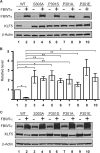
Effects of FBW7α on P301A and P301E KLF5 mutants. A–C, HEK (A and B) and HCT116 (C) cells were transfected with WT, S303A, P301S, P301A, or P301E KLF5 alone or in combination with FBW7α, and lysates were subjected to Western blotting with antibodies against FBW7α, HA (KLF5), and β-actin. A, a representative example of Western blot analysis using protein lysates from HEK cells. B, the relative protein levels of WT KLF5 were set as 1 (lane 1) and the other (lanes 2–10) are shown as relative to this one. Data represent mean ± S.D. (n = 3). *, p < 0.005, **, p < 0.05. C, a representative example of Western blot analysis using protein lysates from HCT116 cells.
FIGURE 8.
FIGURE 8.
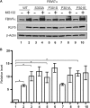
Decrease of FBW7α-mediated proteasomal degradation of P301A and P301E KLF5 mutants. HEK cells were transfected with WT, S303A, P301S, P301A, or P301E KLF5 constructs with FBW7α and treated with MG132 or vehicle. A, example of Western blot analysis using antibodies against FBW7α, HA (KLF5), and β-actin. B, the relative protein levels of WT KLF5 were set as 1 (lane 1), and the others (lanes 2–10) are shown as relative to this one. Data represent mean ± S.D. (n = 3). *, p < 0.05.
FIGURE 9.
FIGURE 9.
A model of the interference of the P301S, P301A, and P301E mutations with FBW7α-mediated proteasomal degradation of KLF5. A, serine 303 of WT KLF5 is phosphorylated by GSK3β, and FBW7α recognizes and binds to phosphorylated serine 303 and marks WT KLF5 for proteasomal degradation. aa, amino acid. B, mutated P301S, P301A, and P301E (P301S(A/E)) residues in KLF5 protein interfere with phosphorylation of serine 303 by GSK3β. Inefficient phosphorylation by GSK3β does not permit binding by FBW7α and inhibits efficacious degradation.

References

    1. McConnell B. B., Ghaleb A. M., Nandan M. O., Yang V. W. (2007) The diverse functions of Kruppel-like factors 4 and 5 in epithelial biology and pathobiology. BioEssays 29, 549–557 - PMC - PubMed
    1. McConnell B. B., Yang V. W. (2010) Mammalian Kruppel-like factors in health and diseases. Physiol. Rev. 90, 1337–1381 - PMC - PubMed
    1. McConnell B. B., Bialkowska A. B., Nandan M. O., Ghaleb A. M., Gordon F. J., Yang V. W. (2009) Haploinsufficiency of Kruppel-like factor 5 rescues the tumor-initiating effect of the ApcMin mutation in the intestine. Cancer Res. 69, 4125–4133 - PMC - PubMed
    1. Nandan M. O., Ghaleb A. M., McConnell B. B., Patel N. V., Robine S., Yang V. W. (2010) Kruppel-like factor 5 is a crucial mediator of intestinal tumorigenesis in mice harboring combined ApcMin and KRASV12 mutations. Mol. Cancer 9, 63. - PMC - PubMed
    1. Nandan M. O., Yoon H. S., Zhao W., Ouko L. A., Chanchevalap S., Yang V. W. (2004) Kruppel-like factor 5 mediates the transforming activity of oncogenic H-Ras. Oncogene 23, 3404–3413 - PMC - PubMed

Publication types

MeSH terms