Review
. 2014 Sep;33(2-3):527-43.
doi: 10.1007/s10555-013-9484-y.

Bone marrow fat: linking adipocyte-induced inflammation with skeletal metastases

Affiliations
Review

Bone marrow fat: linking adipocyte-induced inflammation with skeletal metastases

Aimalie L Hardaway et al. Cancer Metastasis Rev. 2014 Sep.

Abstract

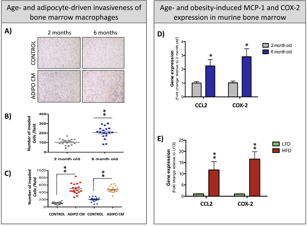
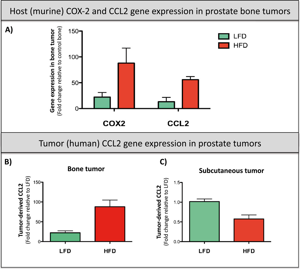
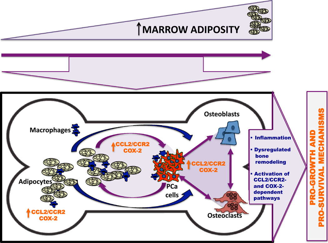
Adipocytes are important but underappreciated components of bone marrow microenvironment, and their numbers greatly increase with age, obesity, and associated metabolic pathologies. Age and obesity are also significant risk factors for development of metastatic prostate cancer. Adipocytes are metabolically active cells that secrete adipokines, growth factors, and inflammatory mediators; influence behavior and function of neighboring cells; and have a potential to disturb local milleu and dysregulate normal bone homeostasis. Increased marrow adiposity has been linked to bone marrow inflammation and osteoporosis of the bone, but its effects on growth and progression of prostate tumors that have metastasized to the skeleton are currently not known. This review focuses on fat-bone relationship in a context of normal bone homeostasis and metastatic tumor growth in bone. We discuss effects of marrow fat cells on bone metabolism, hematopoiesis, and inflammation. Special attention is given to CCL2- and COX-2-driven pathways and their potential as therapeutic targets for bone metastatic disease.


Figures

Fig. 1
Fig. 1
Age and adiposity increase bone marrow macrophage (BMM) invasiveness and induce COX2 and CCL2 expression in the bone marrow, a Images of invasion filters coated with collagen I matrix. Bone marrow macrophages from 2- and 6-month-old FVBN/N5 mice were plated on top of the filter (120,000 cells/filter) and allowed to invade toward control growth medium (control) or medium conditioned by bone marrow adipocytes (Adipo CM). Cells were allowed to invade for 24 h, and filters were fixed and stained with Diff-Quik dye. b, c Quantitation results showing numbers of invaded cells/filter +/− SD. b Comparative analysis of invasiveness of 2- and 6-month-old BMMs under control conditions; c comparative analysis of 2- and 6-month-old BMMs under control and Adipo CM conditions. Three independent biological replicates with six images/filter each were analyzed, d, e Taqman RT-PCR analysis of CCL2 (Macrophage Chemoattractant Protein; Life Technologies probe ID: Mm00441242_m1 and COX-2 (cyclooxygenase-2; Life Technologies probe ID: Mm00478374_m1) expression in murine bone marrow in a context of age (d) and obesity (e). d Two months old (gray) and 6 months old (blue). Data are shown as fold increases relative to 2-month-old bone marrow, e LFD (green )and HFD (red) mice. Data are shown as fold increases relative to LFD mice. All data are normalized to 18S (Life Technologies probe ID:Mm03928990_m1)
Fig. 2
Fig. 2
Host (murine) CCL2 and COX-2 and tumor (human) CCL2 are increased with obesity in PC3 prostate bone tumors. FVB/N/N5, Rag-1−/−mice caged in groups of four were started on either a LFD (10 % calories from fat; Research Diets no. D12450Bi) or a HFD (60 % calories from fat; Research Diets no. D12492i) at 5 weeks of age [152]. Mice were maintained on respective diets for 8 weeks prior to and 6 weeks following the PC3 tumor implantation into bone or subcutaneously (total of 14 weeks). a Taqman RT-PCR analysis of murine (host) CCL2 (Macrophage Chemoattractant Protein; Life Technologies probe ID: Mm00441242_m1) and COX-2 (cyclooxygenase 2; Life Technologies probe ID: Mm00478374_m1) in prostate bone tumors from LFD (green bars) and HFD (red bars) mice. Data were normalized to murine HPRT-1 (Life Technologies probe ID: Mm01545399_m1). b, c Taqman RT PCR analysis of human (tumor)-derived CCL2 (Life Technologies probe ID: Hs00234140_m1) in PC3 bone tumors (b) and PC3 subcutaneous tumors (c). Data were normalized to human HPRT1 (Life Technologies probe ID: Hs02800695_m1). At least three biological replicates were analyzed for each group, and data are shown as fold changes +/− SD
Fig. 3
Fig. 3
Proposed model of adiposity-induced inflammation and its effects on progression of metastatic tumors in bone. The increased amount of marrow fat content is a consequence of age and/or obesity. Prostate cancer cells that have colonized in adipocyte-rich bone marrow niche thrive and progress via CCL2/CCR2- and COX-2-dependent pathways due to (1) increased inflammation driven by adipocyte-macrophage interactions, (2) dysregulated bone remodeling via CCL2 and COX-2 actions on osteoclasts and osteoblasts, and (3) activation of pathways downstream of CCL2/CCR2 and COX-2 axes to promote tumor growth and survival

Similar articles

Cited by

References

    1. Lecka-Czernik B. Marrow fat metabolism is linked to the systemic energy metabolism. Bone. 2011;50(2):534–539. - PMC - PubMed
    1. Lee NK, Sowa H, Hinoi E, Ferron M, Ahn JD, Confavreux C, Dacquin R, Mee PJ, McKee MD, Jung DY, Zhang Z, Kim JK, Mauvais-Jarvis F, Ducy P, Karsenty G. Endocrine regulation of energy metabolism by the skeleton. Cell. 2007;130:456–469. - PMC - PubMed
    1. Weilbaecher KN, Guise TA, McCauley LK. Cancer to bone: a fatal attraction. Nature Reviews. 2011;11:411–425. - PMC - PubMed
    1. Roodman GD. Mechanisms of bone metastasis. New England Journal of Medicine. 2004;350:1655–1664. - PubMed
    1. Coleman RE. Metastatic bone disease: clinical features, pathophysiology and treatment strategies. Cancer Treatment Reviews. 2001;27:165–176. - PubMed

MeSH terms

Substances

LinkOut - more resources