. 2014 Mar;34(3):542-51.
doi: 10.1038/jcbfm.2013.233. Epub 2014 Jan 8.

Effects of the PPAR-α agonist fenofibrate on acute and short-term consequences of brain ischemia

Affiliations

Effects of the PPAR-α agonist fenofibrate on acute and short-term consequences of brain ischemia

Thavarak Ouk et al. J Cereb Blood Flow Metab. 2014 Mar.

Abstract

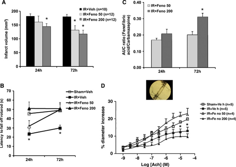
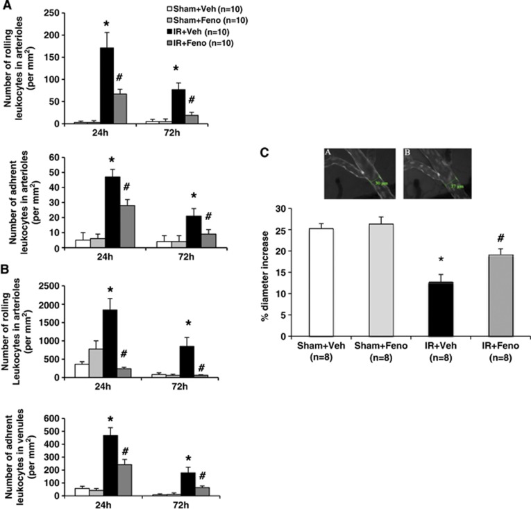
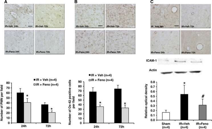
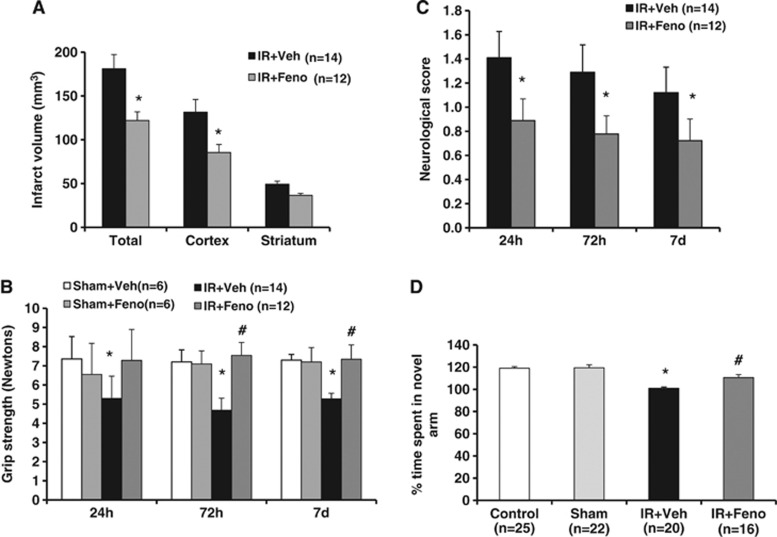
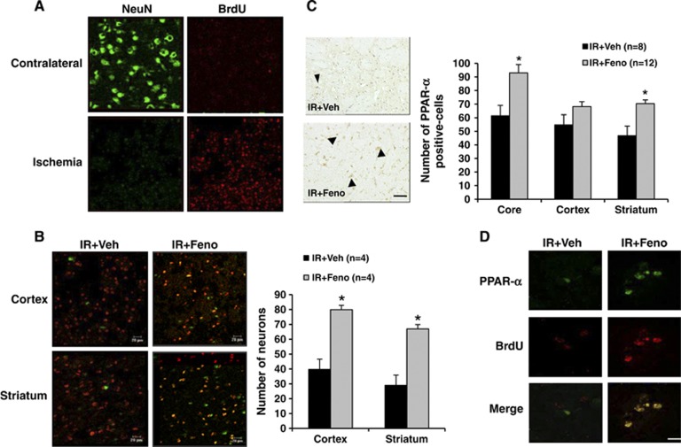
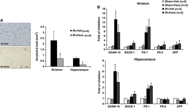
In stroke, there is an imperative need to develop disease-modifying drugs able to (1) induce neuroprotection and vasculoprotection, (2) modulate recovery and brain plasticity, and (3) limit the short-term motor and cognitive consequences. We hypothesized that fenofibrate, a peroxisome proliferator-activated receptor-α (PPAR-α) agonist, could exert a beneficial effect on immediate and short-term poststroke consequences related to its pleiotropic mechanisms. Rats or mice were subjected to focal ischemia to determine the effects of acute treatment by fenofibrate on (i) motor and memory impairment, (2) both cerebral and vascular compartments, (3) inflammation, (4) neurogenesis, and (5) amyloid cascade. We show that fenofibrate administration results in both neuronal and vascular protection and prevents the short-term motor and cognitive poststroke consequences by interaction with several mechanisms. Modulation of PPAR-α generates beneficial effects in the immediate poststroke consequences by mechanisms involving the interactions between polynuclear neutrophils and the vessel wall, and microglial activation. Fenofibrate modulates mechanisms involved in neurorepair and amyloid cascade. Our results suggest that PPAR-α agonists could check the key points of a potential disease-modifying effect in stroke.


Figures

Figure 1
Figure 1
Dose-effect study of fenofibrate. (A) Infarct volumes determined 24 and 72 hours after ischemia were lower in fenofibrate-treated rats (n=12) than in control rats (n=10). *P<0.05 vs. IR+Veh group. (B) The postischemia functional impairment (as evaluated in the rotarod test) was smaller in fenofibrate-treated rats (n=12) than in ischemic animals (n=10) 24 and 72 hours after the induction of cerebral ischemia. *P<0.05 vs. Sham+Veh group. (C) Plasmatic fenofibric acid increased in a dose-dependent manner at 24 and 72 hours. *P<0.05 vs. IR+Feno 50 group. (D) In vitro, the application of increasing concentrations of acetylcholine (ACh) to the middle cerebral artery led to endothelium-dependent relaxation, which was impaired in the aftermath of IR. This dysfunction was abolished in fenofibrate-treated rats at 50 mg/kg per day (n=4). *P<0.05 vs. Sham+Veh group. AUC, area under the curve; IR, ischemia/reperfusion.
Figure 2
Figure 2
Effects of treatment with the peroxisome proliferator-activated receptor-α (PPAR-α) agonist fenofibrate on interaction leukocyte–endothelium during the acute phase of cerebral ischemia and vascular reactivity in vivo. (A) In murine venules, fenofibrate treatment induced from 24 to 72 hours a significant decrease in leukocyte rolling and adhesion (n=10 per group); *P<0.05 vs. Sham+Veh group; #P<0.001 vs. IR+Veh group. (B) In murine arterioles, fenofibrate treatment induced from 24 to 72 hours a significant decrease in leukocyte rolling and adhesion (n=10 per group); *P<0.05 vs. Sham+Veh group; #P<0.001 vs. IR+Veh group. (C) In vivo, acetylcholine (ACh)-induced endothelium-dependent relaxation was determined by videomicroscopy of the murine pial arteries (A: the absence of ACh; B: the presence of ACh). The dysfunction in relaxation usually seen under ischemic conditions was abolished by fenofibrate treatment (n=8 per group). *P<0.05 vs. Sham+Veh group; #P<0.05 vs. IR+Veh group. Scale bar, 100 μm. IR, ischemia/reperfusion.
Figure 3
Figure 3
Effects of treatment with the peroxisome proliferator-activated receptor-α (PPAR-α) agonist fenofibrate on postischemic inflammation in rats. (A) Neutrophil infiltration was decreased after the induction of ischemia by fenofibrate treatment (n=4 per group); *P<0.05 vs. IR+Veh group. Scale bar, 100 μm. (B) Immunohistochemical quantification of the expression of Ox-42 revealed a significant decrease after fenofibrate treatment (n=4 per group); *P<0.05 vs. IR+Veh group. Scale bar, 25 μm. (C) Quantitative evaluation of the intercellular adhesion molecule-1 (ICAM-1) (using immunohistochemistry and western blotting) revealed a significant decrease in the expression of this protein during fenofibrate treatment (n=4 per group) after the induction of ischemia; *P<0.05 vs. Sham group; #P<0.05 vs. IR+Veh group. Scale bar, 100 μm. IR, ischemia/reperfusion.
Figure 4
Figure 4
Effects of delayed treatment with the peroxisome proliferator-activated receptor-α (PPAR-α) agonist fenofibrate on infarct volume, motor level, neurologic deficit, and memory recovery. (A) Delayed administration of fenofibrate enabled a reduction in infarct size. *P<0.05 vs. IR+Veh group. (B) Delayed and prolonged administration of fenofibrate induced motor improvements in grip strength test. *P<0.05 vs. Sham+Veh group; #P<0.05 vs. IR+Veh group. (C) Fenofibrate prevented neurologic deficits induced by ischemia. *P<0.05 vs. IR+Veh group. (D) The postischemia visuo-spatial memory impairment (as evaluated in Y-maze test) was prevented in fenofibrate-treated rats (n=16) than in ischemic animals (n=20). *P<0.05 vs. Sham group; #P<0.05 vs. IR+Veh group. IR, ischemia/reperfusion.
Figure 5
Figure 5
Evaluation 7 days after the induction of cerebral ischemia of the peroxisome proliferator-activated receptor-α (PPAR-α) agonist fenofibrate effects on neurogenesis in rat. (A) Cell proliferation occurred in the ischemic area as evidenced by a significant increase in the number of cells incorporating bromodeoxyuridine (BrdU) compared with contralateral area of the lesion induced by ischemia. Scale bar, 20 μm. (B) Some of these cells differentiate into neurons, as shown by BrdU and NeuN colabelling in the ischemic area. Fenofibrate administration significantly increased cell proliferation and the number of cells colabelled by BrdU and NeuN both in the striatum and in the cortex; *P<0.05 vs. IR+Veh group. Scale bar, 20 μm. (C) After 7 days of fenofibrate treatment, the number of PPAR-α-expressing cells was also significantly higher in the core of ischemia and in the striatum; *P<0.05 vs. IR+Veh group. (D) Fenofibrate administration significantly increased cell proliferation of cells colabelled by bromodeoxyuridine (BrdU) and PPAR-α. Scale bar, 10 μm.
Figure 6
Figure 6
Effects of treatment with the peroxisome proliferator-activated receptor-α (PPAR-α) agonist fenofibrate during the acute phase of cerebral ischemia on stroke-induced amyloid deposition and amyloid-related secretases. (A) Amyloid-β peptide deposition around the boundary of the ischemic focus was decreased 7 days after the induction of ischemia by fenofibrate treatment in the rat (n=4 per group); *P<0.05 vs. IR+Veh group. Scale bar, 25 μm. (B) Analysis of A Disintegrin And Metalloproteinase-10 (ADAM-10), BACE-1, PS-1, PS-2, and APP mRNA levels in whole brain lysates from the striatum or the hippocampus regions showed that ischemia/reperfusion induced a decrease in ADAM-10 and PS-1 mRNA level; *P<0.05 vs. Sham+Veh group. Nevertheless, fenofibrate administration did not significantly downregulate ADAM-10 and PS-1 mRNA while ADAM-10 inhibition was less important in fenofibrate-treated group. IR, ischemia/reperfusion.

References

    1. del Zoppo GJ. Stroke and neurovascular protection. N Engl J Med. 2006;354:553–555. - PubMed
    1. Garcia-Alloza M, Gregory J, Kuchibhotla KV, Fine S, Wei Y, Ayata C, et al. Cerebrovascular lesions induce transient β-amyloid deposition. Brain. 2011;134:3697–3707. - PMC - PubMed
    1. Iadecola C. Neurovascular regulation in the normal brain and in Alzheimer's disease. Nat Rev Neurosci. 2005;5:347–360. - PubMed
    1. Pluta R, Ułamek M, Jablonski M. Consideration of the ischemic basis and treatment of Alzheimer's disease. Folia Neuropathol. 2010;48:11–26. - PubMed
    1. Collino M, Patel NS, Thiemermann C. PPARs as new therapeutic targets for the treatment of cerebral ischaemia/reperfusion injury. Ther Adv Cardiovasc Dis. 2008;2:179–197. - PubMed

Publication types

MeSH terms

LinkOut - more resources