. 2014 Apr;34(4):621-9.
doi: 10.1038/jcbfm.2013.236. Epub 2014 Jan 8.

Pyroptotic neuronal cell death mediated by the AIM2 inflammasome

Affiliations

Pyroptotic neuronal cell death mediated by the AIM2 inflammasome

Stephanie E Adamczak et al. J Cereb Blood Flow Metab. 2014 Apr.

Erratum in

  • Corrigendum.
    [No authors listed] [No authors listed] J Cereb Blood Flow Metab. 2016 Feb;36(2):456. doi: 10.1177/0271678X15620125. J Cereb Blood Flow Metab. 2016. PMID: 26814199 Free PMC article.

Abstract

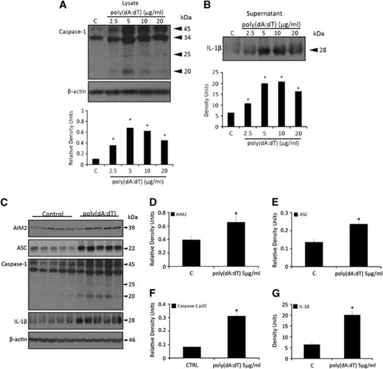
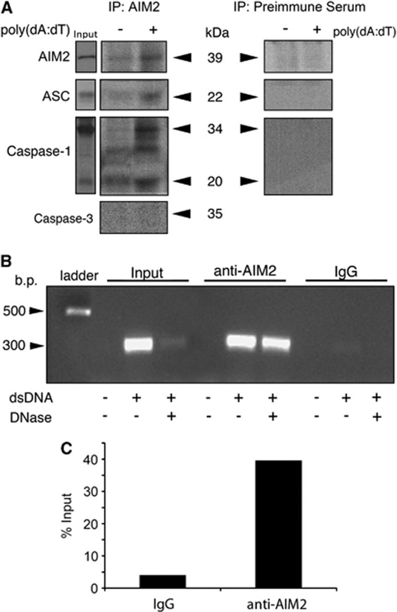
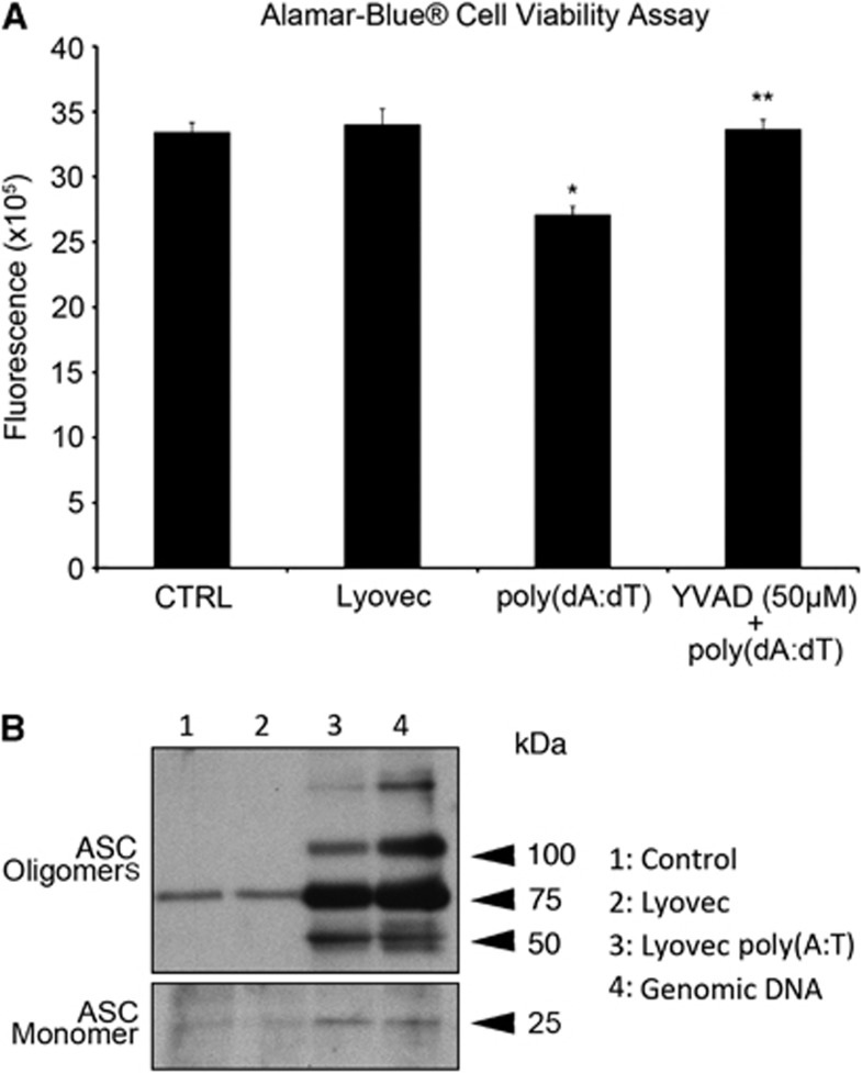
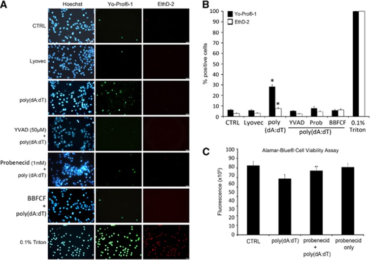
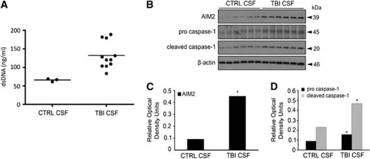
The central nervous system (CNS) is an active participant in the innate immune response to infection and injury. In these studies, we show embryonic cortical neurons express a functional, deoxyribonucleic acid (DNA)-responsive, absent in melanoma 2 (AIM2) inflammasome that activates caspase-1. Neurons undergo pyroptosis, a proinflammatory cell death mechanism characterized by the following: (a) oligomerization of apoptosis-associated speck-like protein containing a caspase recruitment domain (ASC); (b) caspase-1 dependency; (c) formation of discrete pores in the plasma membrane; and (d) release of the inflammatory cytokine interleukin-1β (IL-1β). Probenecid and Brilliant Blue FCF, inhibitors of the pannexin1 channel, prevent AIM2 inflammasome-mediated cell death, identifying pannexin1 as a cell death effector during pyroptosis and probenecid as a novel pyroptosis inhibitor. Furthermore, we show activation of the AIM2 inflammasome in neurons by cerebrospinal fluid (CSF) from traumatic brain injury (TBI) patients and oligomerization of ASC. These findings suggest neuronal pyroptosis is an important cell death mechanism during CNS infection and injury that may be attenuated by probenecid.


Figures

Figure 1
Figure 1
The AIM2 inflammasome ligand, poly(deoxyadenylic-deoxythymidylic) acid sodium salt (poly(dA:dT)), induces processing of caspase-1 and release of mature interleukin-1β (IL-1β) and expression of absent in melanoma 2 (AIM2) inflammasome proteins in embryonic cortical neurons. Immunoblot analysis of neurons stimulated with increasing doses of poly(dA:dT) for 3 hours. Densitometric analysis shows a dose-dependent effect of dsDNA on activation of caspase-1 in lysates (A) and release of IL-1β in supernatant (B). N=5 to 6 per group. Data were normalized to β-actin and are presented as mean+s.e.m. *P<0.05 compared with control. Poly(deoxyadenylic-deoxythymidylic) acid sodium salt induces expression of AIM2 inflammasome proteins. Immunoblots of neuronal lysates stimulated with 5.0 μg/mL poly(dA:dT) for 3 hours (C) show a significant increase in AIM2 (D), ASC (E), cleaved caspase-1 (F), and mature IL-1β (neuronal supernatants) (G) compared with unstimulated controls. β-actin was used as a control for protein loading. Data presented as mean±s.e.m. *P<0.05. N=5 to 6.
Figure 2
Figure 2
Absent in melanoma 2 (AIM2), ASC, and caspase-1 are present in embryonic cortical neurons and mediates the innate immune response to dsDNA. Panel A shows punctate immunoreactivity of caspase-1 in the neurites of poly(deoxyadenylic-deoxythymidylic) acid sodium salt (poly(dA:dT))-stimulated neurons but not in unstimulated neurons. Panel B shows oligomerization of AIM2 foci near the nucleus of dsDNA-stimulated neurons. Both panels A and B show ASC is expressed in the nucleus and cytoplasm of neurons. DAPI, 4′,6-diamidino-2-phenylindole.
Figure 3
Figure 3
Absent in melanoma 2 (AIM2) forms protein–protein interactions with ASC and caspase-1 in embryonic cortical neurons. There is an increase in active caspase-1 immunoprecipitated with anti-AIM2 in lysates stimulated with poly(deoxyadenylic-deoxythymidylic) acid sodium salt (poly(dA:dT)). Input:+poly(dA:dT). (A). In panel B neurons were unstimulated, stimulated with dsDNA (5 μg/mL), or stimulated with dsDNA followed by treatment with DNase I. Lysates (input) were immunoprecipitated with anti-AIM2 or immunoglobulin G (IgG). Electrophoresis of deoxyribonucleic acid (DNA) purified from the precipitated immune complex reveals the 300 bp fragment used to stimulate the neurons was precipitated with anti-AIM2, but not with IgG. The DNA fragment in the input was partly digested by DNase, but still precipitated with anti-AIM2 and not with IgG. Panel C shows % of input pulled down with IgG and anti-AIM2.
Figure 4
Figure 4
Activation of the AIM2 inflammasome induces neuronal pyroptosis. In panel A, embryonic cortical neurons were stimulated with poly(deoxyadenylic-deoxythymidylic) acid sodium salt (poly(dA:dT)) (5 μg/mL; 3 hours) in the presence or absence of YVAD, a caspase-1 inhibitor. There is approximately a 20% reduction in cell viability in neurons stimulated with poly(dA:dT) in the absence of YVAD. Pretreatment with YVAD blocks poly(dA:dT)-induced cell death. LyoVec alone does not decrease cell viability. Data presented as mean+/− s.e.m. *P<0.0001 compared with control (CTRL). **P<0.0001 compared with poly(dA:dT). N=12. In panel B, poly(dA:dT) also induced laddering of ASC, indicating formation of the pyroptosome, a supramolecular assembly of ASC dimers that activates caspase-1 and induces pyroptosis. Lane 4 shows genomic DNA induces oligomerization of ASC as well.
Figure 5
Figure 5
Neuronal pyroptosis is inhibited by probenecid and the pannexin1 inhibitor Brilliant Blue FCF (BB FCF). In panel A, embryonic neonatal cortical neurons were stimulated with poly(deoxyadenylic-deoxythymidylic) acid sodium salt (poly(dA:dT)) in the presence or absence of YVAD, probenecid, or BB FCF, and stained with a small (629 Da) membrane impermeable dye, YO-PRO-1, (green) and a larger (1,293 Da) membrane impermeable dye, ethidium homodimer-2 (Eth-D2), (red) to identify neurons with discrete membrane pores. Hoechst stain (blue) shows total cells in each field. Small, 1 to 2 nm, caspase-1-dependent pores form in a subpopulation (∼20%) of cortical neurons stimulated with poly(dA:dT). Selective inhibition of pannexin1 by either probenecid or BB FCF abolishes pore formation and uptake of the smaller dye after activation of the AIM2 inflammasome. Panel B shows percentage of cells that stained positive for YO-PRO-1 and Eth-D2 counted from four fields per well, six wells per group. Data presented as mean±s.e.m. *P<0.01 compared with control (CTRL). In panel C, embryonic cortical neurons were stimulated with poly(dA:dT) in the presence or absence of 1 mmol/L probenecid. There is approximately a 20% reduction in cell viability in neurons stimulated with poly(dA:dT). Pretreatment with probenecid inhibits poly(dA:dT)-induced cell death. Probenecid alone does not decrease cell viability. Data presented as mean±s.e.m. *P<0.01 compared with CTRL. **P<0.05 compared with poly(dA:dT). N=12.
Figure 6
Figure 6
Traumatic brain injury (TBI) results in release of cell-free nucleic acids into the cerebrospinal fluid (CSF) and assembly of the pyroptosome. Panel A shows dsDNA is elevated in the CSF of TBI patients compared with nontrauma controls (P=0.01). In panel B, immunoblots of neuronal lysates incubated in 50% media–50% CSF from a TBI patient for 24 hours show a significant increase in AIM2 expression (C) and cleavage of caspase-1 (D) compared with neurons incubated in 50% media–50% CSF from a nontrauma control patient. β-actin was used as protein loading control. Data presented as mean±s.e.m. *P<0.0001 compared with control. N=6.

References

    1. Fink SL, Cookson BT. Apoptosis, pyroptosis, and necrosis: mechanistic description of dead and dying eukaryotic cells. Infect Immun. 2005;73:1907–1916. - PMC - PubMed
    1. Martinon F, Burns K, Tschopp J. The inflammasome: a molecular platform triggering activation of inflammatory caspases and processing of proIL-beta. Mol Cell. 2002;10:417–426. - PubMed
    1. de Rivero Vaccari JP, Lotocki G, Alonso OF, Bramlett HM, Dietrich WD, Keane RW. Therapeutic neutralization of the NLRP1 inflammasome reduces the innate immune response and improves histopathology after traumatic brain injury. J Cereb Blood Flow Metab. 2009;29:1251–1261. - PMC - PubMed
    1. de Rivero Vaccari JP, Lotocki G, Marcillo AE, Dietrich WD, Keane RW. A molecular platform in neurons regulates inflammation after spinal cord injury. J Neurosci. 2008;28:3404–3414. - PMC - PubMed
    1. Halle M, Gabrielsen A, Paulsson-Berne G, Gahm C, Agardh HE, Farnebo F, et al. Sustained inflammation due to nuclear factor-kappa B activation in irradiated human arteries. J Am Coll Cardiol. 2010;55:1227–1236. - PubMed

Publication types

MeSH terms