. 2014 Jul;7(4):983-94.
doi: 10.1038/mi.2013.116. Epub 2014 Jan 8.

An altered intestinal mucosal microbiome in HIV-1 infection is associated with mucosal and systemic immune activation and endotoxemia

Affiliations

An altered intestinal mucosal microbiome in HIV-1 infection is associated with mucosal and systemic immune activation and endotoxemia

S M Dillon et al. Mucosal Immunol. 2014 Jul.

Abstract

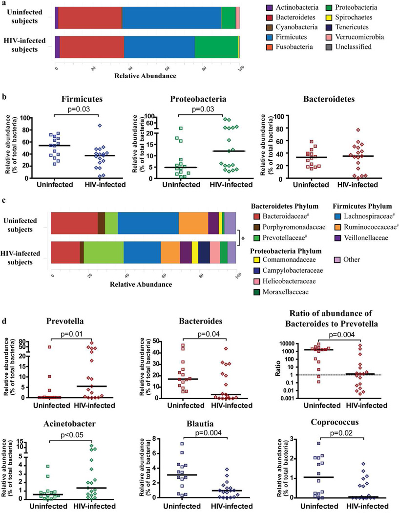
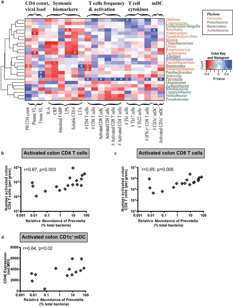
Human immunodeficiency virus-1 (HIV-1) infection disrupts the intestinal immune system, leading to microbial translocation and systemic immune activation. We investigated the impact of HIV-1 infection on the intestinal microbiome and its association with mucosal T-cell and dendritic cell (DC) frequency and activation, as well as with levels of systemic T-cell activation, inflammation, and microbial translocation. Bacterial 16S ribosomal DNA sequencing was performed on colon biopsies and fecal samples from subjects with chronic, untreated HIV-1 infection and uninfected control subjects. Colon biopsies of HIV-1-infected subjects had increased abundances of Proteobacteria and decreased abundances of Firmicutes compared with uninfected donors. Furthermore at the genus level, a significant increase in Prevotella and decrease in Bacteroides was observed in HIV-1-infected subjects, indicating a disruption in the Bacteroidetes bacterial community structure. This HIV-1-associated increase in Prevotella abundance was associated with increased numbers of activated colonic T cells and myeloid DCs. Principal coordinates analysis demonstrated an HIV-1-related change in the microbiome that was associated with increased mucosal cellular immune activation, microbial translocation, and blood T-cell activation. These observations suggest that an important relationship exists between altered mucosal bacterial communities and intestinal inflammation during chronic HIV-1 infection.


Conflict of interest statement

DISCLOSURE: The authors declare no conflict of interest.

Figures

Figure 1
Figure 1. Colonic mucosal dysbiosis in chronic, untreated HIV-1 infection is dominated by increased abundance of Prevotella
Bacterial taxa were identified in colon biopsies from 17 subjects with chronic, untreated HIV-1 infection and 14 uninfected control subjects using bacterial 16S ribosomal DNA sequencing. (a) Stacked bar representing the average relative abundance of the top 9 most abundant phyla within colon biopsies of uninfected and HIV-1 infected subjects with the average percentage of the 3 most dominant phyla shown. Bacteria that were not identified as belonging to any known phylum were termed “Unclassified”. Statistical analysis was performed using the adonis function of the vegan community ecology R package and no overall significant difference existed at the phylum level between uninfected and HIV-1 infected subjects (p=0.11). (b) Comparison of the relative abundance of the Firmicutes, Proteobacteria and Bacteroidetes phyla within uninfected and HIV-1 infected subjects. Values are shown as a fraction of the total bacteria detected within each individual. Lines represent the median value. Statistical analysis was performed using the Mann Whitney U test. (c) Stacked bar plot representing the average relative abundance of the top 10 most abundant bacterial families (from 91 total families detected) within colon biopsies of uninfected and HIV-1 infected subjects with a statistical difference observed in the overall average relative abundance between these two cohorts (*: p=0.02). #Statistical difference observed when the relative abundance of families within uninfected individuals was compared to HIV-1 infected individuals using the Mann-Whitney U test. (d) Plots showing individual relative abundance of various genera that were statistically different between uninfected and HIV-1 infected subjects assessed using the Mann-Whitney U test. Values are shown as a fraction of the total bacteria detected within each individual. Lines represent the median value. The ratio of the relative abundance of Bacteroides to Prevotella was calculated for each individual and plotted as shown. Dotted line designates a ratio of 1.0 (relative abundance of Bacteroides = relative abundance of Prevotella). False discovery rate corrections were not performed and P values are uncorrected.
Figure 2
Figure 2. Dietary habits of uninfected and HIV-1 infected subjects
(a,b) Uninfected and HIV-1 infected subjects completed a short questionnaire at the time of study visit detailing the average number of servings of various diet-related food groups per day or per week. Standardized values are shown as “number of servings per week” for both uninfected (closed symbols) and HIV-1 infected (open symbols) subjects. Lines represent median values. Statistical analysis was performed using the Mann Whitney U test with no comparisons reaching a statistically significant p value (p<0.05). (c,d) Association between the relative abundance of Bacteroides and number of servings of red meat per week in (c) uninfected subjects and (d) HIV-1 infected subjects. Statistical analysis was performed using the Spearman t test.
Figure 3
Figure 3. HIV-1 associated increase in the relative abundance of Prevotella is associated with colonic T cell and mDC activation
Associations between the top 25 most abundant genera within HIV-1 infected subjects with various clinical and immune parameters were assessed. (a) Heatmap representing positive (blue shading) and negative (red shading) associations between CD4 count, plasma and tissue viral load, mucosal T cell frequency, activation, T helper cell (Th)1, Th17 and Th22 and IFN-γ-producing CD8 T cell frequencies as well as CD1c+ mDC frequency and activation. Clustering was performed based on genera associations with the clinical and immune parameters. Genera are color-coded based on their respective phylum. #: number per gram of mucosal tissue; ^Percent of activated CD4+ or CD8+ T cells within viable, CD45+ cells. *p<0.05, Δp<0.06, ϕp<0.07, ψp<0.08. Genera that are altered in HIV-1 infected subjects relative to uninfected subjects are underlined. (b, c and d) Associations between the relative abundance of Prevotella and numbers of activated colon CD4 (b) and CD8 (c) T cells and CD1c+ mDC CD40 expression levels (d) in HIV-1 infected subjects (n=17 for colon T cell activation; n=13 for CD1c+ mDC). Statistical analysis was performed using the Spearman t test. False discovery rate corrections were not performed and P values are uncorrected.
Figure 4
Figure 4. Colonic mucosal dysbiosis is associated with chronic, untreated HIV-1 infection
(a) Principal coordinates (PC) plots demonstrating an association of PC1 values with HIV-1 infection. (b,c) Direct comparisons of PC1 (b) and PC2 (c) scores in uninfected and HIV-1 infected individuals. Lines represent the median value. Statistical analysis was performed using the Mann Whitney U test. (d,e) Heatmaps illustrating associations (positive: blue shading; negative: red shading) between PC1 and bacterial families (d) and CD4 count, plasma and tissue viral load (HIV-infected subjects only), mucosal T cell frequency, activation, T helper cell (Th)1, Th17 and Th22, IFN-γ-producing CD8 T cell frequencies, CD1c+ mDC frequency and activation and peripheral blood (PB) T cell activation (e). Families are color-coded based on their respective phylum. #Number per gram of mucosal tissue; ^Percent of activated CD4+ or CD8+ T cells within viable, CD45+ cells. Statistical analysis was performed using the Spearman t test. *p<0.05. Trending associations are noted as p=0.05. False discovery rate corrections were not performed and P values are uncorrected.

Similar articles

Cited by

References

    1. Kotler DP, Gaetz HP, Lange M, Klein EB, Holt PR. Enteropathy associated with the acquired immunodeficiency syndrome. Annals of internal medicine. 1984;101:421–428. - PubMed
    1. Epple HJ, et al. Acute HIV infection induces mucosal infiltration with CD4+ and CD8+ T cells, epithelial apoptosis, and a mucosal barrier defect. Gastroenterology. 2010;139:1289–1300. - PubMed
    1. Sankaran S, et al. Rapid onset of intestinal epithelial barrier dysfunction in primary human immunodeficiency virus infection is driven by an imbalance between immune response and mucosal repair and regeneration. Journal of virology. 2008;82:538–545. - PMC - PubMed
    1. Brenchley JM, et al. CD4+ T cell depletion during all stages of HIV disease occurs predominantly in the gastrointestinal tract. J Exp Med. 2004;200:749–759. - PMC - PubMed
    1. Brenchley JM, et al. Differential Th17 CD4 T-cell depletion in pathogenic and nonpathogenic lentiviral infections. Blood. 2008;112:2826–2835. - PMC - PubMed

Publication types

MeSH terms