Meta-Analysis
. 2015 Mar;220(2):1031-49.
doi: 10.1007/s00429-013-0698-0. Epub 2014 Jan 8.

Definition and characterization of an extended social-affective default network

Affiliations
Meta-Analysis

Definition and characterization of an extended social-affective default network

Maren Amft et al. Brain Struct Funct. 2015 Mar.

Abstract

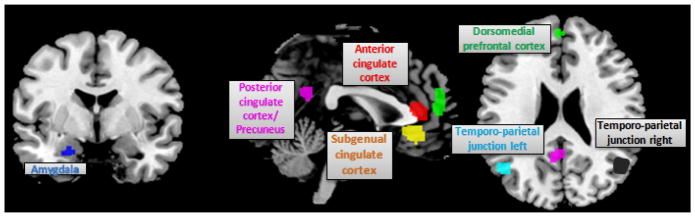
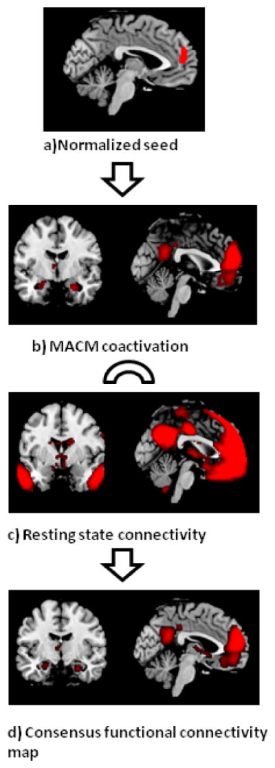
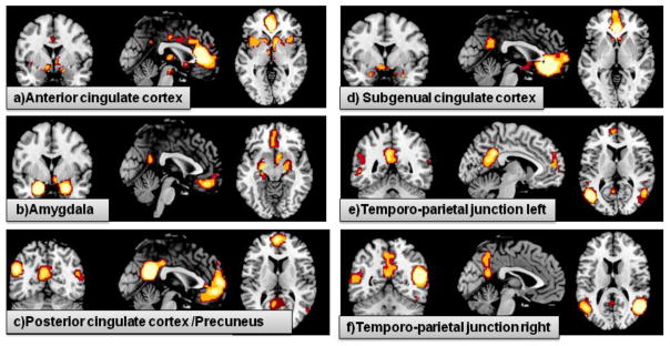
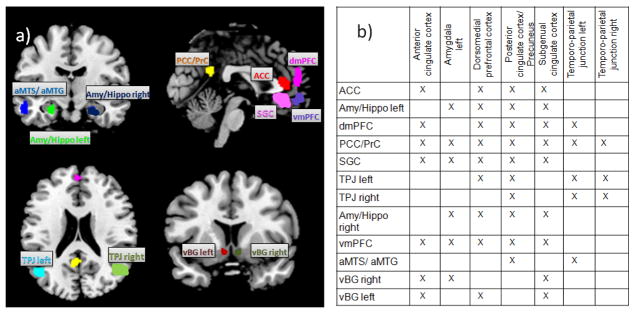
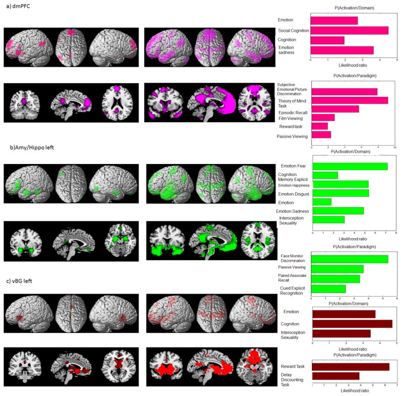
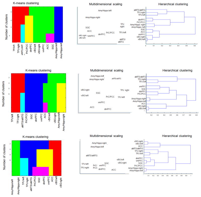
Recent evidence suggests considerable overlap between the default mode network (DMN) and regions involved in social, affective and introspective processes. We considered these overlapping regions as the social-affective part of the DMN. In this study, we established a robust mapping of the underlying brain network formed by these regions and those strongly connected to them (the extended social-affective default network). We first seeded meta-analytic connectivity modeling and resting-state analyses in the meta-analytically defined DMN regions that showed statistical overlap with regions associated with social and affective processing. Consensus connectivity of each seed was subsequently delineated by a conjunction across both connectivity analyses. We then functionally characterized the ensuing regions and performed several cluster analyses. Among the identified regions, the amygdala/hippocampus formed a cluster associated with emotional processes and memory functions. The ventral striatum, anterior cingulum, subgenual cingulum and ventromedial prefrontal cortex formed a heterogeneous subgroup associated with motivation, reward and cognitive modulation of affect. Posterior cingulum/precuneus and dorsomedial prefrontal cortex were associated with mentalizing, self-reference and autobiographic information. The cluster formed by the temporo-parietal junction and anterior middle temporal sulcus/gyrus was associated with language and social cognition. Taken together, the current work highlights a robustly interconnected network that may be central to introspective, socio-affective, that is, self- and other-related mental processes.


Conflict of interest statement

The authors declare no conflict of interest.

Figures

Figure 1
Figure 1. Seed Regions
This figure illustrates the seeds derived meta-analytically by Schilbach et al (2012) as being part of the DMN and additionally are involved in either social cognition or emotion. Seeds have been normalized using BrainMap database. They are displayed on coronal, sagittal and axial sections of the MNI single subject template.
Figure 2
Figure 2. Workflow from normalized region to consensus functional connectivity map of seeds
a Exemplary seed region in the dorso-medial prefrontal cortex b) For MACM-analysis we first identified all experiments in BrainMap database that reported activation within the seed and then tested for convergence across all foci reported in these experiments by using the revised ALE approach. Brain regions that show consistent MACM- coactivation with the dorsomedial prefrontal seed comprise precuneus, posterior cingulate cortex, amygdala/hippocampus, left temporo-parietal junction, anterior cingulate cortex, left ventral basalganglia and ventromedial prefrontal cortex. c) Significant resting-state functional connectivity with the dorsomedial prefrontal cortex was found extensively within the medial frontal lobe (including ventromedial prefrontal cortex, anterior cingulate cortex, subgenual cingulate cortex), as well as with the precuneus, posterior cingulum, thalamus, cerebellum, temporal pole and amygdala/hippocampus. d) The conjunction analysis of the task-dependent (MACM) and task-independent (resting-state) functional connectivity analyses resulted in a consensus functional connectivity map comprising the anterior cingulate cortex, subgenual cingulate cortex, precuneus, ventral basalganglia left, amygdala/hippocampus right, ventromedial prefrontal cortex and left temporo-parietal junction.
Figure 3
Figure 3. Consensus functional connectivity maps
This figure illustrates the conjunction of MACM and resting state connectivity maps. a) Consensus map of anterior cingulate cortex, comprises dorsomedial prefrontal cortex, gyrus cinguli, ventral basalganglia, ventromedial prefrontal cortex, precuneus, subgenual cingulate cortex b) Consensus map of amygdale, comprises contralateral amygdala/hippocampus, precuneus, right ventral basal ganglia, ventromedial prefrontal cortex, subgenual cingulate cortex c) Consensus map of posterior cingulate cortex/precuneus, comprises posterior cingulate cortex, amygdala/hippocampus, anterior cingulate cortex, subgenual cingulate cortex, dorsomedial prefrontal cortex, ventromedial prefrontal cortex, anterior middle temporal sulcus, temporo-parietal junction d) Consensus map of subgenual cingulate cortex, comprises precuneus, amygdala/hippocampus, anterior cingulate cortex, ventromedial prefrontal cortex, ventral basalganglia e) Consensus map of left temporo-parietal junction, comprises dorsomedial prefrontal cortex, precuneus, occipital lobe, right temporo-parietal junction, anterior middle temporal sulcus f) Consensus map of right temporo-parietal junction, comprises left temporo-parietal junction and precuneus
Figure 4
Figure 4. Constituent nodes of the eSAD
a) the 12 eSAD regions resulting from an overlap of multiple consensus maps are displayed on the MNI single subject template. b) Summary of the definition of the eSAD regions (rows) from overlap in the consensus functional connectivity maps of the seed regions from Schilbach et al., 2012 (columns).
Figure 5
Figure 5
Figure 5a–c. Functional characterization, MACM and resting state analysis of the eSAD regions The panel on left column presents results of MACM analysis on surface and orthogonal sections of the MNI template. The one in the middle column illustrates results of resting state analysis. The panel on right side illustrates the functional decoding of the regions. The behavioral domains are shown on top, whereas paradigm classes are provided below.
Figure 6
Figure 6. Clustering of eSAD regions
Illustration of the clustering results based on disparate approaches: non-hierarchical k-means clustering performed at different levels, hierarchical cluster analysis (HC, correlation-distance and complete linkage criteria) as well as multidimensional scaling for the projection of inter-regional differences in 2D space. Top row illustrates clustering referring to function Middle row shows clustering referring to similarities in whole-brain co-activation Bottom row illustrates clustering based on resting state connectivity.

References

    1. Adams RB, Jr, Rule NO, Franklin RG, Jr, Wang E, Stevenson MT, Yoshikawa S, Ambady N. Cross-cultural reading the mind in the eyes: an fMRI investigation. J Cogn Neurosci. 2010;22(1):97–108. doi: 10.1162/jocn.2009.21187. - DOI - PubMed
    1. Adolphs R, Cahill L, Schul R, Babinsky R. Impaired declarative memory for emotional material following bilateral amygdala damage in humans. Learn Mem. 1997;4(3):291–300. - PubMed
    1. Allman JM, Hakeem A, Erwin JM, Nimchinsky E, Hof P. The anterior cingulate cortex. The evolution of an interface between emotion and cognition. Ann N Y Acad Sci. 2001;935:107–117. - PubMed
    1. Amodio DM, Frith CD. Meeting of minds: the medial frontal cortex and social cognition. Nat Rev Neurosci. 2006;7(4):268–277. doi: 10.1038/nrn1884. - DOI - PubMed
    1. Andrews-Hanna JR, Reidler JS, Huang C, Buckner RL. Evidence for the default network’s role in spontaneous cognition. J Neurophysiol. 2010;104(1):322–335. doi: 10.1152/jn.00830.2009. - DOI - PMC - PubMed

Publication types

MeSH terms

LinkOut - more resources