. 2014 Jan 7;2(1):e141.
doi: 10.1038/mtna.2013.68.

RNA Interference by Single- and Double-stranded siRNA With a DNA Extension Containing a 3' Nuclease-resistant Mini-hairpin Structure

Affiliations

RNA Interference by Single- and Double-stranded siRNA With a DNA Extension Containing a 3' Nuclease-resistant Mini-hairpin Structure

Simon J Allison et al. Mol Ther Nucleic Acids. .

Abstract

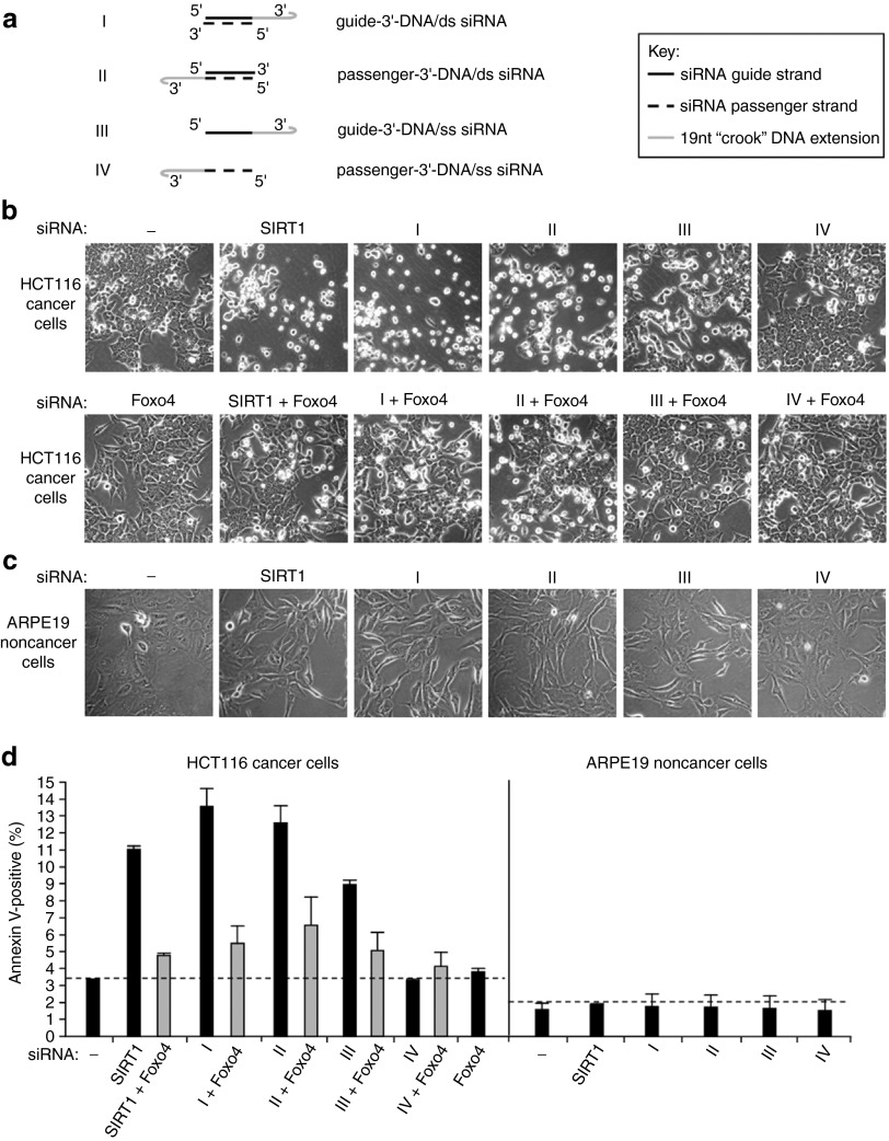
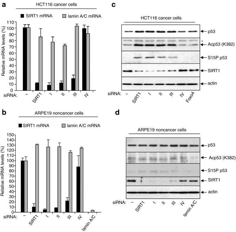
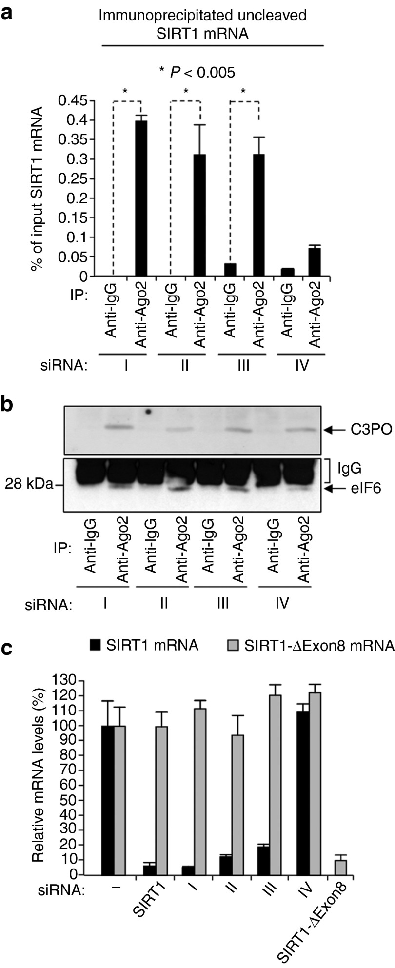
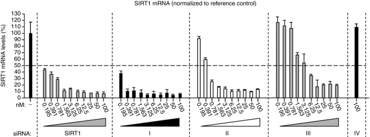
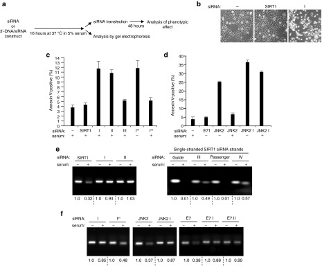
Selective gene silencing by RNA interference (RNAi) involves double-stranded small interfering RNA (ds siRNA) composed of single-stranded (ss) guide and passenger RNAs. siRNA is recognized and processed by Ago2 and C3PO, endonucleases of the RNA-induced silencing complex (RISC). RISC cleaves passenger RNA, exposing the guide RNA for base-pairing with its homologous mRNA target. Remarkably, the 3' end of passenger RNA can accommodate a DNA extension of 19-nucleotides without loss of RNAi function. This construct is termed passenger-3'-DNA/ds siRNA and includes a 3'-nuclease-resistant mini-hairpin structure. To test this novel modification further, we have now compared the following constructs: (I) guide-3'-DNA/ds siRNA, (II) passenger-3'-DNA/ds siRNA, (III) guide-3'-DNA/ss siRNA, and (IV) passenger-3'-DNA/ss siRNA. The RNAi target was SIRT1, a cancer-specific survival factor. Constructs I-III each induced selective knock-down of SIRT1 mRNA and protein in both noncancer and cancer cells, accompanied by apoptotic cell death in the cancer cells. Construct IV, which lacks the SIRT1 guide strand, had no effect. Importantly, the 3'-DNA mini-hairpin conferred nuclease resistance to constructs I and II. Resistance required the double-stranded RNA structure since single-stranded guide-3'-DNA/ss siRNA (construct III) was susceptible to serum nucleases with associated loss of RNAi activity. The potential applications of 3'-DNA/siRNA constructs are discussed.Molecular Therapy-Nucleic Acids (2014) 2, e141; doi:10.1038/mtna.2013.68; published online 7 January 2014.


Figures

Figure 1
Figure 1
Phenotypic effects of 3′-DNA/SIRT1 siRNA constructs I–IV on HCT116 cancer cells and ARPE19 noncancer cells. (a) Schematical representation of the four different 3′-DNA/SIRT1 siRNA constructs that are used in this study and compared throughout to standard unmodified SIRT1 siRNA. I = double-stranded SIRT1 siRNA with a 19 nucleotide “crook” DNA extension on the 3′ end of the siRNA guide strand; II = double-stranded SIRT1 siRNA with the 19nt DNA extension on the 3′ end of the siRNA passenger strand; III = single-stranded SIRT1 siRNA (guide strand) with the 19nt DNA extension on the 3′ end of the siRNA guide strand; IV = single-stranded SIRT1 siRNA (passenger strand) with the DNA extension on the 3′ end of the siRNA passenger strand (b) Phase contrast micrograph images of HCT116 cells 48 hours after transfection with 3′-DNA/SIRT1 siRNA constructs or standard SIRT1 siRNA (upper panel) or following cotransfection of cells with Foxo4 siRNA (lower panel) as indicated. Untreated control = (−). (c) Phase contrast images of ARPE19 cells 72 hours after transfection with the indicated 3′-DNA/SIRT1 siRNA constructs or standard SIRT1 siRNA. (d) Quantification by annexin V-labeling of the apoptotic phenotype induced by 3′-DNA/SIRT1 siRNA constructs 48 hours after transfection of HCT116 cells and its rescue by Foxo4 cosilencing (left). Apoptotic levels in ARPE19 cells as determined by annexin V-labeling 72 hours after transfection of ARPE19 cells (right). Background levels of apoptosis are indicated by the dotted line.
Figure 2
Figure 2
Selective knock-down of SIRT1 mRNA and protein by 3′-DNA/SIRT1 siRNA constructs I–III in HCT116 and ARPE19 cells. Levels of SIRT1 mRNA and lamin A/C mRNA in (a) HCT116 cells and (b) ARPE19 cells 48 hours after transfection with the indicated 3′-DNA/SIRT1 siRNA constructs; quantitative RT-PCR data are shown, mean ± SD of four independent mRNA determinations. Immunoblots showing the effects of the 3′-DNA/SIRT1 siRNA constructs on SIRT1 protein levels in (c) HCT116 cells and (d) ARPE19 cells. Actin shows equivalent total protein loading. Effects on p53 protein and p53 acetylation and phosphorylation at specific sites is also shown (see text for details; *= nonspecific band).
Figure 3
Figure 3
Coimmunoprecipitation of endogenous SIRT1 mRNA with Ago2 protein in HCT116 cells transfected with 3′-DNA/SIRT1 siRNA constructs. (a) Levels of SIRT1 mRNA immunoprecipitated with Ago2 antibody or normal mouse IgG control antibody. Immunoprecipitated SIRT1 mRNA levels are expressed as a percentage of the input SIRT1 mRNA levels. HCT116 cell extracts for the immunoprecipitations were prepared 24 hours after transfection with the indicated 3′-DNA/SIRT1 siRNA constructs (see Materials and Methods). (b) Immunoblots showing coimmunoprecipitation of C3PO and eIF6 with Ago2 in cells transfected with 3′-DNA/SIRT1 siRNA constructs. eIF6 (arrowed) runs just ahead of IgG light chain (indicated). (c) Levels of full-length SIRT1 mRNA and splice variant SIRT1-ΔExon8 mRNA in HCT116 cells 48 hours after transfection with the indicated siRNAs or 3′-DNA/SIRT1 siRNA constructs; quantitative RT-PCR data are shown, mean ± SD of four independent mRNA determinations.
Figure 4
Figure 4
Dose responses for SIRT1 mRNA knock-down by SIRT1 siRNA and 3′-DNA/SIRT1 siRNA constructs. SIRT1 mRNA levels in HCT116 cells 48 hours after transfection with indicated concentrations of the 3′-DNA/SIRT1 siRNA constructs (100 nmol/l to 195 pmol/l, twofold dilution series). SIRT1 mRNA knock-down by standard SIRT1 siRNA was also assessed for comparison of efficacy. mRNA levels determined by quantitative RT-PCR; mean ± SD of four independent mRNA determinations. The SIRT1 mRNA results were normalized to lamin A/C mRNA in each individual sample.
Figure 5
Figure 5
Comparison of the susceptibility of unmodified siRNAs and 3′-DNA/siRNA constructs to degradation by serum nucleases. (a) Schematic indicating the dual experimental approach used to assess the stability of unmodified siRNAs and 3′-DNA/siRNA constructs in serum. Analysis of the phenotypic effect 48 hours after transfection provides a readout of whether RNAi functionality is retained or lost. Visualization by gel electrophoresis allows levels of degradation of the siRNAs or constructs to be directly compared. (b) Phase contrast images of HCT116 cells 48 hours after transfection with serum-preincubated SIRT1 siRNA or serum-preincubated 3′-DNA/SIRT1 siRNA construct I. (c) Quantification by annexin V-labeling of apoptosis induced by SIRT1 siRNA and 3′-DNA/SIRT1 siRNA constructs I–III after 16 hours incubation in 5% serum prior to transfection. Apoptotic levels of HCT116 cells were determined 48 hours after transfection. Mean ± SD of three independent experiments. Induction of apoptosis by a novel 3′-DNA/SIRT1 siRNA construct that has a different 3′ 19 nucleotide DNA extension on the guide strand (Im) was also tested, both with no serum preincubation (−) and after serum preincubation (+). (d) Levels of apoptosis induced in HCT116 cells 48 hours after transfection with unmodified JNK2 siRNA or 3′-DNA/JNK2 siRNA construct I (JNK2 I; see Materials and Methods). The effects of no preincubation in serum (−) and 16 hours preincubation in 5% serum (+) are compared. The effects of transfection of 3′-DNA/E7 siRNA construct I (E7 I; see Materials and Methods) are also shown (no serum preincubation), which failed to induce apoptosis, as expected since HCT116 cells do not express HPV E7 mRNA (see also ref. ). (e) Gel electrophoresis analysis of SIRT1 siRNA and 3′-DNA/SIRT1 siRNA constructs I–IV following incubation of equimolar amounts in no serum (−) or 5% serum (+) for 16 hours. (f) Effect of 16 hours incubation in no serum (−) or 5% serum (+) on levels of the indicated unmodified siRNAs or 3′-DNA/siRNA constructs. The stability of 3′-DNA/SIRT1 siRNA construct I and that of 3′-DNA/SIRT1 siRNA construct Im which differs only in the sequence of the 19 nucleotide DNA extension were compared. Unmodified JNK2 siRNA versus 3′-DNA/JNK2 siRNA construct I (JNK2 I); unmodified E7 siRNA versus 3′-DNA/E7 siRNA constructs I and II (E7 I; E7 II; see Materials and Methods).

References

    1. Siomi H, Siomi MC. On the road to reading the RNA-interference code. Nature. 2009;457:396–404. - PubMed
    1. Carthew RW, Sontheimer EJ. Origins and Mechanisms of miRNAs and siRNAs. Cell. 2009;136:642–655. - PMC - PubMed
    1. Liu J, Carmell MA, Rivas FV, Marsden CG, Thomson JM, Song JJ, et al. Argonaute2 is the catalytic engine of mammalian RNAi. Science. 2004;305:1437–1441. - PubMed
    1. Song JJ, Smith SK, Hannon GJ, Joshua-Tor L. Crystal structure of Argonaute and its implications for RISC slicer activity. Science. 2004;305:1434–1437. - PubMed
    1. Rivas FV, Tolia NH, Song JJ, Aragon JP, Liu J, Hannon GJ, et al. Purified Argonaute2 and an siRNA form recombinant human RISC. Nat Struct Mol Biol. 2005;12:340–349. - PubMed