

Review
doi: 10.1161/STROKEAHA.113.002491.
Epub 2014 Jan 7.
Evaluating intracranial atherosclerosis rather than intracranial stenosis
Affiliations
- PMID: 24399377
- PMCID: PMC3957430
- DOI: 10.1161/STROKEAHA.113.002491
Item in Clipboard
Review
Evaluating intracranial atherosclerosis rather than intracranial stenosis
Stroke.
2014 Feb.
No abstract available
Keywords: collateral circulation; fractional flow reserve, myocardial; intracranial arteriosclerosis; neuroimaging.
Figures
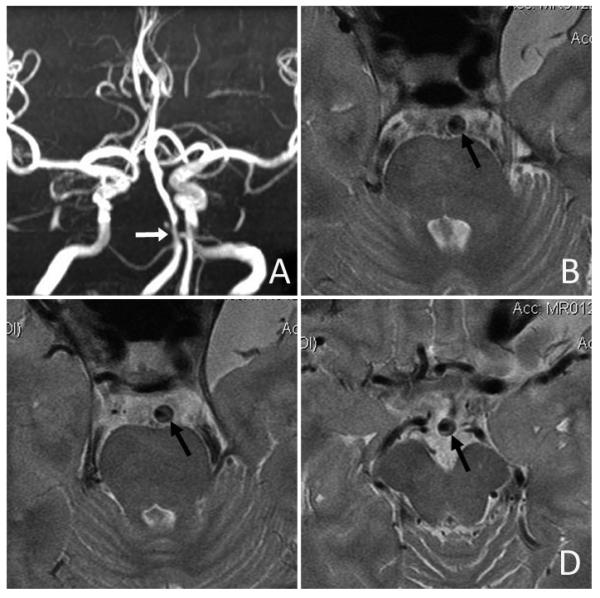
Intracranial plaque and arterial wall imaging by high-resolution MRI. An ICAS lesion located at proximal basilar artery with severe luminal stenosis was identified on time-of-flight MRA (white arrow in Panel A). High-resolution MRI revealed an eccentric atherosclerotic plaque along the anterolateral and posterolateral walls of basilar artery (black arrows in Panels B, C and D). (Courtesy of Professor WH Xu of Peking Union Medical College Hospital, Beijing, China.)
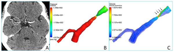
CTA source image (A) showing a right middle cerebral artery stenosis, and the reconstructed CFD models illustrating pressure (B) and flow velocity (C) changes across the lesion. Decreased pressure (B) and increased flow velocity (C) in situ and downstream to the lesion are highlighted with arrows on the CFD models.
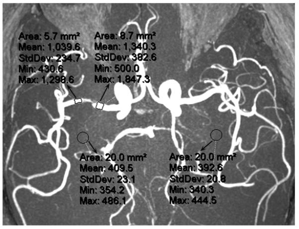
The method for measurement of SIR of an ICAS on a MRA maximum intensity projection. SIR of the lesion at right middle cerebral artery is calculated as the ratio of mean signal intensities distal (1,039.6) and proximal (1,340.3) to the lesion, adjusted by the mean background signal intensity (401.1; mean of 409.5 and 392.6), which is (1,039.6 – 401.1) / (1,340.3 – 401.1) = 0.68.
Similar articles
-
Noninvasive imaging is improving but digital subtraction angiography remains the gold standard.Neurology. 2007 Jun 12;68(24):2057-8. doi: 10.1212/01.wnl.0000268580.86336.af. Neurology. 2007. PMID: 17562825 No abstract available.
-
Velocity criteria for intracranial stenosis revisited: an international multicenter study of transcranial Doppler and digital subtraction angiography.Stroke. 2011 Dec;42(12):3429-34. doi: 10.1161/STROKEAHA.111.621235. Epub 2011 Sep 29. Stroke. 2011. PMID: 21960567
-
Transcranial color-coded duplex sonography, magnetic resonance angiography, and computed tomography angiography: methods, applications, advantages, and limitations.J Clin Ultrasound. 1995 Feb;23(2):89-111. doi: 10.1002/jcu.1870230205. J Clin Ultrasound. 1995. PMID: 7699104 Review.
-
Significance of good collateral compensation in symptomatic intracranial atherosclerosis.Cerebrovasc Dis. 2012;33(6):517-24. doi: 10.1159/000337332. Epub 2012 Apr 25. Cerebrovasc Dis. 2012. PMID: 22538868
-
MR arteriography of intracranial circulation.AJR Am J Roentgenol. 1998 Dec;171(6):1469-78. doi: 10.2214/ajr.171.6.9843273. AJR Am J Roentgenol. 1998. PMID: 9843273 Review. No abstract available.
Cited by
-
Functional assessment of cerebral artery stenosis: A pilot study based on computational fluid dynamics.J Cereb Blood Flow Metab. 2017 Jul;37(7):2567-2576. doi: 10.1177/0271678X16671321. Epub 2016 Jan 1. J Cereb Blood Flow Metab. 2017. PMID: 27702878 Free PMC article.
-
High-resolution magnetic resonance imaging features of time-of-flight magnetic resonance angiography signal loss and its relevance to ischemic stroke.Quant Imaging Med Surg. 2024 Sep 1;14(9):6820-6829. doi: 10.21037/qims-24-329. Epub 2024 Aug 29. Quant Imaging Med Surg. 2024. PMID: 39281140 Free PMC article.
-
Transcranial Doppler Ultrasonography as a Diagnostic Tool for Cerebrovascular Disorders.Front Hum Neurosci. 2022 Apr 29;16:841809. doi: 10.3389/fnhum.2022.841809. eCollection 2022. Front Hum Neurosci. 2022. PMID: 35572008 Free PMC article. Review.
-
Noninvasive quantification of cerebrovascular pressure changes using 4D Flow MRI.Magn Reson Med. 2021 Dec;86(6):3096-3110. doi: 10.1002/mrm.28928. Epub 2021 Aug 25. Magn Reson Med. 2021. PMID: 34431550 Free PMC article.
-
Association of Lp-PLA2 Mass and Aysmptomatic Intracranial and Extracranial Arterial Stenosis in Hypertension Patients.PLoS One. 2015 Jun 22;10(6):e0130473. doi: 10.1371/journal.pone.0130473. eCollection 2015. PLoS One. 2015. PMID: 26098634 Free PMC article.
References
-
- Qureshi AI, Feldmann E, Gomez CR, Johnston SC, Kasner SE, Quick DC, et al. Intracranial atherosclerotic disease: an update. Ann Neurol. 2009;66:730–738. - PubMed
-
- Chimowitz MI, Lynn MJ, Howlett-Smith H, Stern BJ, Hertzberg VS, Frankel MR, et al. Comparison of warfarin and aspirin for symptomatic intracranial arterial stenosis. N Engl J Med. 2005;352:1305–1316. - PubMed
-
- Feldmann E. Diagnosis and quantitation of intracranial stenosis. J Neuroimaging. 2009;19(Suppl 1):22S–24S. - PubMed
-
- Kasner SE, Chimowitz MI, Lynn MJ, Howlett-Smith H, Stern BJ, Hertzberg VS, et al. Predictors of ischemic stroke in the territory of a symptomatic intracranial arterial stenosis. Circulation. 2006;113:555–563. - PubMed
Publication types
MeSH terms
Grants and funding
LinkOut - more resources
Full Text Sources
Other Literature Sources
Medical
