. 2014 Feb;5(1):113-22.
doi: 10.1007/s13244-013-0302-4. Epub 2014 Jan 8.

Magnetic resonance imaging of meningiomas: a pictorial review

Affiliations

Magnetic resonance imaging of meningiomas: a pictorial review

J Watts et al. Insights Imaging. 2014 Feb.

Abstract

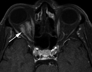
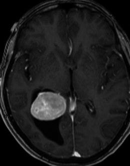
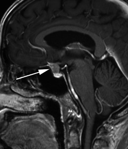
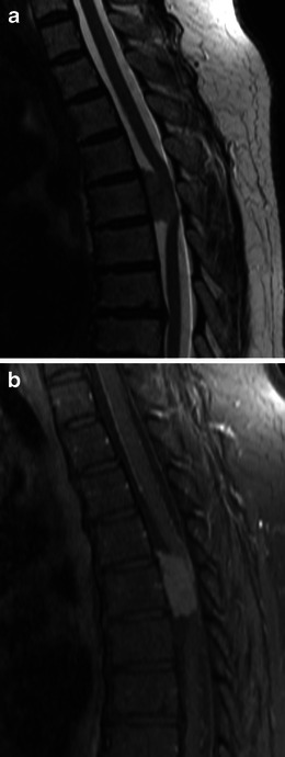
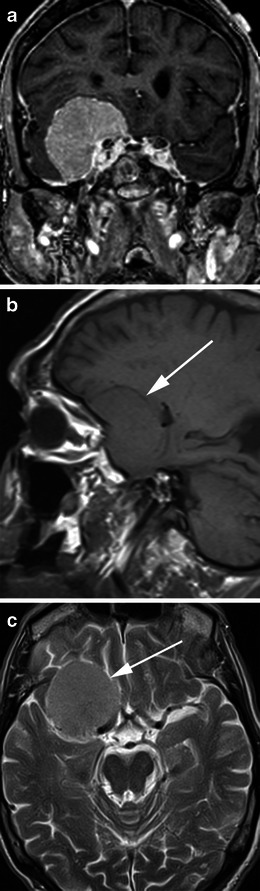
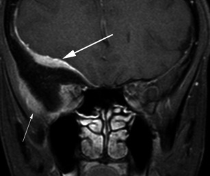
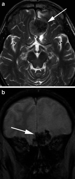
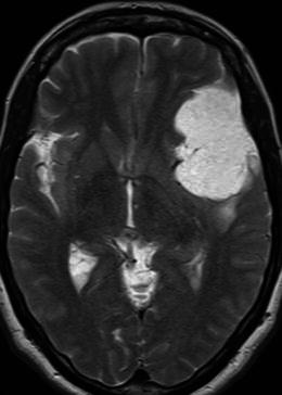
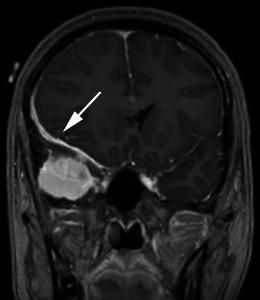
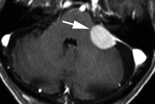
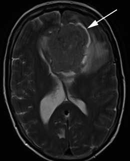
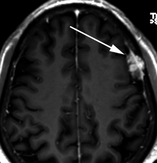
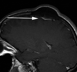
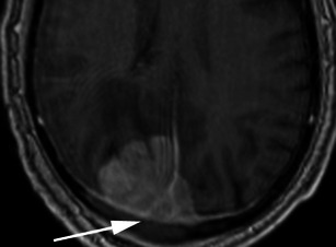
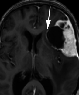
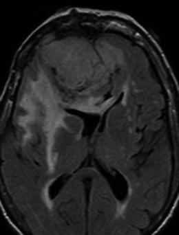
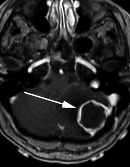
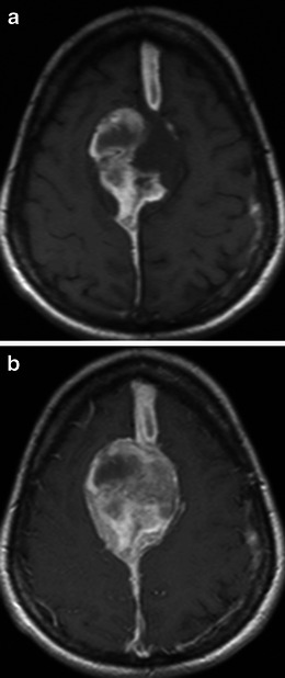
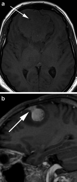
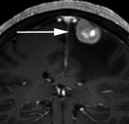
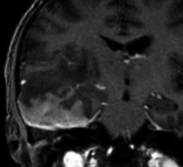
Meningiomas are the most common non-glial tumour of the central nervous system (CNS). There are a number of characteristic imaging features of meningiomas on magnetic resonance imaging (MRI) that allow an accurate diagnosis, however there are a number of atypical features that may be diagnostically challenging. Furthermore, a number of other neoplastic and non-neoplastic conditions may mimic meningiomas. This pictorial review discusses the typical and atypical MRI features of meningiomas and their mimics.

Teaching points: There are several characteristic features of meningiomas on MRI that allow an accurate diagnosis Some meningiomas may display atypical imaging characteristics that may be diagnostically challenging Routine MRI sequences do not reliably distinguish between benign and malignant meningiomas Spectroscopy and diffusion tensor imaging may be useful in the diagnosis of malignant meningiomas A number of conditions may mimic meningiomas; however, they may have additional differentiating features.


Figures

Fig. 1
Fig. 1
Post-contrast axial T1-weighted sequence in a 64-year-old man with proptosis demonstrates a homogeneously enhancing, intraconal mass within the right orbit (arrow) that partially encircles the right optic nerve. Smooth dural thickening and enhancement extends posteriorly to the optic canal. The imaging appearance is consistent with a meningioma
Fig. 2
Fig. 2
Post-contrast axial T1-weighted sequence in a 62-year-old woman with headaches demonstrates a large, homogeneously enhancing mass within the trigone of the right lateral ventricle associated with ventricular dilatation. Histology was that of a meningioma (grade I)
Fig. 3
Fig. 3
Post-contrast sagittal T1-weighted sequence in a 52-year-old woman with reduced vision in the left eye demonstrates a homogeneously enhancing suprasellar mass (arrow) extending inferiorly into the superior aspect of the sella where it contacts the brightly enhancing anterior pituitary. The mass results in mild elevation of the left side of the optic chiasm
Fig. 4
Fig. 4
a Sagittal T2-weighted sequence in a 68-year-old woman with mild weakness and dysaesthesia of the left leg demonstrates an intradural, extramedullary mass anteriorly at T5, which displaces the spinal cord posteriorly, resulting in severe narrowing of the central spinal canal. b Post-contrast sagittal T1-weighted sequence in the same patient as a demonstrates homogeneous enhancement. The histological diagnosis at surgery was a meningioma
Fig. 5
Fig. 5
a Post-contrast coronal T1-weighted sequence in an 80-year-old woman with a 1-month history of confusion demonstrates a well-circumscribed, homogenously enhancing mass within the floor right middle cranial fossa with a broad dural tail that inferomedially contacts the right cavernous sinus. The right temporal lobe is compressed superiorly by the mass without invasion. The lesion was proven at surgery to be a meningioma. b Sagittal T1-weighted sequence in the same patient as a demonstrates the mass (arrow) to be isointense to grey matter and to extend anteriorly along the posterior aspect of the anterior cranial fossa floor. c Axial T2-weighted sequence in the same patient as a demonstrates the mass (arrow) to be mildly, homogeneously hyperintense to grey matter
Fig. 6
Fig. 6
Post-contrast coronal T1-weighted sequence in a 33-year-old woman who presents with right proptosis demonstrates extensive dural thickening and enhancement along the greater wing of sphenoid consistent with an en plaque meningioma (large arrow). The lesion results in marked hyperostosis of the greater wing of sphenoid with resultant indentation of the right temporal lobe. Further enhancing tumour is identified external to the calvarium between the greater wing of sphenoid and the temporalis muscle (small arrow)
Fig. 7
Fig. 7
a Axial T2-weighted sequence in a 75-year-old man being investigated for Bell palsy demonstrates a heterogeneous, extra axial mass in the midline of the olfactory groove, left more than right, in keeping with a meningioma (arrow) associated with mild oedema within the left frontal lobe. There are intrinsic areas of low signal intensity in keeping with calcification that do not enhance on post contrast sequences. b Coronal T2* sequence in the same patient as a demonstrates susceptibility effect (arrow) within the mass in keeping with the presence of calcification
Fig. 8
Fig. 8
Axial T2-weighted sequence in a 55-year-old woman with an 8-year history of syncopal attacks demonstrates a large, extra-axial mass over the inferolateral left frontal lobe and anterior temporal lobe that is markedly hyperintense, slightly more so than CSF. There is mild vasogenic oedema present posterior to the mass. Histology was consistent with a microcystic meningioma
Fig. 9
Fig. 9
Post-contrast coronal T1-weighted sequence in a 35-year-old woman with headaches demonstrates a homogenously enhancing extra axial mass within the right middle cranial fossa with a broad dural tail (arrow) extending along the sphenoid wing and around the lateral aspect of the right frontal lobe. The mass results in subfalcine herniation to the left by 7 mm and effacement of the right lateral ventricle. Histology at surgery was a meningioma (grade 1)
Fig. 10
Fig. 10
Post-contrast axial T1-weighted sequence in an 83-year-old man with dizziness and falls demonstrates a homogeneously enhancing, extra-axial mass within the left cerebellopontine angle (arrow) with a broad dural attachment and a dural tail extending anterior to the left internal acoustic meatus and posteriorly to the sigmoid sinus. The mass overlies the left internal acoustic meatus without intracanalicular extension. Appearance and histology are consistent with a meningioma (grade I)
Fig. 11
Fig. 11
Axial T2-weighted sequence in a 57-year-old woman with confusion demonstrates a large extra-axial mass centred on the falx that is isointense to grey matter with a prominent surrounding CSF signal intensity cleft between the mass and the adjacent frontal cortex, best appreciated around the left aspect (arrow). The diagnosis was meningioma (grade I) at histology
Fig. 12
Fig. 12
Post-contrast axial T1-weighted MRI in a 73-year-old man with headaches demonstrates a mildly heterogeneously enhancing extra-axial mass over the left lateral convexity (arrow) consistent with a meningioma at histology that invades the inner table and diploic space of the left frontal bone. The outer table is contacted and thinned without breach
Fig. 13
Fig. 13
Post-contrast sagittal T1-weighted sequence in a 49-year-old woman with a long-standing palpable lump demonstrates a pronounced hyperostotic focus within the left parietal bone with the bulk protruding extracranially. Immediately below the bone lesion there is a thin sheet of homogenously enhancing extra-axial soft tissue (arrow) consistent with an en plaque meningioma that was proved at surgery
Fig. 14
Fig. 14
Post-contrast axial T1-weighted sequence in a 79-year-old man demonstrates a large, homogeneously enhancing extra axial mass within the right occipital lobe that extends into and expands the posterior aspect of the superior sagittal sinus (arrow) and crosses the midline into the paramedian left occipital lobe. Posteriorly, the mass extends into the adjacent skull with erosion of the outer table. Histology at surgery was a meningioma (grade III)
Fig. 15
Fig. 15
Post-contrast axial T1-weighted sequence in an 80-year-old woman with confusion demonstrates a solid and cystic, extra-axial mass overlying the left frontal lobe with a fine rim of enhancement (arrow) around the largest cystic component consistent with a peritumoural cyst. The homogeneously enhancing solid component demonstrated the typical isointensity to grey matter on the T1- and T2-weighted sequences of a meningioma
Fig. 16
Fig. 16
Axial FLAIR sequence in an 81-year-old woman with confusion and falls demonstrates a large extra-axial mass that is isointense to grey matter and is predominantly located within the right frontal lobe but crosses the midline falx anteriorly to indent the left frontal lobe. It induces substantial vasogenic oedema within the right frontal lobe, around the right basal ganglia and through the genu of the corpus callosum to the left forceps minor
Fig. 17
Fig. 17
Post-contrast axial T1-weighted sequence in a 68-year-old woman with headache and ataxia demonstrates a ring enhancing mass within the left posterior cranial fossa (arrow) with a broad dural base posterolaterally. There is surrounding vasogenic oedema with midline shift and effacement of the fourth ventricle. Histology at surgery was a meningioma with central necrosis secondary to infarction
Fig. 18
Fig. 18
a Axial T1-weighted sequence in a 45-year-old woman with a 5-day history of headaches demonstrates a large, extra-axial mass centred on the falx that bulges laterally with convex margins to indent the paramedian frontal lobes bilaterally. There is heterogeneous hyperintensity within the mass consistent with intratumoural haemorrhage with subdural haemorrhage extending anteriorly and posteriorly away from the mass along the falx. Histology at surgery was a meningioma (grade I). b Post-contrast axial T1-weighted sequence in the same patient as a demonstrates mildly heterogeneous enhancement of the non-haemorrhagic component of the mass
Fig. 19
Fig. 19
a Axial T1-weighted sequence in a 48-year-old man with presumed meningioma on CT demonstrates a large, extra-axial mass in the anterior cranial fossa within the interhemispheric fissure resulting in marked indentation of the medial aspect of the frontal lobes. The mass is predominantly isointense to grey matter and a number of flow voids are visualized around the periphery of the mass and centrally (arrow) in keeping with prominent vascularity. Histology at surgery was a haemangiopericytoma. b Post-contrast sagittal T1-weighted sequence in a 32-year-old man with a history of seizure post fall demonstrates an avidly enhancing, mildly heterogeneous, extra-axial mass indenting the right frontoparietal junction superiorly with a prominent pedicle (arrow) attached to the dura. A peripheral low signal intensity rim represents surrounding cortical grey matter and mild vasogenic oedema. Histology at surgery was a haemangiopericytoma
Fig. 20
Fig. 20
Post-contrast coronal T1-weighted sequence in a 70-year-old woman with headaches demonstrates a mildly heterogeneously enhancing mass indenting the right precentral gyrus (arrow) with a broad dural attachment and marked surrounding vasogenic oedema. The lesion was hyperintense to grey matter on the T2-weighted sequence. Histology at surgery was adenocarcinoma consistent with a breast primary
Fig. 21
Fig. 21
Post-contrast coronal T1-weighted sequence in a 63-year-old man with a remote history of follicular lymphoma demonstrates an extra-axial mass within the right middle cranial fossa with a broad dural attachment and dural thickening with intra axial extension into the right temporal lobe. There is homogeneous enhancement of the dural component and mildly heterogeneous enhancement of the intra-axial component. There is surrounding oedema and mass effect. Appearances could represent a meningioma with intra-axial extension, however were found to be lymphoma at histology

Similar articles

Cited by

References

    1. Toh CH, et al. Differentiation between classic and atypical meningiomas with use of diffusion tensor imaging. AJNR Am J Neuroradiol. 2008;29(9):1630–1635. doi: 10.3174/ajnr.A1170. - DOI - PMC - PubMed
    1. Whittle IR, et al. Meningiomas. Lancet. 2004;363(9420):1535–1543. doi: 10.1016/S0140-6736(04)16153-9. - DOI - PubMed
    1. O’Leary S, et al. Atypical imaging appearances of intracranial meningiomas. Clin Radiol. 2007;62(1):10–17. doi: 10.1016/j.crad.2006.09.009. - DOI - PubMed
    1. Nagar VA, et al. Diffusion-weighted MR imaging: diagnosing atypical or malignant meningiomas and detecting tumor dedifferentiation. AJNR Am J Neuroradiol. 2008;29(6):1147–1152. doi: 10.3174/ajnr.A0996. - DOI - PMC - PubMed
    1. Buetow MP, Buetow PC, Smirniotopoulos JG. Typical, atypical, and misleading features in meningioma. Radiographics. 1991;11(6):1087–1106. doi: 10.1148/radiographics.11.6.1749851. - DOI - PubMed

LinkOut - more resources