Review
. 2013 Dec 24:7:887.
doi: 10.3389/fnhum.2013.00887.

Recovery after brain injury: mechanisms and principles

Affiliations
Review

Recovery after brain injury: mechanisms and principles

Randolph J Nudo. Front Hum Neurosci. .

Abstract

The past 20 years have represented an important period in the development of principles underlying neuroplasticity, especially as they apply to recovery from neurological injury. It is now generally accepted that acquired brain injuries, such as occur in stroke or trauma, initiate a cascade of regenerative events that last for at least several weeks, if not months. Many investigators have pointed out striking parallels between post-injury plasticity and the molecular and cellular events that take place during normal brain development. As evidence for the principles and mechanisms underlying post-injury neuroplasticity has been gleaned from both animal models and human populations, novel approaches to therapeutic intervention have been proposed. One important theme has persisted as the sophistication of clinicians and scientists in their knowledge of neuroplasticity mechanisms has grown: behavioral experience is the most potent modulator of brain plasticity. While there is substantial evidence for this principle in normal, healthy brains, the injured brain is particularly malleable. Based on the quantity and quality of motor experience, the brain can be reshaped after injury in either adaptive or maladaptive ways. This paper reviews selected studies that have demonstrated the neurophysiological and neuroanatomical changes that are triggered by motor experience, by injury, and the interaction of these processes. In addition, recent studies using new and elegant techniques are providing novel perspectives on the events that take place in the injured brain, providing a real-time window into post-injury plasticity. These new approaches are likely to accelerate the pace of basic research, and provide a wealth of opportunities to translate basic principles into therapeutic methodologies.

Keywords: axonal sprouting; motor cortex; motor learning; recovery; stroke; traumatic brain injury.


Figures

Figure 1
Figure 1
Representation of distal forelimb movements in primary motor cortex (area 4) of a squirrel monkey. Under ketamine sedation, movements were evoked by intracortical microstimulation at each of 321 sites (small white dots) located approximately 250 μm apart. The distal forelimb representation is comprised of digit (red), wrist (w/fa; green), forearm (green) movements, as well as combinations of single-joint movements (yellow). This fractionated pattern of movement representations is due to the intermingling of corticospinal neurons that project to different subsets of motor neurons (Milliken et al., 2013).
Figure 2
Figure 2
Representation of distal forelimb representations in motor cortex after digit skill training as defined by intracortical microstimulation. Digit areas (red) expand after only 12 days of training. Combination movements that reflect the individual kinematics that the monkey employs also expand their representations. (A) Pre-training map. (B) Post-training map. (C) Still images of squirrel monkey retrieving food pellets from small wells (Nudo et al., 1996a).
Figure 3
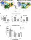
Figure 3
Differential effects of skill vs. use. (A) ICMS-derived motor map (digit, red; wrist, green; elbow/shoulder, light blue) of a rat that learned a skilled reaching movement. (B) ICMS-derived motor map of a rat that learned to press a bar. The two forelimb areas are outlined in white. The caudal forelimb area (CFA) is separated from the rostral forelimb area (RFA) by a band of head/neck representations (yellow). The hindlimb area (HLA) is shown in dark blue and nonresponsive sites in gray. (C) Note the enlarged digit and wrist/forearm representations in the skilled reaching condition (SRC), and enlarged should representation in the unskilled reaching condition (URC, bar press). (D) In the CFA, synapses per neuron were significantly increased (*p < 0.05), but no changes occurred in RFA or HLA (Kleim et al., 2002a).
Figure 4
Figure 4
Reorganization of the rat premotor cortex after controlled cortical impact in the primary motor cortex. (A) Coronal section through the primary motor cortex (caudal forelimb area, or CFA) of a rat approximately one month after a controlled cortical impact. Impactor tip dimension and shape is shown in the inset. (B) Behavioral performance on a single-pellet retrieval task before and after the injury (*p < 0.05). (C) Alteration in motor maps in the rat premotor cortex (rostral forelimb area, or RFA) approximately one month after a controlled cortical impact. In the premotor area that was spared by the lesion, digit representations contracted, while proximal representations expanded. This suggests that the behavioral recovery that was observed was due to compensatory kinematic patterns rather than true recovery (Nishibe et al., 2010).
Figure 5
Figure 5
Functional map changes in forelimb (sFL) and hindlimb (sHL) somatosensory cortex after a focal infarct in mouse. Thalamic projections (arrows) and intracortical connections (double arrows) are also shown. (A) Normal somatosensory representation of sFL and sHL. (B) Within hours after focal infarct (gray), yellow areas show reduced sensory specificity, responding to both FL and HL stimulation. (C) Over the ensuing weeks, growth-promoting processes are triggered. Local axonal sprouting (double-headed arrows), dendritic spine expansion, and synaptogenesis occurs in the peri-infarct cortex. (D) Several weeks after stroke, specificity in sensory responses returns. Neurons that were formerly responsive to stimulation of hindlimb become responsive to forelimb stimulation (Murphy and Corbett, 2009).
Figure 6
Figure 6
Rewiring of corticocortical connections after ischemic infarct. (A) In normal healthy squirrel monkeys, the primary motor cortex (M1) has dense reciprocal connections with both the premotor cortex (PMv, PMd, SMA) as well as the primary somatosensory cortex (S1) and the second somatosensory area (S2). (B) In addition to M1, the ventral premotor cortex (PMv) has dense connections with a rostral area called pre-PMv. PMv has moderate connections with S2, but negligible connections with S1. (C) Several weeks after an ischemic infarct in M1, axons originating in PMv can be seen making sharp bends and avoiding the infarct area, as shown in this tract-tracing study. (D) A low-magnification plot of axons within the section show that the axons originating from PMv course around the central sulcus. Substantial terminal bouton labeling (not shown) appears in S1 (areas 1 and 2). The blue line in (B) signifies the de novo pathway that forms after the lesion (Dancause et al., 2005).
Figure 7
Figure 7
Effects of disuse on motor maps in the absence of injury. The preferred forelimbs of normal, healthy adult squirrel monkeys were placed in soft, restrictive casts for periods up to 5 months. ICMS mapping studies showed a progressive decrease in digit representation and a progressive increase in wrist/forearm representation. These effects were reversible after removal of the cast. These studies demonstrate that disuse has a substantial impact on motor cortex representations independent of the injury-induced disuse and neuropathological changes associated with stroke or traumatic injury (Milliken et al., 2013).

References

    1. Allred R. P., Jones T. A. (2008). Maladaptive effects of learning with the less-affected forelimb after focal cortical infarcts in rats. Exp. Neurol. 210, 172–181 10.1016/j.expneurol.2007.10.010 - DOI - PMC - PubMed
    1. Allred R. P., Maldonado M. A., Hsu J.e., Jones T. A. (2005). Training the “less-affected” forelimb after unilateral cortical infarcts interferes with functional recovery of the impaired forelimb in rats. Restor. Neurol. Neurosci. 23, 297–302 - PubMed
    1. Aroniadou V. A., Keller A. (1993). The patterns and synaptic properties of horizontal intracortical connections in the rat motor cortex. J. Neurophysiol. 70, 1553–1569 - PubMed
    1. Biernaskie J., Szymanska A., Windle V., Corbett D. (2005). Bi-hemispheric contribution to functional motor recovery of the affected forelimb following focal ischemic brain injury in rats. Eur. J. Neurosci. 21, 989–999 10.1111/j.1460-9568.2005.03899.x - DOI - PubMed
    1. Birkenmeier R. L., Prager E. M., Lang C. E. (2010). Translating animal doses of task-specific training to people with chronic stroke in 1-hour therapy sessions: a proof-of-concept study. Neurorehabil. Neural Repair 24, 620–635 10.1177/1545968310361957 - DOI - PMC - PubMed