. 2014 Jan 6;9(1):e84346.
doi: 10.1371/journal.pone.0084346. eCollection 2014.

A neutralization epitope in the hepatitis C virus E2 glycoprotein interacts with host entry factor CD81

Affiliations

A neutralization epitope in the hepatitis C virus E2 glycoprotein interacts with host entry factor CD81

Zhong Zhao et al. PLoS One. .

Abstract

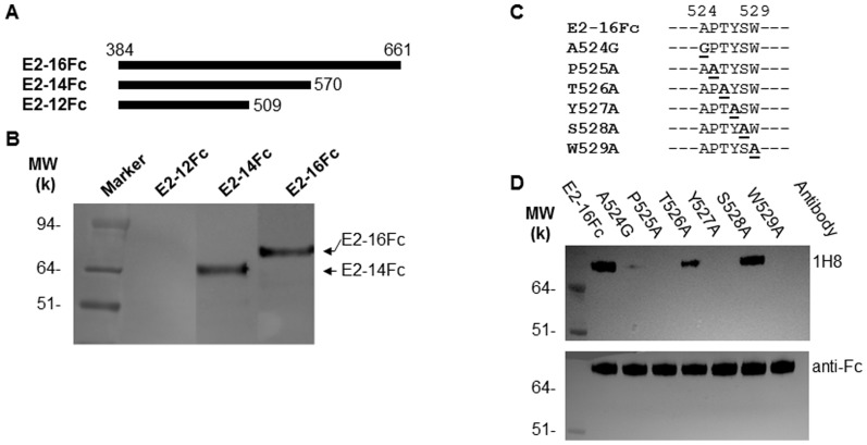
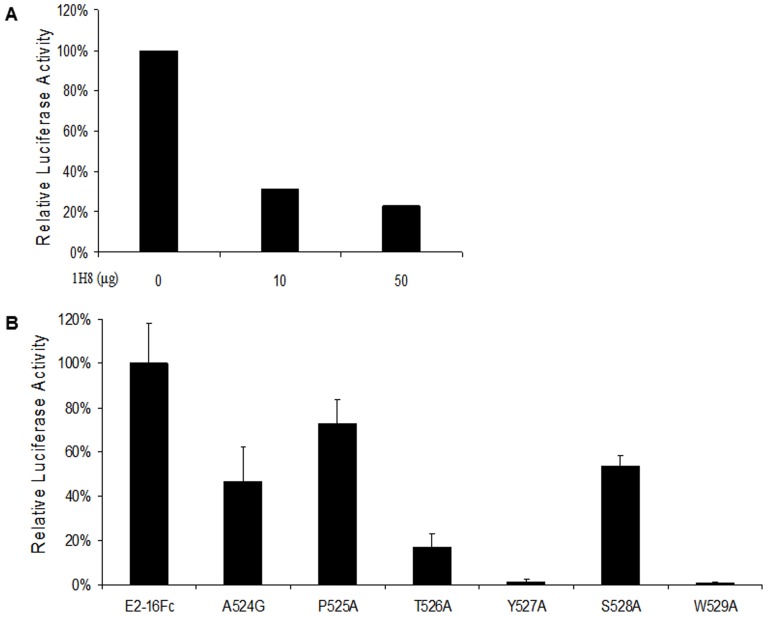
The identification of a specific immunogenic candidate that will effectively activate the appropriate pathway for neutralizing antibody production is fundamental for vaccine design. By using a monoclonal antibody (1H8) that neutralizes HCV in vitro, we have demonstrated here that 1H8 recognized an epitope mapped between residues A524 and W529 of the E2 protein. We also found that the epitope residues A524, P525, Y527 and W529 were crucial for antibody binding, while the residues T526, Y527 and W529 within the same epitope engaged in the interaction with the host entry factor CD81. Furthermore, we detected "1H8-like" antibodies, defined as those with amino acid-specificity similar to 1H8, in the plasma of patients with chronic HCV infection. The time course study of plasma samples from Patient H, a well-characterized case of post-transfusion hepatitis C, showed that "1H8-like" antibodies could be detected in a sample collected almost two years after the initial infection, thus confirming the immunogenicity of this epitope in vivo. The characterization of this neutralization epitope with a function in host entry factor CD81 interaction should enhance our understanding of antibody-mediated neutralization of HCV infections.


Conflict of interest statement

Competing Interests: The authors have declared that no competing interests exist.

Figures

Figure 1
Figure 1. Effect of 1H8 on neutralization of the HCV genotype 2a virus in vitro.
Hybridoma cell culture supernatant containing approximately 16.67 µg/mL of antibody 1H8 was collected and used in neutralization assay in a 3-fold dilution series . Monoclonal mouse anti-E2 antibody 2C1 supernatant was used as a positive control. Percent neutralization was calculated based on an irrelevant mouse anti-CD4 antibody GK1.5 supernatant .
Figure 2
Figure 2. Epitope mapping by screening of random peptide phage display libraries.
The amino acid sequences of phage clusters identified after three rounds of screening three random peptide phage display libraries with 1H8. The peptides sequences derived from the phage clones are shown in comparison with a partial linear sequence of the E2, whose sequence is derived from the HCV H77 strain. The key residues for the 1H8 binding are indicated.
Figure 3
Figure 3. Effect of truncation and mutation of HCV E2 protein on the binding to 1H8.
(A) A schematic diagram of the wild-type HCV E2 protein (based on the H77 strain sequence) and its truncated forms that were used in this study. (B) A Western blot analysis was performed to determine the reactivity of mAb 1H8 with the proteins indicated. The secreted forms of these E2 proteins were produced after transient transfection of Huh7 cells. For the Western blots, 1H8 (1∶1000 dilution) and an HRP-conjugated anti-mouse IgG (1∶3000 dilution) were used as the primary and secondary antibodies, respectively. (C) Site-directed mutagenesis of the E2-16Fc construct was performed to determine the key residues for the binding of 1H8. The residues at positions 525, 526, 527, 528 and 529 were replaced by alanine one at a time. At position 524, the alanine was replaced by a glycine. A hyphen indicates an amino acid that is identical to that of the H77 strain sequence. (D) Western blot was performed by using 1H8 (top panel) versus anti-human IgG1 Fc (bottom panel) to determine the effect of the mutations on the binding of 1H8.
Figure 4
Figure 4. Recognition of a linear peptide from HCV E2 by 1H8.
(A) Biotin-conjugated peptides containing amino acids from 520 to 533 of the E2 protein were chemically synthesized to represent the linear epitope of 1H8. Specific substitutions at positions 528 and 529 were introduced (in bold letters), one at the time. A hyphen represents an amino acid that is identical to that of the H77 strain sequence. (B) Detection of the binding of 1H8 to the epitope peptides in an ELISA. A biotin-linked peptide (200 ng) was coated onto a well of 96-well plate which was pre-coated with streptavidin. 1H8, at a 1∶4000 dilution, was used as the primary antibody. HRP-linked anti-mouse antibody was used as the secondary antibody. The data were obtained from at least three independent experiments. The x-axis indicates the peptide used in the ELISA. The y-axis indicates the resulting absorbance at 450 nm, representing the specific binding of 1H8 to the peptide.
Figure 5
Figure 5. The role of amino acids of the 1H8 binding site in the CD81 interaction of the E2 protein.
(A) E2-16Fc was incubated with the Protein A pre-coated 96-well plates at 100 µL/well at room temperature for 1 hour. 30 µL supernatants containing 2.5×10⁶ units of luciferase activity of CD81-Luc was added to each well in the presence or absence of 1H8, and incubated for an additional hour. After an extensive washing, the luciferase activity was measured. The x-axis indicates the 1H8 used in the assay. The y axis indicates the relative luciferase activity, representing the direct interaction between E2-16Fc and CD81-Luc. (B) The interaction of E2-16Fc and its mutants with CD81-Luc was determined under similar experimental conditions as described in (A). The y-axis indicates the relative luciferase activity, representing the direct interaction of E2-16Fc or its mutant with CD81-Luc.
Figure 6
Figure 6. Effect of naturally occurring variants of the E2 protein with specific residue substitutions in the linear epitope on the 1H8 binding and the CD81 interaction.
(A) The residues 524–529 derived from the E2 protein of H77 strain, i.e., E2-16Fc, is indicated, which serves as a reference sequence. The bold letter shows the presence of a specific residue at that position. The hyphen indicates any possible amino acids at these positions although the residues at positions of 525, 526,527 and 529 are highly conserved among the strains (Table 1). The prevalence of the strains containing a specific residue (bold letter) in the ViPR database is shown. (B) The reactivity of 1H8 to the E2 protein with an indicated substitution was determined in a Western blot analysis. The secreted forms of these E2 proteins were produced in Huh7 cells. 1H8 (1∶1000 dilution) and an HRP-conjugated anti-mouse IgG (1∶3000 dilution) were used as the primary and secondary antibodies, respectively. The detection with anti-Fc (1∶3000 dilution) was served as a control. (C) Determination of the interaction between the E2-16Fc variants and CD81-Luc in a luciferase-based binding assay. The x-axis indicates the different constructed E2-16Fc used in the assay. The y axis indicates the relative luciferase activity, representing the direct interaction between E2-16Fc and CD81-Luc.
Figure 7
Figure 7. Time course of epitope-specific antibody production in Patient H.
Biotin-linked peptides containing the 1H8 binding site and the specific mutation of the binding site were incubated with streptavidin-coated 96-well plates at 200 ng/well. The plasma samples obtained from Patient H were diluted 1∶800 for the ELISA. An unrelated peptide was used as a negative control for the assay. The data were obtained from at least three independent experiments. The x-axis indicates the peptide used in the ELISA and the time when the sample was collected. The y-axis indicates the absorbance at 450 nm, representing the binding activity of the “1H8-like” antibodies in the samples from Patient H.

References

    1. Centers for Disease Control and Prevention (2012) CDC Health Information for International Travel (Oxford Univ Press, New York).
    1. Lauer GM, Walker BD (2001) Hepatitis C virus infection. N Engl J Med 345: 41–52. - PubMed
    1. Alter HJ, Liang TJ (2012) Hepatitis C: The end of the beginning and possibly the beginning of the end. Ann Intern Med 156: 317–318. - PMC - PubMed
    1. Stoll-Keller F, Barth H, Fafi-Kremer S, Zeisel MB, Baumert TF (2009) Development of hepatitis C virus vaccines: challenges and progress. Expert Rev Vaccines 8: 333–345. - PMC - PubMed
    1. Halliday J, Klenerman P, Barnes E (2011) Vaccination for hepatitis C virus: closing in on an evasive target. Expert Rev Vaccines 10: 659–672. - PMC - PubMed

Publication types

MeSH terms