

Modeling single ventricle physiology: review of engineering tools to study first stage palliation of hypoplastic left heart syndrome
- PMID: 24400277
- PMCID: PMC3864195
- DOI: 10.3389/fped.2013.00031
Modeling single ventricle physiology: review of engineering tools to study first stage palliation of hypoplastic left heart syndrome
Abstract
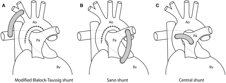
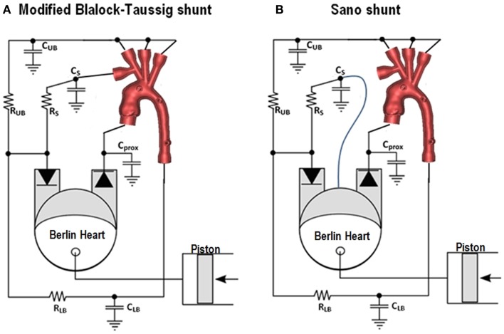
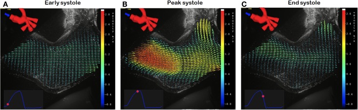
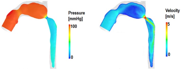
First stage palliation of hypoplastic left heart syndrome, i.e., the Norwood operation, results in a complex physiological arrangement, involving different shunting options (modified Blalock-Taussig, RV-PA conduit, central shunt from the ascending aorta) and enlargement of the hypoplastic ascending aorta. Engineering techniques, both computational and experimental, can aid in the understanding of the Norwood physiology and their correct implementation can potentially lead to refinement of the decision-making process, by means of patient-specific simulations. This paper presents some of the available tools that can corroborate clinical evidence by providing detailed insight into the fluid dynamics of the Norwood circulation as well as alternative surgical scenarios (i.e., virtual surgery). Patient-specific anatomies can be manufactured by means of rapid prototyping and such models can be inserted in experimental set-ups (mock circulatory loops) that can provide a valuable source of validation data as well as hydrodynamic information. Such models can be tuned to respond to differing the patient physiologies. Experimental set-ups can also be compatible with visualization techniques, like particle image velocimetry and cardiovascular magnetic resonance, further adding to the knowledge of the local fluid dynamics. Multi-scale computational models include detailed three-dimensional (3D) anatomical information coupled to a lumped parameter network representing the remainder of the circulation. These models output both overall hemodynamic parameters while also enabling to investigate the local fluid dynamics of the aortic arch or the shunt. As an alternative, pure lumped parameter models can also be employed to model Stage 1 palliation, taking advantage of a much lower computational cost, albeit missing the 3D anatomical component. Finally, analytical techniques, such as wave intensity analysis, can be employed to study the Norwood physiology, providing a mechanistic perspective on the ventriculo-arterial coupling for this specific surgical scenario.
Keywords: Norwood procedure; computational modeling; experimental modeling; shunting; single ventricle.
Figures

References
-
- Hennein HA, Bove EL. Hypoplastic Left Heart Syndrome. Armonk, NY: Futura Publishing Company; (2002).
Publication types
Grants and funding
LinkOut - more resources
Full Text Sources
Other Literature Sources
Research Materials