Review
. 2013 Dec;14(12):823-38.
doi: 10.1038/nrn3618.

Temporal fate specification and neural progenitor competence during development

Review

Temporal fate specification and neural progenitor competence during development

Minoree Kohwi et al. Nat Rev Neurosci. 2013 Dec.

Abstract

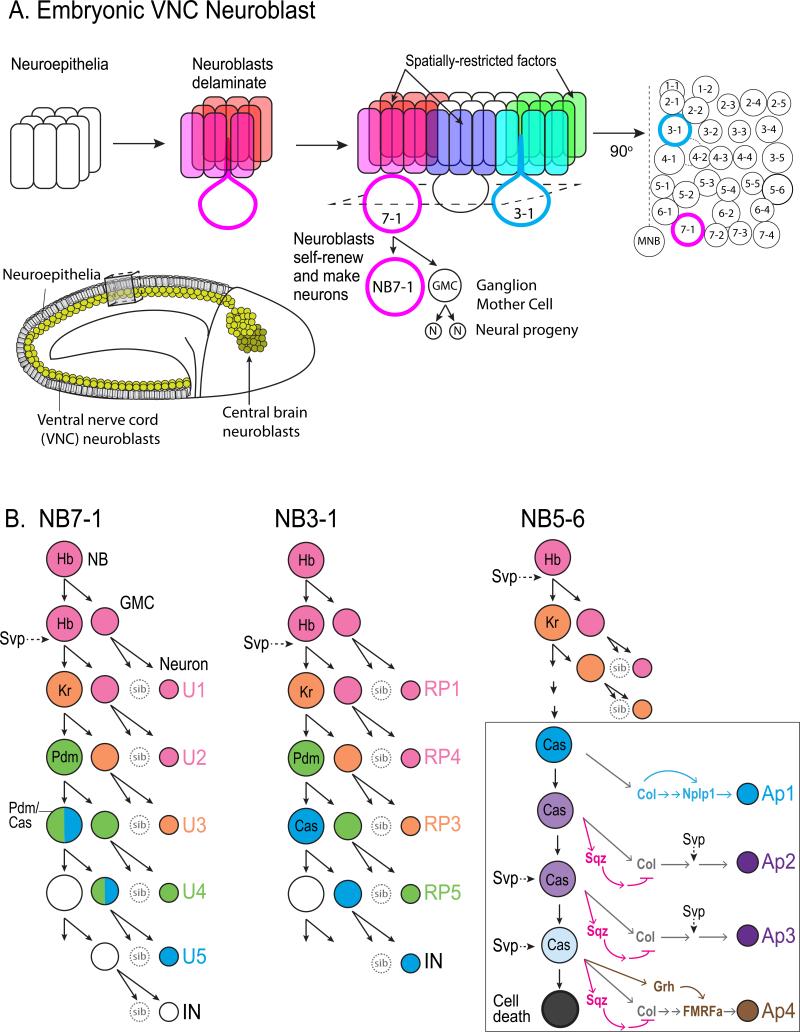
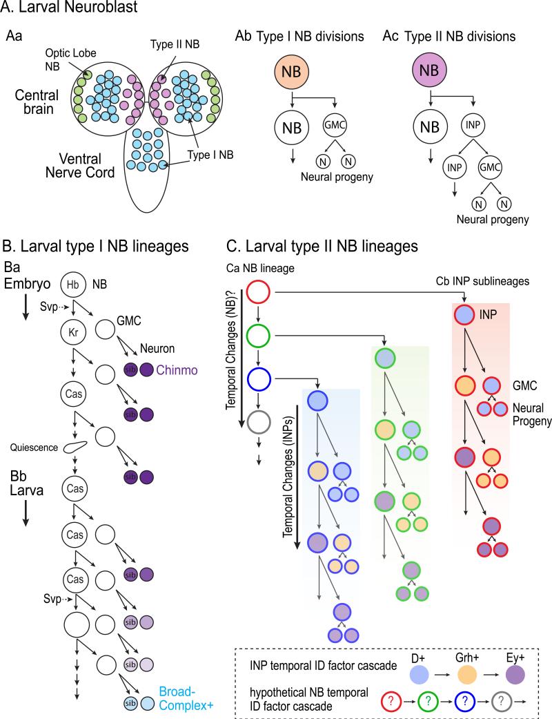
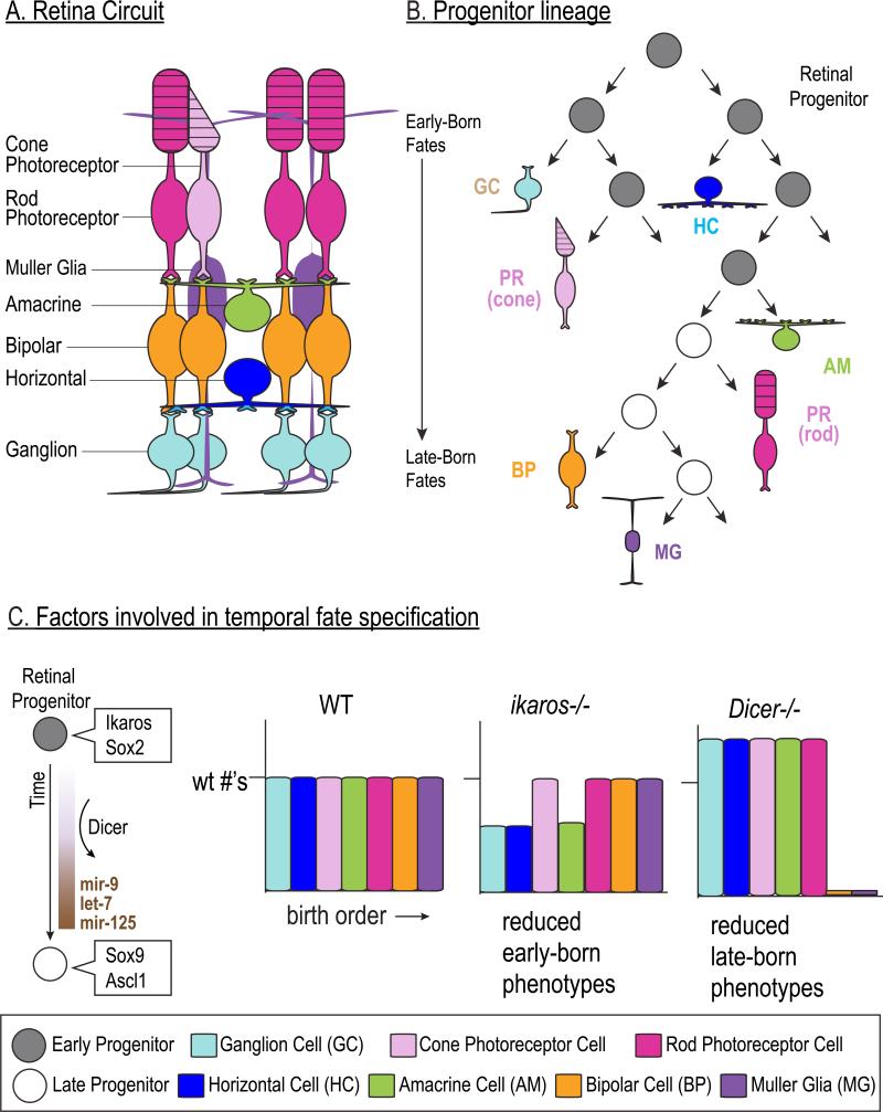
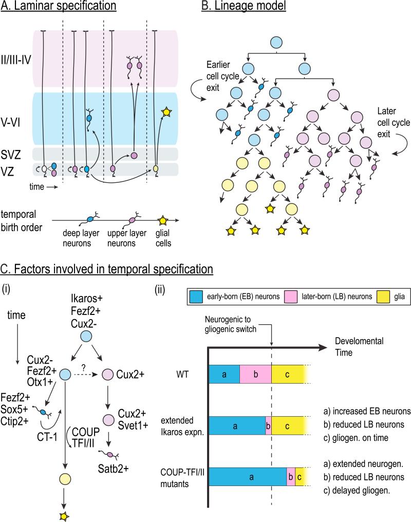
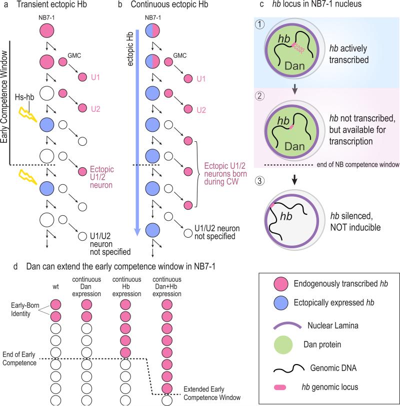
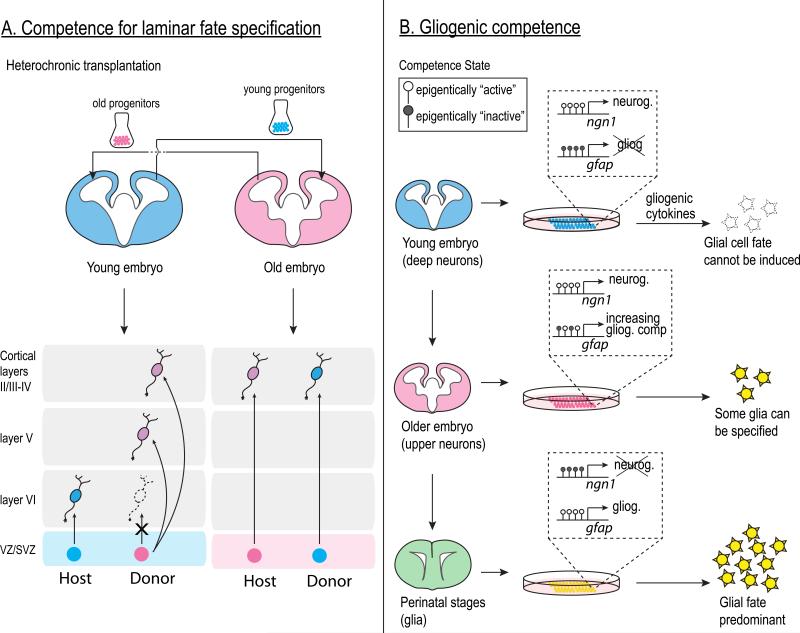
The vast diversity of neurons and glia of the CNS is generated from a small, heterogeneous population of progenitors that undergo transcriptional changes during development to sequentially specify distinct cell fates. Guided by cell-intrinsic and -extrinsic cues, invertebrate and mammalian neural progenitors carefully regulate when and how many of each cell type is produced, enabling the formation of functional neural circuits. Emerging evidence indicates that neural progenitors also undergo changes in global chromatin architecture, thereby restricting when a particular cell type can be generated. Studies of temporal-identity specification and progenitor competence can provide insight into how we could use neural progenitors to more effectively generate specific cell types for brain repair.


Figures

Figure 1
Figure 1. Neurogenesis in Drosophila embryo ventral nerve cord neuroblast lineages
A. (a1) Neuroblasts are specified and subsequently extruded (delaminated) from the neuroepithelia at the onset of neurogenesis. (a2) Each individual neuroblast can be uniquely identified based on its stereotyped position within the nerve cord and its expression of spatially restricted factors. (a3) Neuroblasts are named according to their row and column within the neuroblast “grid.” Thus, NB7-1 (red) is present in the seventh row and occupies the first column position, whereas NB3-1 (blue) is positioned in the third row. (a4) Each neuroblast undergoes a series of asymmetric divisions that give rise to a self-renewed neuroblast and a differentiating ganglion mother cell (GMC). The GMC divides again to generate two neural progeny. The inset shows a Drosophila embryo at stage 9, at the onset of neuroblast delamination. Neuroblasts are shown in yellow along the ventral nerve cord just beneath epithelial layer. b-d. The lineages of NB7-1, NB3-1, and NB5-6 neuroblasts are shown. Each sequentially expresses the temporal identity factors Hunchback (Hb), Kruppel (Kr), Pou domain protein (Pdm), and Castor (Cas) and give rise to a unique lineage of neural progeny in a stereotyped birth order. The COUP-family nuclear receptor, Seven-up (Svp) is transiently expressed in neuroblasts to regulate timing of temporal fate determinant expression. D, inset. At the end of the NB5-6 lineage, Castor initiates a subtemporal transcriptional cascade by activating Squeeze (Sqz, red rectangle) and Grainyhead (Grh, green rectangle). The delay in the accumulation of Sqz and Grh as NB5-6 divides results in distinct neuronal subtypes generated during the Castor window. At the end of the lineage, NB5-6 undergoes cell death.
Figure 2
Figure 2. Neurogenesis in Drosophila larval central brain neuroblast lineages
A. (Aa) The larval central brain harbors roughly 100 neuroblasts in addition to the optic lobe neuroblasts (green). These are divided into type I (blue) and type II (red) neuroblasts. (Ab) Type I neuroblasts of the central brain and ventral nerve cord (blue) divide similarly to embryonic ventral nerve cord neuroblasts shown in Figure 1 by generating a ganglion mother cell (GMC) that divides to produce two neural progeny. (Ac) Type II neuroblasts divide to give rise to an intermediate progenitor (INP) that undergoes additional “neuroblast-like” asymmetric divisions, thus greatly amplifying the number of neural progeny. B. Type I neuroblast lineages at the larval stages express Cas and typically give rise to a series of Chinmo-expressing neurons followed by a series of Broad-Complex-expressing neurons. Chinmo expression is downregulated in the postmitotic progeny in a gradient, with early-born neurons expressing the highest Chinmo levels. (Ba) While Svp regulates the Hb-to-Kr transition in the embryo, (Bb) it is re-expressed in larval neuroblasts to regulate the temporal transition from Chinmo expression to Broad-Complex expression. C. INPs generated from a Type II neuroblast sequentially express Dichaete (D, blue), Grainyhead (Grh, orange), and Eyeless (Ey, purple) to temporally specify distinct neural progeny. Given that neural progeny born early in the neuroblast lineage are different from those born later, it is likely that the neuroblast itself undergoes temporal transitions that are inherited by the INPs (hypothetical NB temporal identity factors are depicted by colored circle outlines).
Figure 3
Figure 3. Temporal fate specification in mammalian retina
A. Schematic illustration of the main cells types in the retina and their organization within the retinal circuit. The retina is comprised of six major classes of neurons and one type of glia (the Muller Glia). B. Retinal progenitors give rise to these distinct cell types in an overlapping but sequential order. Ganglion cells are generated first by early retinal progenitors (light blue) and bipolar cells (orange) and Muller glia (purples) are born last from late progenitors. C. Several molecular factors are expressed in either early (gray) or late (white) progenitors and can determine the temporal phenotypes of the progeny. Dicer is required for the expression of several microRNAs that regulate the temporal transition of the progenitors to produce late-born cell fates. In the mouse retina lacking the transcription factor Ikaros, there is a decrease in the number of cells with early-born fates, although the cone photoreceptors (pink bar) are not affected. In contrast, in mouse retina lacking Dicer, a key enzyme involved in microRNA processing, there is a loss of the late-born cell fates.
Figure 4
Figure 4. Temporal fate specification in mammalian cortex
A. The six layers of the mammalian cortex are generated by the sequential production of distinct types of neurons that migrate to progressively more superficial layers in an “inside-out” fashion. Deep-layer neurons (blue) are born first from the ventricular zone (VZ) radial glia. Subsequently, upper-layer neurons (red) are born from a subset of VZ radial glia as well as the intermediate progenitors in the subventricular zone (SVZ) that are born from the VZ progenitors. Finally, glia (green) are born after the neurogenic period ends. B. Progenitors that give rise to deep layer neurons (blue) exit the cell cycle earlier than the progenitors that primarily give rise to the upper layer neurons (red). C. Factors that function in laminar cell fate are shown on the left. A multipotent progenitor gives rise to more restricted lineages that preferentially generate deep (Cux2-negative) or superficial (Cux2-expressing) cortical neurons. COUP-TFI and II as well as extrinsic signals like CT-1 act on progenitors to switch from neurogenesis to gliogenesis. It is currently unknown whether all Cux2-expressing SVZ progenitors that generate upper layer neurons are derived from the Cux2-expressing VZ progenitors, or whether these are separate progenitor pools. Extension of Ikaros expression in cortical progenitors or loss of COUP-TF results in an expansion of early-born cortical phenotypes (a, blue) at the expense of later-born phenotypes (b, red). However, extension of Ikaros affects the balance of neuronal fates, but does not affect timing of gliogenesis (c, green).
Figure 5
Figure 5. Reorganization of the neuroblast genome regulates competence transition in Drosophila embryos
A. The Hb temporal identity factor is expressed in NB7-1 of the fly embryonic nerve cord for the first two divisions (red), giving rise to the U1/U2 early born motoneurons, which maintain Hb expression. A transient pulse of ectopic Hb (blue) can induce the neuroblast to produce an ectopic U1/U2 neuron up to the fifth division (a). These first five divisions are called the “early competence window.” After this window ends (dotted line), NB7-1 is no longer competent to respond to ectopic Hb and cannot specify early-born neuronal fate. B. If ectopic Hb (blue) is continuously expressed in the neuroblast, only the postmitotic progeny born during the early competence window, and not those born after, will activate endogenous hb transcription (red). C. (c1) During the first two divisions when hb is actively transcribed, the hb genomic locus is positioned in the nuclear interior. (c2) During the subsequent three divisions, the hb gene is transcriptionally inactive, but is still positioned in the nuclear interior and is amenable for activation in the progeny. (c3) At the end of the five division competence window, the hb locus becomes repositioned to the nuclear lamina where it is permanently silenced and is no longer inducible. The nuclear factor Dan (green) is expressed in the neuroblast during the early competence window (c1 and c2), and its downregulation (c3) is required for hb gene repositioning to the lamina. D. Dan can extend the NB7-1 early competence window. Continuous expression of Hb alone results in the specification of early-born identity only during the early competence window. Continuous expression of Hb and Dan together results in prolonged NB7-1 competence to specify early-born identity.
Figure 6
Figure 6. Competence transitions during mammalian neurogenesis
A. Cortical progenitors lose competence to specify early-born neuronal phenotypes over time. Heterochronic transplantation experiments, in which neural progenitors isolated from one developmental stage (donor) is placed in a similar environment of a different stage (host), show that early progenitors (blue) transplanted into an older host can give rise to later-born phenotypes (red); however older progenitors (red) transplanted into the young embryo do not give rise to early born (layer VI) phenotypes. B. Changes in chromatin structure at neuronal and gliogenic genes as development progresses contribute to the neurogenic to gliogenic competence transition in the embryo. In early progenitors, regulatory DNA sequences of key gliogenic genes (such as GFAP) are hypermethylated and silenced. In older progenitors, these DNA regions become hypomethylated and are now competent for transcriptional activation. Neural progenitors cultured from older embryos (red and green embryos) are more competent to respond to gliogenic signals to give rise to glial cells (green stars).

References

    1. Jessell TM. Neuronal specification in the spinal cord: inductive signals and transcriptional codes. Nature reviews. Genetics. 2000;1:20–29. doi:10.1038/35049541. - PubMed
    1. Pearson BJ, Doe CQ. Specification of temporal identity in the developing nervous system. Annu Rev Cell Dev Biol. 2004;20:619–647. doi:10.1146/annurev.cellbio.19.111301.115142. - PubMed
    1. Broadus J, et al. New neuroblast markers and the origin of the aCC/pCC neurons in the Drosophila central nervous system. Mech Dev. 1995;53:393–402. doi:0925477395004548 [pii] - PubMed
    1. Doe CQ, Technau GM. Identification and cell lineage of individual neural precursors in the Drosophila CNS. Trends Neurosci. 1993;16:510–514. - PubMed
    1. Isshiki T, Pearson B, Holbrook S, Doe CQ. Drosophila neuroblasts sequentially express transcription factors which specify the temporal identity of their neuronal progeny. Cell. 2001;106:511–521. doi:S0092-8674(01)00465-2 [pii] - PubMed

Publication types

MeSH terms