

Digital image analysis of endoscopic ultrasonography is helpful in diagnosing gastric mesenchymal tumors
- PMID: 24400772
- PMCID: PMC3890630
- DOI: 10.1186/1471-230X-14-7
Digital image analysis of endoscopic ultrasonography is helpful in diagnosing gastric mesenchymal tumors
Abstract
Background: Endoscopic ultrasonography (EUS) is a valuable imaging tool for evaluating subepithelial lesions in the stomach. However, there are few studies on differentiation between gastrointestinal stromal tumors (GISTs) and benign mesenchymal tumors, such as leiomyoma or schwannoma, with the use of EUS. In addition, there are limitations in the analysis of the characteristic features of such tumors due to poor interobserver agreement as a result of subjective interpretation of EUS images. Therefore, the aim of this study was to evaluate the role of digital image analysis in distinguishing the features of GISTs from those of benign mesenchymal tumors on EUS.
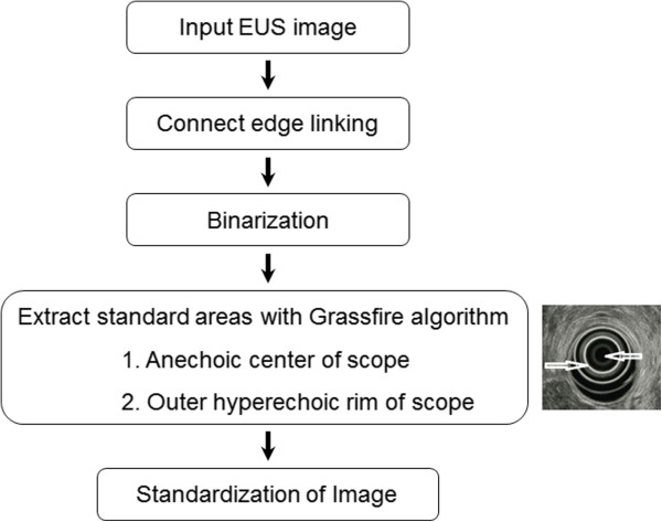
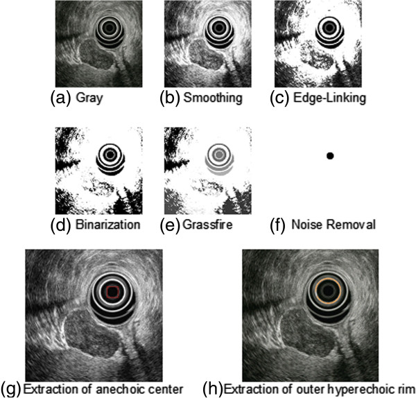
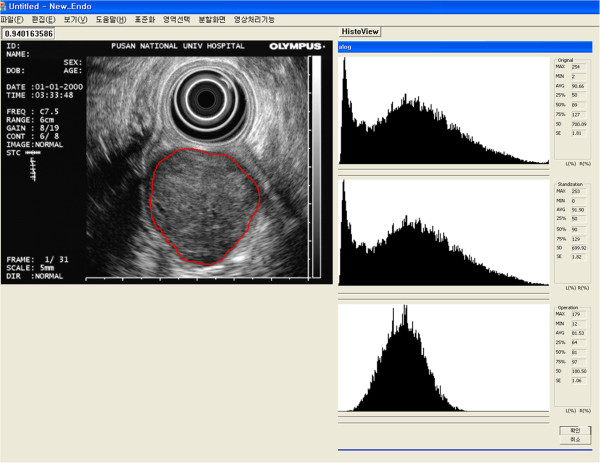
Methods: We enrolled 65 patients with histopathologically proven gastric GIST, leiomyoma or schwannoma on surgically resected specimens who underwent EUS examination at our endoscopic unit from January 2007 to September 2010. After standardization of the EUS images, brightness values including the mean (Tmean), indicative of echogenicity, and the standard deviation (TSD), indicative of heterogeneity, in the tumors were analyzed.
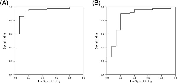
Results: The Tmean and TSD were significantly higher in GIST than in leiomyoma and schwannoma (p < 0.001). However, there was no significant difference in the Tmean or TSD between benign and malignant GISTs. The sensitivity and specificity were almost optimized for differentiating GIST from leiomyoma or schwannoma when the critical values of Tmean and TSD were 65 and 75, respectively. The presence of at least 1 of these 2 findings in a given tumor resulted in a sensitivity of 94%, specificity of 80%, positive predictive value of 94%, negative predictive value of 80%, and accuracy of 90.8% for predicting GIST.
Conclusions: Digital image analysis provides objective information on EUS images; thus, it can be useful in diagnosing gastric mesenchymal tumors.
Figures
References
-
- Sarlomo-Rikala M, Kovatich AJ, Barusevicius A, Miettinen M. CD117: a sensitive marker for gastrointestinal stromal tumors that is more specific than CD34. Mod Pathol. 1998;11:728–734. - PubMed
-
- Blay JY, Bonvalot S, Casali P, Choi H, Debiec-Richter M, Dei Tos AP, Emile JF, Gronchi A, Hogendoorn PC, Joensuu H, Le Cesne A, McClure J, Maurel J, Nupponen N, Ray-Coquard I, Reichardt P, Sciot R, Stroobants S, van Glabbeke M, van Oosterom A, Demetri GD. Consensus meeting for the management of gastrointestinal stromal tumors. Report of the GIST Consensus Conference of 20–21 March 2004, under the auspices of ESMO. Ann Oncol. 2005;16:566–578. doi: 10.1093/annonc/mdi127. - DOI - PubMed
-
- Miettinen M, Sobin LH, Lasota J. Gastrointestinal stromal tumors of the stomach: a clinicopathologic, immunohistochemical, and molecular genetic study of 1765 cases with long-term follow-up. Am J Surg Pathol. 2005;29:52–68. doi: 10.1097/01.pas.0000146010.92933.de. - DOI - PubMed
Publication types
MeSH terms
LinkOut - more resources
Full Text Sources
Other Literature Sources
Medical
