. 2014 Dec;21(12):882-93.
doi: 10.1111/jvh.12225. Epub 2014 Jan 9.

Sequences in the terminal protein and reverse transcriptase domains of the hepatitis B virus polymerase contribute to RNA binding and encapsidation

Affiliations

Sequences in the terminal protein and reverse transcriptase domains of the hepatitis B virus polymerase contribute to RNA binding and encapsidation

F Cao et al. J Viral Hepat. 2014 Dec.

Abstract

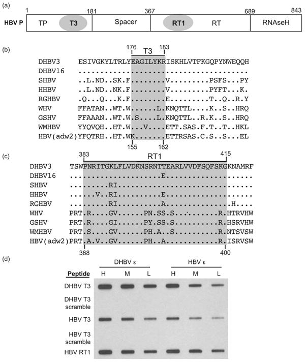
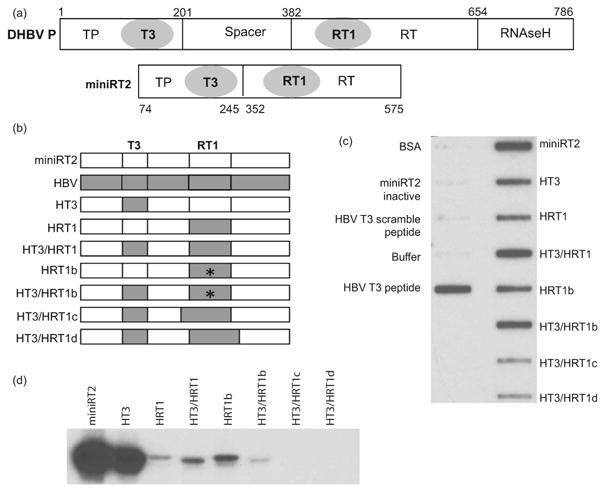
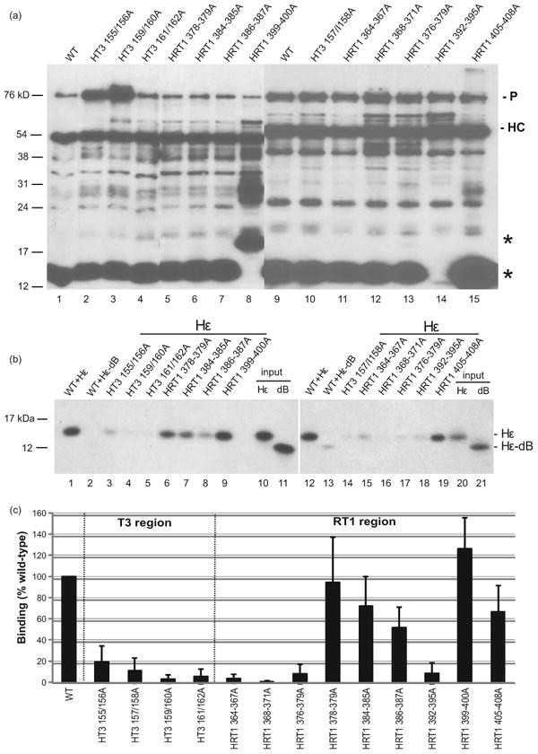
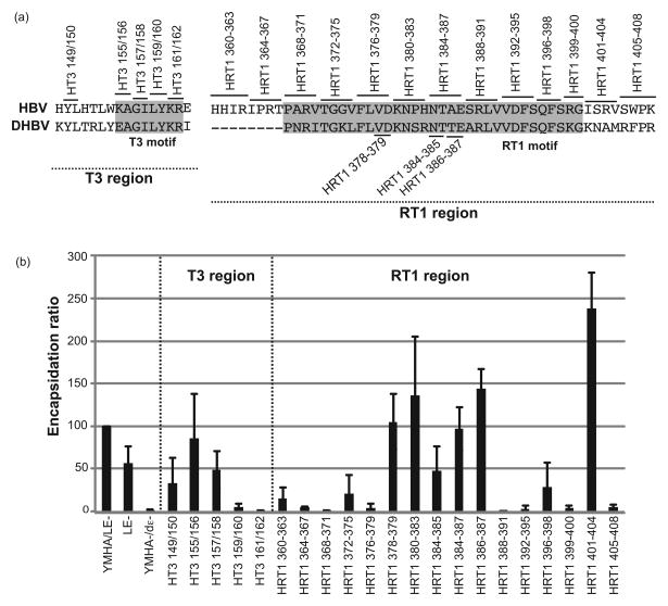
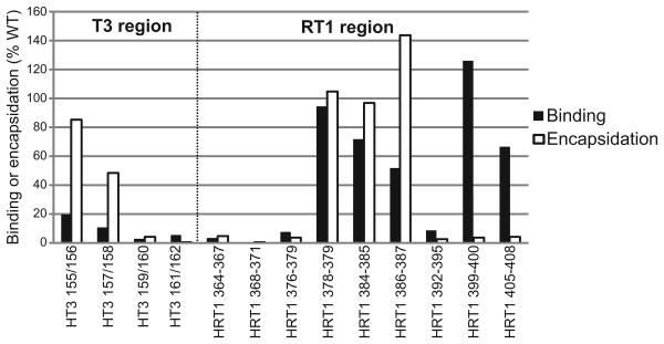
Hepatitis B virus (HBV) antiviral therapy is plagued by limited efficacy and resistance to most nucleos(t)ide analog drugs. We have proposed that the complex RNA binding mechanism of the HBV reverse transcriptase (P) may be a novel target for antivirals. We previously found that RNA binds to the duck HBV (DHBV) P through interactions with the T3 and RT1 motifs in the viral terminal protein and reverse transcriptase domains, respectively. Here, we extended these studies to HBV P. HBV T3 and RT1 synthetic peptides bound RNA in a similar manner as did analogous DHBV peptides. The HBV T3 motif could partially substitute for DHBV T3 during RNA binding and DNA priming by DHBV P, whereas replacing RT1 supported substantial RNA binding but not priming. Substituting both the HBV T3 and RT1 motifs restored near wild-type levels of RNA binding but supported very little priming. Alanine-scanning mutations to the HBV T3 and RT1 motifs blocked HBV ε RNA binding in vitro and pgRNA encapsidation in cells. These data indicate that both the HBV T3 and RT1 motifs contain sequences essential for HBV ε RNA binding and encapsidation of the RNA pregenome, which is similar to their functions in DHBV. Small molecules that bind to T3 and/or RT1 would therefore inhibit encapsidation of the viral RNA and block genomic replication. Such drugs would target a novel viral function and would be good candidates for use in combination with the nucleoside analogs to improve efficacy of antiviral therapy.

Keywords: DNA priming; RNA binding; encapsidation; hepatitis B virus.


Figures

Figure 1
Figure 1. HBV T3 and RT1 sequences bind nucleic acids non-specifically
(a) The HBV polymerase. The terminal protein (TP), spacer, reverse transcriptase (RT), and RNAseH domains and the T3 and RT1 motifs are shown. (b) The T3 motif. Sequences of P flanking the T3 motif are shown for avian (DHBV through RGHBV) and mammalian (WHV through HBV) hepadnaviruses. The T3 motif is shaded. (c) The RT1 motif. The definition of this motif has been expanded by two residues at the N-terminus relative to the numbering used in Badtke et al. (40). (d) T3 and RT1 peptide RNA binding assay. HBV and DHBV T3 and RT1 peptides were bound to a nitrocellulose filter in a slot-blot apparatus, 32P-labeled HBV or DHBV ε RNA was passed through the filter in 3 concentrations (H, M, L), the filter was washed, and bound RNA was detected by autoradiography. DHBV and HBV T3-scramble are negative control peptides in which the sequences have been scrambled.
Figure 2
Figure 2. Function of the HBV T3 and RT1 motifs in DHBV P
(a) The DHBV polymerase and its miniRT2 truncation derivative. The terminal protein (TP), spacer, reverse transcriptase (RT), and RNAseH domains are shown. The amino acid positions for the domain and truncation boundaries are for DHBV P. (b) Structure of chimeric DHBV mRT2 derivatives carrying HBV T3 and/or RT1 motifs. * denotes the P382S/H383R mutations in the RT1 region. (c) In vitro RNA binding assay. Binding of purified miniRT2 derivatives to 32P-labeled DRF+ RNA was determined using a filter-binding assay analogous to the peptide:RNA binding assay in Fig. 1. (d) In vitro DNA priming assay. Equal amounts of miniRT2 chimeric constructs were incubated with DHBV ε and [α32P]dGTP and the products were resolved by SDS-PAGE. DNA priming was detected as covalent attachment of 32P to the proteins.
Figure 3
Figure 3. In vitro RNA binding activity of full-length HBV with mutant T3 and RT1 motifs
(a) Accumulation of HBV P mutants. HBV P derivatives were expressed in transfected 293T cells, immunoprecipitated with anti-FLAG antibodies, resolved by SDS-PAGE, and detected by western blotting using the M2 anti-FLAG antibody; the exposure of the left gel was shorter to limit saturation of the more intense bands. The position of HBV P (P) and the antibody heavy chain (HC) are indicated. * denotes the position of an N-terminal fragment of the 3xFLAG-tagged wild-type P. (b) The immunoaffinity-purified HBV P derivatives were incubated with 32P-labeled wild-type Hε or mutant Hε-dB RNA and co-precipitated products were resolved by SDS–PAGE. Input representing 0.5% of the indicated ε RNA added to each binding reaction mixture is in lanes 10, 11, 20, and 21. (c) Bound 32P-labeled ε RNA signals were quantified via phosphorimaging and compared to the binding of wild-type P to Hε RNA. The data represent the mean ± one standard deviation from at least three independent experiments.
Figure 4
Figure 4. The HBV T3 and RT1 motifs contain sequences essential for encapsidation
(a) Alignment of amino acids sequence from the DHBV3 and HBV T3 and RT1 regions; shaded regions represent the T3 and RT1 motifs. Alanine-scanning mutations were introduced at the indicated positions in the HBV T3 and RT1 motifs in context of the viral genome. (b) Encapsidation assay. Huh7 cells were transfected with the HBV genomic expression vectors, HBV core particles were isolated, total cytoplasmic RNA and encapsidated RNAs were isolated, and RNA levels were measured by quantitative TaqMan PCR. The encapsidation ratios for all mutants were normalized to the ratio observed with the HBV(YMHA/LE-) control as determined in the same assay. The data are represented as the average ± one standard deviation from three to five independent experiments.
Figure 5
Figure 5. Comparison of the effect of mutating HBV T3 and RT1 on RNA binding in vitro and encapsidation in vivo
All values are normalized to binding to wild-type HBV ε or encapsidation by wild-type HBV P.

Similar articles

Cited by

References

    1. Seeger C, Zoulim F, Mason WS. Hepadnaviruses. In: Knipe DM, Howley P, Griffin DE, Lamb RA, Martin MA, Roizman B, et al., editors. Fields Virology 5. Philadelphia: Lippincott Williams & Wilkins; 2007. pp. 2977–3029.
    1. Shepard CW, Simard EP, Finelli L, Fiore AE, Bell BP. Hepatitis B virus infection: epidemiology and vaccination. EpidemiolRev. 2006;28:112–25. - PubMed
    1. Ganem D, Prince AM. Hepatitis B virus infection--natural history and clinical consequences. NEnglJ Med. 2004;350(11):1118–29. - PubMed
    1. Lavanchy D. Hepatitis B virus epidemiology, disease burden, treatment, and current and emerging prevention and control measures. J Viral Hepat. 2004;11(2):97–107. - PubMed
    1. Sorrell MF, Belongia EA, Costa J, Gareen IF, Grem JL, Inadomi JM, et al. National Institutes of Health Consensus Development Conference Statement: management of hepatitis B. AnnInternMed. 2009;150(2):104–10. - PubMed

Publication types