. 2014 Jan 9;4(1):1.
doi: 10.1186/2110-5820-4-1.

Lung ultrasound in the critically ill

Affiliations

Lung ultrasound in the critically ill

Daniel A Lichtenstein. Ann Intensive Care. .

Abstract

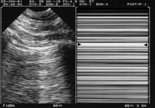
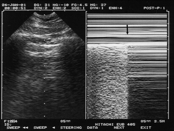
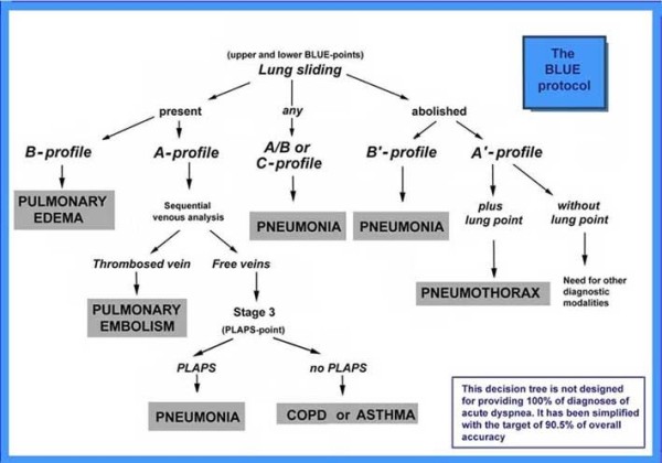
Lung ultrasound is a basic application of critical ultrasound, defined as a loop associating urgent diagnoses with immediate therapeutic decisions. It requires the mastery of ten signs: the bat sign (pleural line), lung sliding (yielding seashore sign), the A-line (horizontal artifact), the quad sign, and sinusoid sign indicating pleural effusion, the fractal, and tissue-like sign indicating lung consolidation, the B-line, and lung rockets indicating interstitial syndrome, abolished lung sliding with the stratosphere sign suggesting pneumothorax, and the lung point indicating pneumothorax. Two more signs, the lung pulse and the dynamic air bronchogram, are used to distinguish atelectasis from pneumonia. All of these disorders were assessed using CT as the "gold standard" with sensitivity and specificity ranging from 90% to 100%, allowing ultrasound to be considered as a reasonable bedside "gold standard" in the critically ill. The BLUE-protocol is a fast protocol (<3 minutes), which allows diagnosis of acute respiratory failure. It includes a venous analysis done in appropriate cases. Pulmonary edema, pulmonary embolism, pneumonia, chronic obstructive pulmonary disease, asthma, and pneumothorax yield specific profiles. Pulmonary edema, e.g., yields anterior lung rockets associated with lung sliding, making the "B-profile." The FALLS-protocol adapts the BLUE-protocol to acute circulatory failure. It makes sequential search for obstructive, cardiogenic, hypovolemic, and distributive shock using simple real-time echocardiography (right ventricle dilatation, pericardial effusion), then lung ultrasound for assessing a direct parameter of clinical volemia: the apparition of B-lines, schematically, is considered as the endpoint for fluid therapy. Other aims of lung ultrasound are decreasing medical irradiation: the LUCIFLR program (most CTs in ARDS or trauma can be postponed), a use in traumatology, intensive care unit, neonates (the signs are the same than in adults), many disciplines (pulmonology, cardiology…), austere countries, and a help in any procedure (thoracentesis). A 1992, cost-effective gray-scale unit, without Doppler, and a microconvex probe are efficient. Lung ultrasound is a holistic discipline for many reasons (e.g., one probe, perfect for the lung, is able to scan the whole-body). Its integration can provide a new definition of priorities. The BLUE-protocol and FALLS-protocol allow simplification of expert echocardiography, a clear advantage when correct cardiac windows are missing.


Figures

Figure 1
Figure 1
Areas of investigation and the BLUE-points. Two hands placed this way (size equivalent to the patient’s hands, upper hand touching the clavicle, thumbs excluded) correspond to the location of the lung, and allow three standardized points to be defined. The upper-BLUE-point is at the middle of the upper hand. The lower-BLUE-point is at the middle of the lower palm. The PLAPS-point is defined by the intersection of: a horizontal line at the level of the lower BLUE-point; a vertical line at the posterior axillary line. Small probes, such as this Japanese microconvex one (1992), allow positioning posterior to this line as far as possible in supine patients, providing more sensitive detection of posterolateral alveolar or pleural syndromes (PLAPS). The diaphragm is usually at the lower end of the lower hand. Extract from “Whole body ultrasonography in the critically ill” (2010 Ed, Chapter 14), with kind permission of Springer Science.
Figure 2
Figure 2
Normal lung surface. Left: Scan of the intercostal space. The ribs (vertical arrows). Rib shadows are displayed below. The pleural line (upper, horizontal arrows), a horizontal hyperechoic line, half a centimeter below the rib line in adults. The proportions are the same in neonates. The association of ribs and pleural line make a solid landmark called the bat sign. The pleural line indicates the parietal pleura in all cases. Below the pleural line, this horizontal repetition artifact of the pleural line has been called the A-line (lower, small horizontal arrows). The A-line indicates that air (gas more precisely) is the component visible below the pleural line. Right: M-mode reveals the seashore sign, which indicates that the lung moves at the chest wall. The seashore sign therefore indicates that the pleural line also is the visceral pleura. Above the pleural line, the motionless chest wall displays a stratified pattern. Below the pleural line, the dynamics of lung sliding show this sandy pattern. Note that both images are strictly aligned, of importance in critical settings. Both images, i.e., lung sliding plus A-lines make the A-profile (when found at the anterior chest wall). They give basic information on the level of capillary pressure. Extract from “Whole body ultrasonography in the critically ill” (2010 Ed, Chapter 14), with kind permission of Springer Science.
Figure 3
Figure 3
Pleural effusion. Left and middle: minute pleural effusion at the PLAPS-point. Below the pleural line, a line regular and roughly parallel to the pleural line can be seen: the lung line, indicating the visceral pleura (arrows). This line, together with the pleural line and the shadow of the ribs, display a kind of quad: the quad sign. Right: M-mode shows a movement of the lung line (white arrows) toward the pleural line (black arrows) on inspiration—the sinusoid sign, indicating also a free pleural effusion, and a viscosity enabling the use of small caliper needle if thoracentesis is envisaged. E, expiration. Quantitative data: this effusion found at the PLAPS-point has an expiratory thickness of roughly 13 mm, i.e., an expectedly small volume (study in progress). A 15-mm distance is our minimum required for safe diagnostic or therapeutic puncture, allowing to simplify the problem of modeling the real volume of an effusion (Ref. 14). Extract from “Whole body ultrasonography in the critically ill” (2010 Ed, Chapter 15), with kind permission of Springer Science.
Figure 4
Figure 4
Lung consolidation. Two signs of lung consolidation. Left: a massive consolidation (probe at the PLAPS-point) invades the whole left lower lobe. No aerated lung tissue is present, and no fractal sign can be generated. The deep border is at the mediastinal line (arrows). The pattern is tissue-like, similar to the spleen (S). The thickness of this image is roughly 10 cm, a value incompatible with a pleural effusion. Image acquired using an ADR-4000 and a sectorial probe (1982 mobile technology) Right: a middle lobe consolidation, which does not invade the whole lobe. This generates a shredded, fractal boundary between the consolidation and the underlying aerated lung (arrows): the quite specific shred (or fractal) sign. Such an anterior consolidation generates the C-profile in the BLUE-protocol. Compare with the regular lung line of Figure 3. Note the blurred letters due to multiple transfers of this image. Quantitative data: a reasonable thickness at the right image is 5.5 cm, giving an index of 5.5 corresponding to a 165-mL consolidation, roughly. In the left image, the 10-cm depth would correspond to a volume of roughly 1 L. Adapted from “Whole body ultrasonography in the critically ill” (2010 Ed, Chapter 16), with kind permission of Springer Science.
Figure 5
Figure 5
Interstitial syndrome and the lung rockets. Two examples of interstitial syndrome. Left: four or five B-lines (see precise description in the text) are visible, called lung rockets (here septal rockets correlating with thickened subpleural interlobular septa). Middle: twice as many B-lines, called ground-glass rockets. Two examples of pulmonary edema (with ground glass areas on CT on the middle figure). Right: Z-lines for comparison. These parasites are ill-defined, short, and do not erase A-lines (arrows), among several criteria. Extract from “Whole body ultrasonography in the critically ill” (2010 Ed, Chapter 17), with kind permission of Springer Science.
Figure 6
Figure 6
Pneumothorax and the stratosphere sign. Left: same pattern as in Figure 2, i.e., pleural line with A-lines, indicating gas below the pleural line. Not visible on the left image, lung sliding is totally absent. Right: here on M-mode, the abolition of lung sliding is visible through the stratosphere sign (which replaces the seashore sign) and indicates total absence of motion. This suggests pneumothorax as a possible cause (see others in text). Arrows: location of the pleural line. The combination of abolished lung sliding with A-lines, at the anterior chest wall, is the A’-profile of the BLUE-protocol (as opposed to the A-profile, where lung sliding is present, ruling out pneumothorax). Extract from “Whole body ultrasonography in the critically ill” (2010 Ed, Chapter 18), with kind permission of Springer Science.
Figure 7
Figure 7
Pneumothorax and the lung point. A specific sign of pneumothorax. Real-time mode allows detection of the inspiratory increase in volume of the collapsed lung. When reaching the chest wall where the probe is laid, it makes a sudden change in the ultrasound image, from an A’-profile to an A- or B-profile usually. The change is sudden because (using an appropriate equipment, without average filters or time lag mainly) ultrasound is a highly sensitive method, able to detect subtle changes, such as the difference between free gas and alveolar gas. The left image shows the pleural line just before the visceral pleura appears. The right image shows (arrow) the very moment the visceral pleura has touched the parietal pleural. This sign has been called lung point (it can be seen along a line, but one point is sufficient for the diagnosis). Video visible at CEURF.net. Extract from “Whole body ultrasonography in the critically ill” (2010 Ed, Chapter 18), with kind permission of Springer Science.
Figure 8
Figure 8
The BLUE-protocol decision tree. This decision tree, slightly modified from the original article (Chest 2008;134:117–125), with the permission of Chest, indicates a way proposed for immediate diagnosis of the main causes of acute respiratory failure, using a lung and venous ultrasound approach.

Similar articles

Cited by

References

    1. Jardin F, Farcot JC, Boisante L, Curien N, Margairaz A, Bourdarias JP. Influence of positive end-expiratory pressure on left ventricle performance. New Engl J Med. 1981;4(7):387–392. doi: 10.1056/NEJM198102123040703. - DOI - PubMed
    1. Dénier A. Les ultrasons, leur application au diagnostic. Presse Med. 1946;4:307–308.
    1. Slasky BS, Auerbach D, Skolnick ML. Value of portable real-time ultrasound in the intensive care unit. Crit Care Med. 1983;4:160–164. doi: 10.1097/00003246-198303000-00002. - DOI - PubMed
    1. Weinberger SE, Drazen JM. Harrison’s principles of internal medicine. 16. New York: McGraw-Hill; 2005. Diagnostic procedures in respiratory diseases; pp. 1505–1508.
    1. Desai SR, Hansel DM. Lung imaging in the adult respiratory distress syndrome: current practice and new insights. Intensive Care Med. 1997;4:7–15. doi: 10.1007/s001340050284. - DOI - PubMed