

Micropatterned multicolor dynamically adhesive substrates to control cell adhesion and multicellular organization
- PMID: 24401172
- PMCID: PMC3983373
- DOI: 10.1021/la404037s
Micropatterned multicolor dynamically adhesive substrates to control cell adhesion and multicellular organization
Abstract
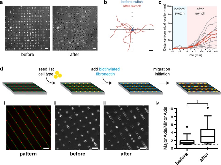
We present a novel technique to examine cell-cell interactions and directed cell migration using micropatterned substrates of three distinct regions: an adhesive region, a nonadhesive region, and a dynamically adhesive region switched by addition of a soluble factor to the medium. Combining microcontact printing with avidin-biotin capture chemistry, we pattern nonadhesive regions of avidin that become adhesive through the capture of biotinylated fibronectin. Our strategy overcomes several limitations of current two-color dynamically adhesive substrates by incorporating a third, permanently nonadhesive region. Having three spatially and functionally distinct regions allows for the realization of more complex configurations of cellular cocultures as well as intricate interface geometries between two cell populations for diverse heterotypic cell-cell interaction studies. We can now achieve spatial control over the path and direction of migration in addition to temporal control of the onset of migration, enabling studies that better recapitulate coordinated multicellular migration and organization in vitro. We confirm that cellular behavior is unaltered on captured biotinylated fibronectin as compared to printed fibronectin by examining the cells' ability to spread, form adhesions, and migrate. We demonstrate the versatility of this approach in studies of migration and cellular cocultures, and further highlight its utility by probing Notch-Delta juxtacrine signaling at a patterned interface.
Figures


Comment in
- Nanomedicine (Lond). 2014 Apr;9(5):573-6
Similar articles
-
Micropatterned dynamically adhesive substrates for cell migration.Langmuir. 2010 Nov 16;26(22):17733-8. doi: 10.1021/la102955m. Epub 2010 Oct 1. Langmuir. 2010. PMID: 20886900
-
Directing cell migration using micropatterned and dynamically adhesive polymer brushes.Acta Biomater. 2014 Jun;10(6):2415-22. doi: 10.1016/j.actbio.2014.01.029. Epub 2014 Feb 6. Acta Biomater. 2014. PMID: 24508539
-
Regulation of collective cell polarity and migration using dynamically adhesive micropatterned substrates.Acta Biomater. 2021 May;126:291-300. doi: 10.1016/j.actbio.2021.03.032. Epub 2021 Mar 16. Acta Biomater. 2021. PMID: 33741539
-
Matrix-Immobilized BMP-2 on Microcontact Printed Fibronectin as an in vitro Tool to Study BMP-Mediated Signaling and Cell Migration.Front Bioeng Biotechnol. 2015 May 11;3:62. doi: 10.3389/fbioe.2015.00062. eCollection 2015. Front Bioeng Biotechnol. 2015. PMID: 26029690 Free PMC article.
-
Integrins in cell adhesion and signaling.Hum Cell. 1996 Sep;9(3):181-6. Hum Cell. 1996. PMID: 9183647 Review.
Cited by
-
Rapid Subtractive Patterning of Live Cell Layers with a Microfluidic Probe.J Vis Exp. 2016 Sep 15;(115):54447. doi: 10.3791/54447. J Vis Exp. 2016. PMID: 27685165 Free PMC article.
-
Image-Based Profiling of Patient-Derived Pancreatic Tumor-Stromal Cell Interactions Within a Micropatterned Tumor Model.Technol Cancer Res Treat. 2018 Jan 1;17:1533033818803632. doi: 10.1177/1533033818803632. Technol Cancer Res Treat. 2018. PMID: 30348057 Free PMC article.
-
Serial Passaging Affects Stromal Cell Mechanosensitivity on Hyaluronic Acid Hydrogels.Macromol Biosci. 2024 Jan;24(1):e2300110. doi: 10.1002/mabi.202300110. Epub 2023 Oct 10. Macromol Biosci. 2024. PMID: 37747449 Free PMC article.
-
Smart Biointerfaces via Click Chemistry-Enabled Nanopatterning of Multiple Bioligands and DNA Force Sensors.ACS Appl Mater Interfaces. 2024 May 1;16(17):21534-21545. doi: 10.1021/acsami.4c00831. Epub 2024 Apr 18. ACS Appl Mater Interfaces. 2024. PMID: 38634566 Free PMC article.
-
Microcontact peeling as a new method for cell micropatterning.PLoS One. 2014 Jul 25;9(7):e102735. doi: 10.1371/journal.pone.0102735. eCollection 2014. PLoS One. 2014. PMID: 25062030 Free PMC article.
References
-
- Singhvi R.; Kumar A.; Lopez G. P.; Stephanopoulos G. N.; Wang D. I.; Whitesides G. M.; Ingber D. E. Engineering cell shape and function. Science 1994, 264, 696–698. - PubMed
-
- Kane R. S.; Takayama S.; Ostuni E.; Ingber D. E.; Whitesides G. M. Patterning proteins and cells using soft lithography. Biomaterials 1999, 20, 2363–2376. - PubMed
-
- Mrksich M. A surface chemistry approach to studying cell adhesion. Chem. Soc. Rev. 2000, 29, 267–273.
-
- Whitesides G. M.; Ostuni E.; Takayama S.; Jiang X.; Ingber D. E. Soft lithography in biology and biochemistry. Annu. Rev. Biomed. Eng. 2001, 3, 335–373. - PubMed
-
- Xia Y.; Whitesides G. M. Soft lithography. Annu. Rev. Mater. Sci. 1998, 28, 153–184.
Publication types
MeSH terms
Substances
Grants and funding
LinkOut - more resources
Full Text Sources
Other Literature Sources
