. 2014 Feb;124(2):656-69.
doi: 10.1172/JCI70090. Epub 2014 Jan 9.

Neuronal ferritin heavy chain and drug abuse affect HIV-associated cognitive dysfunction

Neuronal ferritin heavy chain and drug abuse affect HIV-associated cognitive dysfunction

Jonathan Pitcher et al. J Clin Invest. 2014 Feb.

Abstract

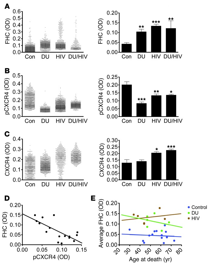
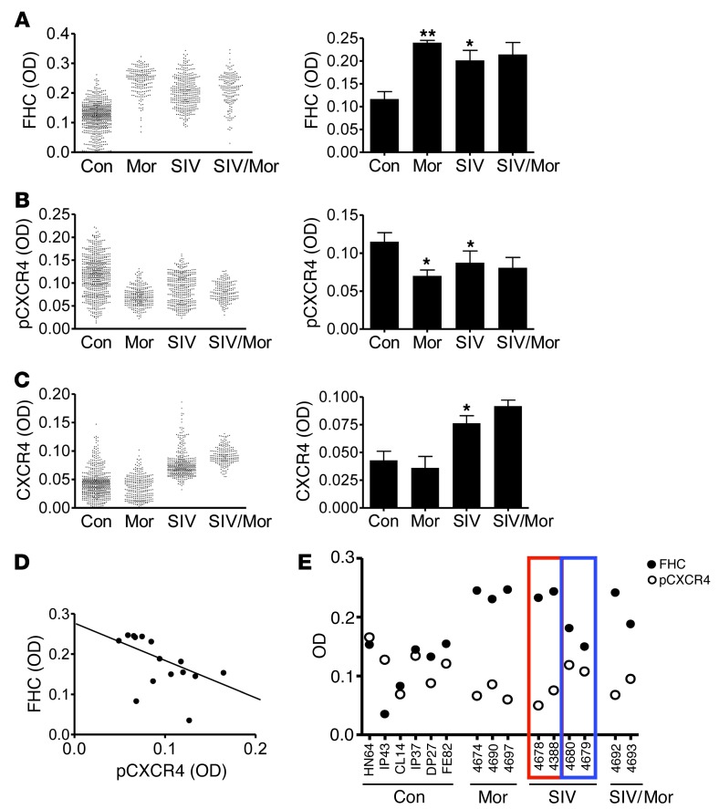
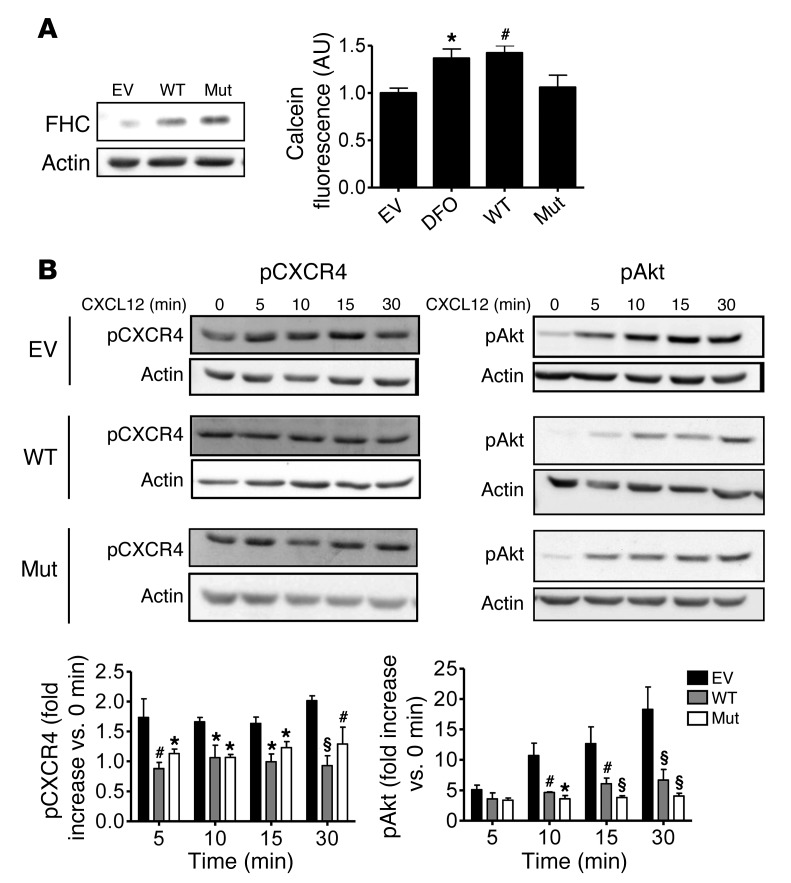
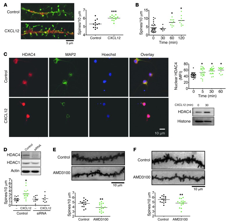
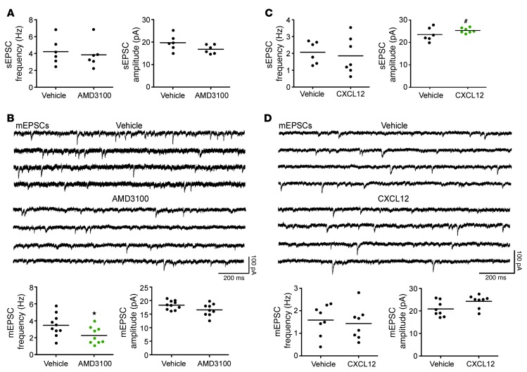
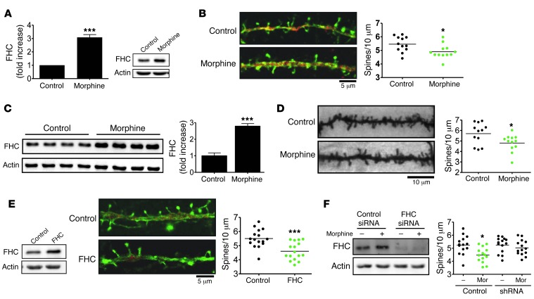
Interaction of the chemokine CXCL12 with its receptor CXCR4 promotes neuronal function and survival during embryonic development and throughout adulthood. Previous studies indicated that μ-opioid agonists specifically elevate neuronal levels of the protein ferritin heavy chain (FHC), which negatively regulates CXCR4 signaling and affects the neuroprotective function of the CXCL12/CXCR4 axis. Here, we determined that CXCL12/CXCR4 activity increased dendritic spine density, and also examined FHC expression and CXCR4 status in opiate abusers and patients with HIV-associated neurocognitive disorders (HAND), which is typically exacerbated by illicit drug use. Drug abusers and HIV patients with HAND had increased levels of FHC, which correlated with reduced CXCR4 activation, within cortical neurons. We confirmed these findings in a nonhuman primate model of SIV infection with morphine administration. Transfection of a CXCR4-expressing human cell line with an iron-deficient FHC mutant confirmed that increased FHC expression deregulated CXCR4 signaling and that this function of FHC was independent of iron binding. Furthermore, examination of morphine-treated rodents and isolated neurons expressing FHC shRNA revealed that FHC contributed to morphine-induced dendritic spine loss. Together, these data implicate FHC-dependent deregulation of CXCL12/CXCR4 as a contributing factor to cognitive dysfunction in neuroAIDS.


Figures

Figure 1
Figure 1. HIV infection and opiate drug use associate with increased FHC and decreased pCXCR4 within neurons of the prefrontal cortex in human HIV-infected and drug-using patients.
OD of FHC (A), pCXCR4 (active) (B), and total CXCR4 (C) within MAP2+ neurons was quantified among control (n = 7–14), drug-using only (DU; n = 5–7), HIV only (HIV; n = 5–16), and drug-using HIV (DU/HIV; n = 3–8) patients. Approximately 100 neurons were analyzed for each patient. Mean OD for each neuron and average neuronal OD for each patient group are shown. (D) Plotting each patient’s average FHC OD to that of pCXCR4 revealed a significant inverse correlation, indicative of a negative relationship between FHC expression and CXCR4 activation (n = 16; Pearson r = –0.724; P = 0.002). (E) No significant relationship was found between average neuronal FHC OD and age at death for any patient group. *P < 0.05, **P < 0.01, ***P < 0.001.
Figure 2
Figure 2. SIV infection and morphine treatment associate with increased FHC and decreased pCXCR4 within neurons of the prefrontal cortex in rhesus macaques.
The OD of FHC (A), pCXCR4 (active) (B), and total CXCR4 (C) within MAP2+ neurons was quantified among control (n = 7), morphine-treated only (Mor; n = 3), SIV-infected only (SIV; n = 4), and morphine-treated, SIV-infected (SIV/Mor; n = 2) macaques. 100 neurons were analyzed per animal. Mean OD for each neuron and average neuronal OD for each treatment group are shown. Note that statistical analysis excluded morphine-treated SIV animals (n = 2). (D) Plotting each animal’s average FHC OD to that of pCXCR4 revealed a significant inverse correlation, indicative of a negative relationship between FHC expression and CXCR4 activation (n = 15; Pearson r = –0.547; P = 0.035). (E) Among the 4 SIV-infected macaques, FHC and pCXCR4 levels were apparently stratified: 2 animals expressed higher levels of FHC and lower levels of pCXCR4 (red outline), and 2 animals expressed lower levels of FHC and higher levels of pCXCR4 (blue outline). Clinical data identified the animals with greater FHC as rapid progressors that retained an elevated viral load after infection, suggesting a relationship between FHC induction and disease severity. *P < 0.05, **P < 0.01.
Figure 3
Figure 3. WT FHC and mutant FHC 222 similarly inhibit CXCR4 signaling in HOS cells.
(A) WT FHC, mutant FHC 222 FHC (Mut; non–iron-binding), or EV was transfected into HOS cells (left), and the specific inability of mutant FHC 222 to bind iron was confirmed by calcein assay (right), a measure of free intracellular iron levels. Calcein fluorescence was increased by treatment with DFO or by overexpressing WT FHC, but not mutant FHC 222. (B) Effects of these proteins on CXCL12 signaling were compared by treating cells with 20 nM CXCL12 for various times. WT FHC– and mutant FHC 222–expressing cells demonstrated similar reductions in CXCL12-induced Akt and CXCR4 activation (n = 4). *P < 0.05, #P < 0.01, §P < 0.001 versus empty vector. See complete unedited blots in the supplemental material.
Figure 4
Figure 4. CXCL12 regulates dendritic spine density in vitro and in vivo.
(A) Rat cortical neurons were cultured for 21 days in the presence of a glial feeder layer. CXCL12 treatment (20 nM, 3 hours) increased dendritic spine density (n = 12). (B) Rat cortical neurons were cultured for 21 days as a pure neuronal population. CXCL12 treatment (20 nM) time-dependently increased dendritic spines (n = 7–29). (C) CXCL12 treatment (20 nM) also increased nuclear HDAC4 levels in pure neuronal cultures, as visualized by immunocytochemistry at 30 minutes and quantified over time (n = 12–34). Similar results were obtained by Western blot using nuclear extracts from neurons. (D) After 8 days in culture, transfection of a pure neuronal population with siRNA specifically decreased HDAC4, but not HDAC1, 36 hours after transfection. siRNA-mediated HDAC4 deficiency completely blocked the effects of CXCL12 on dendritic spines (n = 8–17). (E) The CXCR4 antagonist AMD3100 (1 μg i.c.v.) was injected into the left lateral ventricle of 3-week-old rats. 6 hours after injection, animals were sacrificed, and layer II/III pyramidal neurons were analyzed, revealing reduced total dendritic spine density (n = 15). (F) This effect of AMD3100 was similarly confirmed in a prolonged treatment model: implantation of 3-week-old rats with osmotic pumps delivering constant levels of AMD3100 (0.75 μg/h) for 4 days decreased total dendritic spine density (n = 15). *P < 0.05; **P < 0.01; ***P < 0.001. Scale bars: 5 μm (A); 10 μm (C, E, and F). See complete unedited blots in the supplemental material.
Figure 5
Figure 5. Regulation of the CXCR4 axis alters the frequency and amplitude of EPSCs of layer II/III pyramidal neurons.
(A) AMD3100 injection i.c.v. had no effect on sEPSC frequency or amplitude (sample data not shown). (B) Sample traces of mEPSCs recorded at –70 mV in the presence of tetrodotoxin (1 μM) and picrotoxin (100 μM) in layer II/III pyramidal neurons from vehicle- and AMD3100-exposed animals. Quantification revealed AMD3100 exposure significantly decreased mEPSC frequency, but not amplitude. (C) Incubation of prefrontal cortical slices with CXCL12 (20 nM, 3 hours) induced no change in sEPSC frequency, but significantly increased sEPSC amplitude (sample data not shown). (D) Sample traces of mEPSCs recorded at –70 mV in the presence of tetrodotoxin (1 μM) and picrotoxin (100 μM) in layer II/III pyramidal neurons from vehicle- and CXCL12-exposed slices. Quantification revealed CXCL12 exposure had no significant effect on mEPSC frequency or amplitude. *P < 0.04; #P < 0.03.
Figure 6
Figure 6. Morphine regulates dendritic spine density through effects on neuronal FHC.
(A and B) Rat cortical neurons cultured for 21 days were treated with morphine (1 μm, 24 hours), which both (A) increased FHC levels (n = 4) and (B) decreased dendritic spine density (n = 12). (C and D) Similarly, 3-week-old rats treated with sustained-release morphine pellets for 4 days exhibited (C) increased brain FHC levels (n = 4) and (D) decreased total dendritic spine density (n = 12). (E) FHC overexpression in the absence of morphine also decreased dendritic spine density, as measured in cultured rat cortical neurons transfected with a FHC-expressing plasmid or empty vector (n = 16). (F) The ability of morphine to regulate dendritic spine density was blocked in FHC-deficient neurons, as shown using FHC-specific or scrambled control shRNA. *P < 0.05; ***P < 0.001. Scale bars: 5 μm (B and E); 10 μm (D). See complete unedited blots in the supplemental material.

Similar articles

Cited by

References

    1. Tiveron MC, Cremer H. CXCL12/CXCR4 signalling in neuronal cell migration. Curr Opin Neurobiol. 2008;18(3):237–244. doi: 10.1016/j.conb.2008.06.004. - DOI - PubMed
    1. Shimizu S, Khan MZ, Hippensteel RL, Parkar A, Raghupathi R, Meucci O. Role of the transcription factor E2F1 in CXCR4-mediated neurotoxicity and HIV neuropathology. Neurobiol Dis. 2007;25(1):17–26. doi: 10.1016/j.nbd.2006.08.004. - DOI - PMC - PubMed
    1. Khan MZ, Brandimarti R, Musser BJ, Resue DM, Fatatis A, Meucci O. The chemokine receptor CXCR4 regulates cell-cycle proteins in neurons. J Neurovirol. 2003;9(3):300–314. - PMC - PubMed
    1. Khan MZ, Brandimarti R, Shimizu S, Nicolai J, Crowe E, Meucci O. The chemokine CXCL12 promotes survival of postmitotic neurons by regulating Rb protein. Cell Death Differ. 2008;15(10):1663–1672. doi: 10.1038/cdd.2008.95. - DOI - PMC - PubMed
    1. Nicolai J, Burbassi S, Rubin J, Meucci O. CXCL12 inhibits expression of the NMDA receptor’s NR2B subunit through a histone deacetylase-dependent pathway contributing to neuronal survival. Cell Death Dis. 2010;1:e33. doi: 10.1038/cddis.2010.10. - DOI - PMC - PubMed

Publication types

MeSH terms