. 2014 Feb;124(2):801-11.
doi: 10.1172/JCI70291. Epub 2014 Jan 9.

Prenatal retinoid deficiency leads to airway hyperresponsiveness in adult mice

Prenatal retinoid deficiency leads to airway hyperresponsiveness in adult mice

Felicia Chen et al. J Clin Invest. 2014 Feb.

Abstract

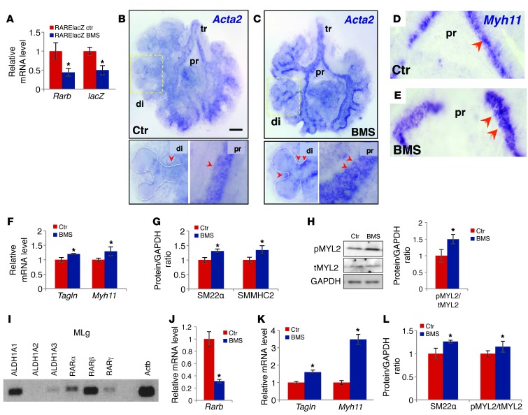
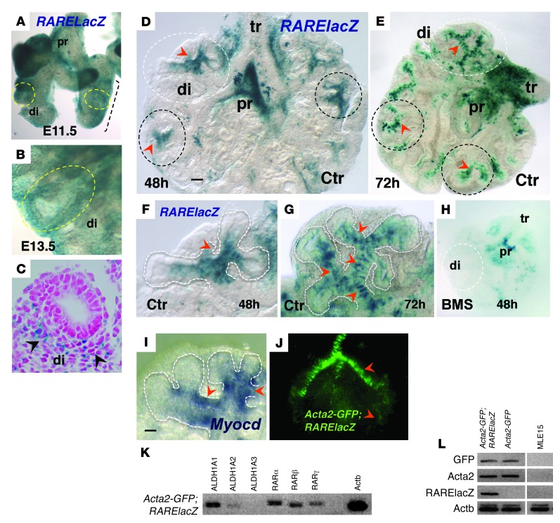
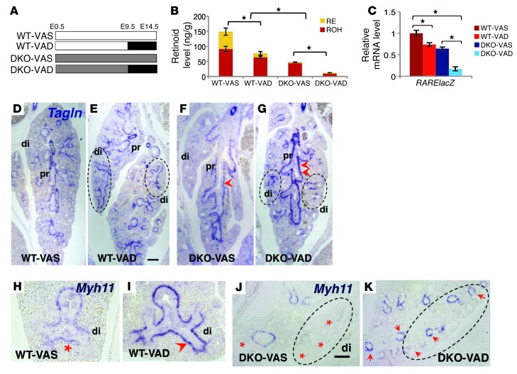
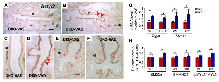
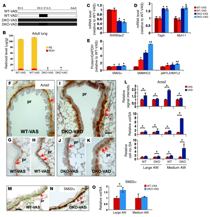
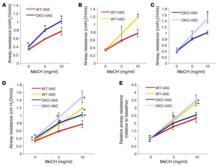
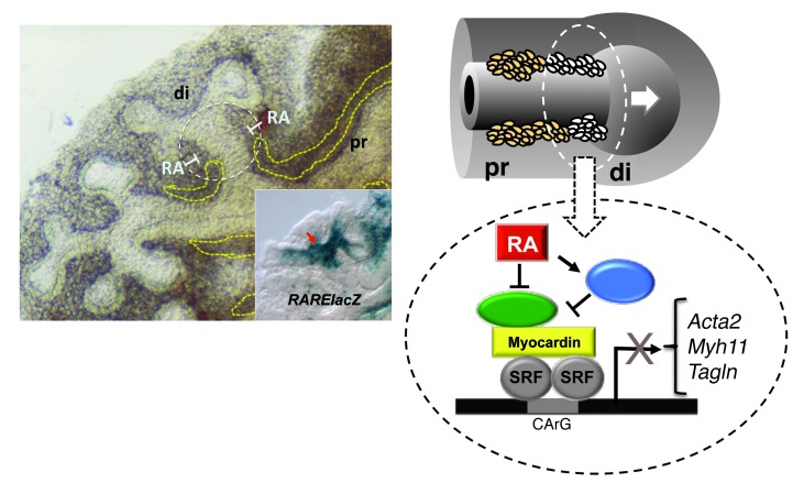
There is increasing evidence that vitamin A deficiency in utero correlates with abnormal airway smooth muscle (SM) function in postnatal life. The bioactive vitamin A metabolite retinoic acid (RA) is essential for formation of the lung primordium; however, little is known about the impact of early fetal RA deficiency on postnatal lung structure and function. Here, we provide evidence that during murine lung development, endogenous RA has a key role in restricting the airway SM differentiation program during airway formation. Using murine models of pharmacological, genetic, and dietary vitamin A/RA deficiency, we found that disruption of RA signaling during embryonic development consistently resulted in an altered airway SM phenotype with markedly increased expression of SM markers. The aberrant phenotype persisted postnatally regardless of the adult vitamin A status and manifested as structural changes in the bronchial SM and hyperresponsiveness of the airway without evidence of inflammation. Our data reveal a role for endogenous RA signaling in restricting SM differentiation and preventing precocious and excessive SM differentiation when airways are forming.


Figures

Figure 1
Figure 1. Disruption of RA signaling leads to aberrant expression of SM markers in mouse embryonic lung explant (AH) and MLg cell (IL) cultures.
BMS-treated RARElacZ lungs (48 hours): significant downregulation of Rarb and RARElacZ (A) and increased expression of Tagln and Myh11 (F) and their products SM22α and SMMHC2, respectively) (G). (BE) Whole-mount ISH of Acta2 (B and C) and Myh11 (D and E) increased signals in the mesenchyme of proximal (pr) and distal (di) airways in BMS-treated lungs compared with those of controls (arrowheads). Strong ectopic Acta2 expression was seen in stalks of distal buds in BMS-treated lungs. (H) Increased ratio of pMYL2/tMYL2 in BMS-treated lungs. (I) PCR detection of RA pathway components in MLg cells: RA-synthesizing enzymes (ALDH1A1, -2, and -3) and RA receptors (RARα, RARβ, and RARγ). (JL) BMS treatment (24 hours) disrupted RA signaling (downregulation of Rarb) and increased expression of SM marker genes (Tagln and Myh11) and products (SM22α) and of the pMYL2/tMYL2 ratio. (A, F, I, and K) PCR and (G, H, and L) Western blot analysis. n = 3–4 per condition. *P < 0.05. tr, trachea. Scale bar: 200 μm.
Figure 2
Figure 2. RA is active at sites of SM differentiation in developing distal airways.
(AC) X-gal staining of E11.5 (A) and E13.5 (B and C) RARElacZ lungs; mesenchymal signals were prominent in the distal lung (di, circled). (C) Cross section of E13.5 lung. (DH) X-gal staining of cultured lung (48 and 72 hours). Strong local RA activity in the mesenchyme at the stalks of forming buds was seen at 48 hours (D and F, arrowheads; circled areas). At 72 hours, the pattern was more prominent, with multiple areas of RARElacZ labeling the mesenchyme associated with newly formed distal airways (E and G). Signals declined or were undetectable in more proximal branches, although they were present in trachea and main bronchi, being abolished by BMS treatment (H). Myocardin (Myocd) signals (ISH) were enriched in areas of strong RA activity (I, F, and G, arrowheads). (JL) Lung culture from Acta2-GFP;RARElacZ mice showing GFP signals associated with airway SM (J). SM cells isolated by FACS expressed RA pathway components and activated RA signaling (K and L, PCR; negative controls: cells from Acta-GFP, non-RARElacZ, and MLE15 line). Scale bars: 55 μm (D) and 45 μm (I). n = 3–5 per condition for all experiments.
Figure 3
Figure 3. VAD in utero results in aberrant airway SM differentiation in E14.5 lungs.
(A) Diagram of experimental design. (B) HPLC measurements of total retinoid (RE and ROH) in E14.5 lung homogenates (ng/g) from WT and DKO mice in VAS and VAD groups. (C) qPCR of RARElacZ expression in lung homogenates (all mice were bred into a RARElacZ line). (DK) ISH of Tagln (DG) and Myh11 (HK) showing increased expression of SM markers in the proximal airways of VAD lungs (arrowheads), most prominent in DKO VAD lungs (G). Ectopic expression extended to the most distal airways (circled regions) in the VAD groups (red asterisks denote no or low signals). n = 6 per condition. *P < 0.05 compared with the VAS group. Scale bars: 150 μm (E) and 180 μm (J).
Figure 4
Figure 4. Aberrant airway SM differentiation in VAD E14.5 lungs.
(AF) Acta2 IHC in DKO lungs: an increased severity of the phenotype was seen in VAD lungs compared with that in VAS lungs. Stronger Acta2 signals were observed in the proximal airways, and ectopic Acta2 expression was seen in the distal airways of DKO-VAD lung. qPCR (G) and Western blot analysis (H) of SM markers and the pMYL2/tMYL2 ratio in lung homogenates confirm the aberrant SM differentiation when RA signaling was disrupted in vivo. n = 6 per condition. *P < 0.05 compared with the VAS group. Scale bars: 135 μm (A) and 45 μm (E).
Figure 5
Figure 5. Consequences of prenatal VAD in the SM phenotype of the adult lung.
(A) Diagram of experimental design. (B) HPLC measurements of retinoids in the lung homogenates of adult mice: no difference between VAS and VAD groups, but significantly lower levels in DKO compared with WT mice. (CE) Significantly lower levels of RARElacZ (C) correlating with Tagln and Myh11 upregulation (D) by qPCR in DKO lungs compared with WT. (E) Significant increase in SM22α, SMMHC2, and pMYL2/tMYL2 ratio levels in lung homogenates of WT-VAD, DKO-VAS, and DKO-VAD mice compared with those in WT-VAS lung homogenates by Western blot analysis. IHC of SM markers in adult proximal airways (FK: Acta2; MN: Sm22α) showing increased expression in VAD groups (double arrows). Quantitative analysis of Acta2 (L) and Sm22α (O) IHC: signal intensity, relative volume of staining per SA, and relative number of labeled cells per SA in proximal airways (large and medium size, see Methods) suggest a significant increase in SM mass in VAD lungs relative to VAS lungs. Data represent the mean ± SEM, n = 3 per condition. *P < 0.05 compared with WT-VAS; #P < 0.05 compared with DKO-VAS. Scale bars: 30 μm (I) and 20 μm (M). AW, airway.
Figure 6
Figure 6. Prenatal VAD results in increased airway responsiveness in adulthood. flexiVent analysis of airway resistance in a nonchallenged state and in response to methacholine (MeCH).
(A) Airway resistance of DKO-VAS adult mice was higher than that of WT-VAS adult mice at baseline and at all doses of methacholine. (B and C) VAD mice showed increased response to methacholine compared with that of VAS groups in both WT (B) and DKO (C) mice. (D) Both WT and DKO mice subjected to a prenatal VAD diet had the highest response to methacholine at 10 mg/ml. (E) Relative airway resistance (absolute resistance normalized by baseline value): differences between DKO and WT mice were minimized, but differences between VAS and VAD mice persisted. *P < 0.05 compared with WT-VAS (n = 4 DKO-VAS; n = 3 for other conditions).
Figure 7
Figure 7. Proposed mechanism of SM regulation by endogenous RA.
During branching of the developing airways, a program of differentiation of SM takes place in the distal mesenchyme at the newly formed stalks of the lung buds (white circled area). RA signaling is activated in these areas, as evidenced by locally enriched RARElacZ expression (inset) at these sites. Endogenous RA regulates this SM program, restricting SM gene expression and preventing excessive formation of SM while airways are branching. As new generations of airways arise, the process is reiterated, and the RA restriction of the SM program becomes less crucial proximally (left panel, dashed yellow lines). RA fine-tunes the SM gene expression program, likely by inhibiting the expression of a key activator of SM transcription (in green) or by inducing the expression of the transcriptional repressor (in blue), ultimately resulting in the downregulation of SM gene expression.

Similar articles

Cited by

References

    1. West KP, Howard GR, Sommer A. Vitamin A and infection: public health implications. Annu Rev Nutr. 1989;9:63–86. doi: 10.1146/annurev.nu.09.070189.000431. - DOI - PubMed
    1. West KP., Jr Extent of vitamin A deficiency among preschool children and women of reproductive age. J Nutr. 2002;132(9 Suppl):2857S–2866S. - PubMed
    1. Williams SR. Nutrition and Diet Therapy: Instructor’s Manual and Text Book. Maryland Heights, Missouri, USA: Mosby; 1997.
    1. Chambon P. The retinoid signaling pathway: molecular and genetic analyses. Semin Cell Biol. 1994;5(2):115–125. doi: 10.1006/scel.1994.1015. - DOI - PubMed
    1. Clagett-Dame M, DeLuca HF. The role of vitamin a in mammalian reproduction and embryonic development. Annu Rev Nutr. 2002;22:347–381. doi: 10.1146/annurev.nutr.22.010402.102745E. - DOI - PubMed

Publication types

MeSH terms