. 2014;13(5):801-6.
doi: 10.4161/cc.27726. Epub 2014 Jan 8.

Evidence for eIF2α phosphorylation-independent effects of GSK2656157, a novel catalytic inhibitor of PERK with clinical implications

Affiliations

Evidence for eIF2α phosphorylation-independent effects of GSK2656157, a novel catalytic inhibitor of PERK with clinical implications

Jothilatha Krishnamoorthy et al. Cell Cycle. 2014.

Abstract

The endoplasmic reticulum (ER)-resident protein kinase PERK is a major component of the unfolded protein response (UPR), which promotes the adaptation of cells to various forms of stress. PERK phosphorylates the α subunit of the translation initiation factor eIF2 at serine 51, a modification that plays a key role in the regulation of mRNA translation in stressed cells. Several studies have demonstrated that the PERK-eIF2α phosphorylation pathway maintains insulin biosynthesis and glucose homeostasis, facilitates tumor formation and decreases the efficacy of tumor treatment with chemotherapeutic drugs. Recently, a selective catalytic PERK inhibitor termed GSK2656157 has been developed with anti-tumor properties in mice. Herein, we provide evidence that inhibition of PERK activity by GSK2656157 does not always correlate with inhibition of eIF2α phosphorylation. Also, GSK2656157 does not always mimic the biological effects of the genetic inactivation of PERK. Furthermore, cells treated with GSK2656157 increase eIF2α phosphorylation as a means to compensate for the loss of PERK. Using human tumor cells impaired in eIF2α phosphorylation, we demonstrate that GSK2656157 induces ER stress-mediated death suggesting that the drug acts independent of the inhibition of eIF2α phosphorylation. We conclude that GSK2656157 might be a useful compound to dissect pathways that compensate for the loss of PERK and/or identify PERK pathways that are independent of eIF2α phosphorylation.

Keywords: PERK/PEK kinase; endoplasmic reticulum stress; mRNA translation; pharmacological inhibitors; protein phosphorylation; translation initiation factor eIF2; tumorigenesis; unfolded protein response.


Figures

None
Figure 1. Analysis of the effects of GSK2656157 on cells with conditionally active PERK or PKR. (A) Schematic representation of the GyrB system. The regulatory domain (RD) of either PKR (i.e., dsRNA-binding domain) or PERK (i.e., lumenal domain) was replaced by the GyrB domain, which was fused to the kinase domain (KD) of each eIF2α kinase. Coumermycin mediates the dimerization of the chimera kinase leading to its activation and induction of eIF2α phosphorylation. (B and C) GyrB.PERK cells were left untreated (B, lane 1) or treated with 100 ng/ml coumermycin for 6 h in the absence (B, lane 2; C, lane 1) or presence of the indicated concentrations of the PERK inhibitor (PERKi) (C, lanes 2–6). (D) GyrB.PKR cells were left untreated (lane 1) or treated with 100 ng/ml coumermycin for 6 h in the absence (lanes 2 and 3) or presence of the indicated concentrations of PERKi (lanes 4–8). (B–D) Cells extracts (50 μg of protein) were subjected to immunoblot analyses for the indicated proteins.
None
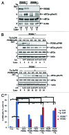
Figure 2. Control of eIF2α phosphorylation by GSK2656157 in HT1080 cells. (A) Parental HT1080 cells were treated with the indicated concentrations of PERKi in the absence (−) or presence (+) of 1 μM thapsigargin (TG). As control untreated cells (lanes 1, 9, and 13) or cells treated 1 μM TG in the absence of PERKi (lanes 2, 10, and 14) were used. (B) Parental HT1080 cells were left untreated (lane 1) or treated with the indicated concentrations of the PERKi for 24 h (lanes 3–7). As control for eIF2α phosphorylation, cells were treated with 1 μM TG for 2 h (lane 2). (A and B) Cells extracts (50 μg of protein) were subjected to immunoblot analyses for the indicated proteins.
None
Figure 3. Characterization of the biochemical properties of GSK2656157 in PERK+/+ and PERK−/− MEFs. (A) Immortalized PERK+/+ and PERK−/− MEFs were left untreated (lanes 1 and 3) or treated with 1 μM of TG for 2 h (lanes 2 and 4). (B) PERK+/+ and PERK−/− MEFs were left untreated (lanes 1 and 8) or treated with 1 μM of TG in the absence (lanes 2 and 9) or presence of the indicated concentrations of PERKi for 2 h (lanes 3–7 and 10–14). (A and B) Protein extracts (50 μg) were subjected to immunoblot analyses for the indicated proteins. (C) PERK+/+ and PERK−/− MEFs untreated (control) or treated with 1 μM TG in the absence or presence of the indicated concentrations of PERKi for 24 h. As control, cells treated with the indicated concentrations of PERKi in the absence of TG for 24 h. Cell death was measured by the percentage of cells in sub-G1 population by FACS analysis. Histograms represent the quantification of 3 independent experiments.
None
Figure 4. Characterization of the biological properties of GSK2656157 in tumor cells exposed to ER stress. (A) Cell extracts (50 μg of protein) from wild-type (WT) and knock-in (KI) HT1080 cells untreated or treated with 1 μM thapsigargin (TG) for 2 h were subjected to western blot analysis for the indicated proteins. Note the delayed migration of the HA-eIF2αS51A in knock-in (KI) cells (lanes 3 and 4) compared with endogenous eIF2α in wild-type (WT) cells (lanes 1 and 2). (B) HT1080 WT and KI cells were treated with 1 μM TG for either 24 h. (C) Cells were treated with 1 μM TG either in the absence or presence of the indicated concentrations of PERKi for 18 h. As control, cells were treated with the same concentrations of PERKi in the absence of TG for 18 h. (B and C) Cell death was assessed by the percentage of cells in sub-G1 population by FACS analysis. Histograms represent the quantification of 3 independent experiments.

References

    1. Schröder M, Kaufman RJ. The mammalian unfolded protein response. Annu Rev Biochem. 2005;74:739–89. doi: 10.1146/annurev.biochem.73.011303.074134. - DOI - PubMed
    1. Ron D, Walter P. Signal integration in the endoplasmic reticulum unfolded protein response. Nat Rev Mol Cell Biol. 2007;8:519–29. doi: 10.1038/nrm2199. - DOI - PubMed
    1. Ma Y, Hendershot LM. The role of the unfolded protein response in tumour development: friend or foe? Nat Rev Cancer. 2004;4:966–77. doi: 10.1038/nrc1505. - DOI - PubMed
    1. Ron D. Translational control in the endoplasmic reticulum stress response. J Clin Invest. 2002;110:1383–8. - PMC - PubMed
    1. Raven JF, Koromilas AE. PERK and PKR: old kinases learn new tricks. Cell Cycle. 2008;7:1146–50. doi: 10.4161/cc.7.9.5811. - DOI - PubMed

Publication types