. 2014 Jan 9:9:11.
doi: 10.1186/1748-717X-9-11.

Adequacy of inhale/exhale breathhold CT based ITV margins and image-guided registration for free-breathing pancreas and liver SBRT

Affiliations

Adequacy of inhale/exhale breathhold CT based ITV margins and image-guided registration for free-breathing pancreas and liver SBRT

Wensha Yang et al. Radiat Oncol. .

Abstract

Purpose: To evaluate use of breath-hold CTs and implanted fiducials for definition of the internal target volume (ITV) margin for upper abdominal stereotactic body radiation therapy (SBRT). To study the statistics of inter- and intra-fractional motion information.

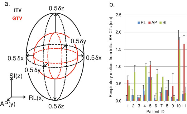
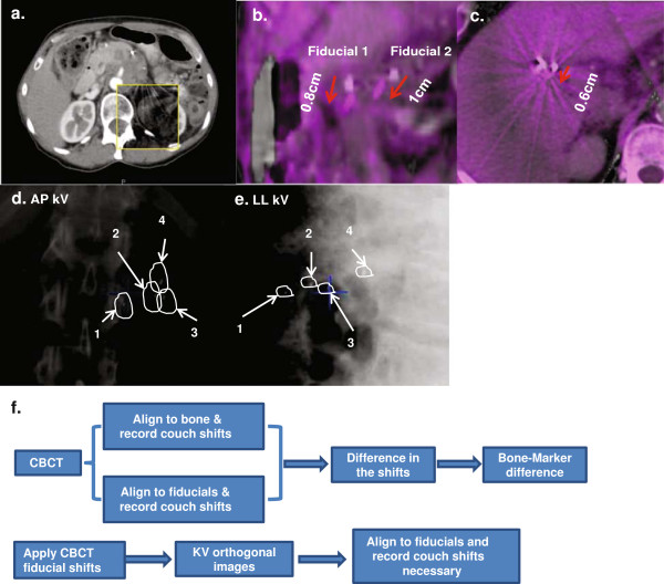
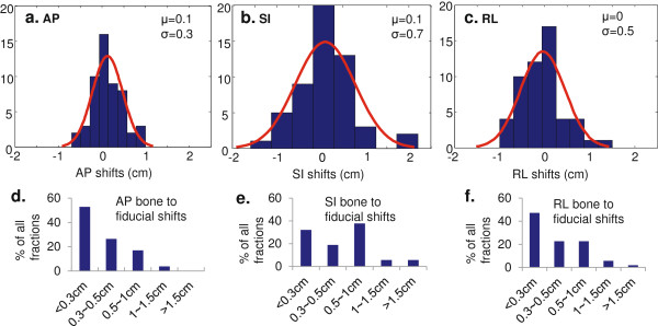
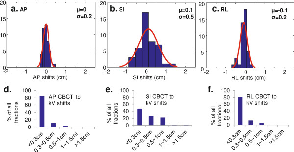
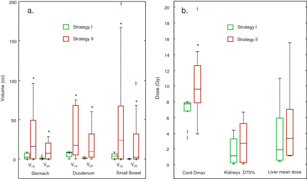
Methods and materials: 11 patients treated with SBRT for locally advanced pancreatic cancer (LAPC) or liver cancer were included in the study. Patients underwent fiducial implantation, free-breathing CT and breath-hold CTs at end inhalation/exhalation. All patients were planned and treated with SBRT using volumetric modulated arc therapy (VMAT). Two margin strategies were studied: Strategy I uses PTV = ITV + 3 mm; Strategy II uses PTV = GTV + 1.5 cm. Both CBCT and kV orthogonal images were taken and analyzed for setup before patient treatments. Tumor motion statistics based on skeletal registration and on fiducial registration were analyzed by fitting to Gaussian functions.

Results: All 11 patients met SBRT planning dose constraints using strategy I. Average ITV margins for the 11 patients were 2 mm RL, 6 mm AP, and 6 mm SI. Skeletal registration resulted in high probability (RL = 69%, AP = 4.6%, SI = 39%) that part of the tumor will be outside the ITV. With the 3 mm ITV expansion (Strategy 1), the probability reduced to RL 32%, AP 0.3%, SI 20% for skeletal registration; and RL 1.2%, AP 0%, SI 7% for fiducial registration. All 7 pancreatic patients and 2 liver patients failed to meet SBRT dose constraints using strategy II. The liver dose was increased by 36% for the other 2 liver patients that met the SBRT dose constraints with strategy II.

Conclusions: Image guidance matching to skeletal anatomy is inadequate for SBRT positioning in the upper abdomen and usage of fiducials is highly recommended. Even with fiducial implantation and definition of an ITV, a minimal 3 mm planning margin around the ITV is needed to accommodate intra-fractional uncertainties.


Figures

Figure 1
Figure 1
GTV to ITV margin expansion and respiratory fiducial motion for all patients. a. GTV(red) to ITV(black( expansion strategy. b. The motion range evaluated from breath-hold CTs for 11 patients (1–7 are pancreas patients, 8–11 are liver patients), with averages 0.6 (0–1.8) cm AP, 0.6 (0.1-1.5) cm SI, and 0.2 (0.1-0.7) cm RL.
Figure 2
Figure 2
Example images of fiducial motion and DRR to kV registration; flow chart of pre-treatment image registration. a. A typical pre-RT CBCT registered to planning CT using skeletal landmarks. b. Fiducial displacement from planning CT to CBCT with skeletal alignment for a pancreas patient. c. Fiducial displacement from planning CT to CBCT with skeletal alignment for a liver patient. d-e. an example of kV orthogonal images with fiducial matching to the contour derived from DRR. f. Flow chart describes how the imaging guidance was performed before the treatment and how the shifts were resolved.
Figure 3
Figure 3
Statistics for bone to fiducial shifts. a-c. Histograms of bone-marker discrepancies for AP, SI, and RL directions, respectively. Gaussian fits of the histograms give average (μ) and standard deviation (σ) values in each of the three directions. d-f. Bar plots of % of fractions that has bone-marker shifts in <0.3 cm, 0.3 - 0.5 cm, 0.5 - 1 cm, 1 - 1.5 cm, and >1.5 cm for AP, SI and RL directions.
Figure 4
Figure 4
Statistics for CBCT to kV shifts. a-c. Histograms of differences between CBCT and kV 2D images for AP, SI, and RL directions, respectively. Gaussian fits of the histograms determine the average (μ) and standard deviation (σ) for each of the three directions. d-f. Bar plots of% of fractions that has CBCT to kV shifts in <0.3 cm, 0.3 - 0.5 cm, 0.5 - 1 cm, 1 - 1.5 cm, and >1.5 cm for AP, SI and RL directions.
Figure 5
Figure 5
Box plots of normal tissue doses for two strategies. All cases satisfied 90 - 95% of PTV receiving the prescription dose. a. V15 and V20 for stomach, duodenum and bowel; b. maximum dose for cord, D75% for total kidneys and mean dose for liver. *statistically significant with p < 0.05

References

    1. Tward DJ, Siewerdsen JH, Daly MJ. et al.Soft-tissue detectability in cone-beam CT: evaluation by 2AFC tests in relation to physical performance metrics. Med Phys. 2007;34(11):4459–4471. doi: 10.1118/1.2790586. - DOI - PubMed
    1. Whitfield G, Jain P, Green M. et al.Quantifying motion for pancreatic radiotherapy margin calculation. Radiother Oncol. 2012;103(3):360–366. doi: 10.1016/j.radonc.2012.02.012. - DOI - PubMed
    1. Haripotepornkul NH, Nath SK, Scanderbeg D. et al.Evaluation of intra- and inter-fraction movement of the cervix during intensity modulated radiation therapy. Radiother Oncol. 2011;98(3):347–351. doi: 10.1016/j.radonc.2010.11.015. - DOI - PubMed
    1. Katz AW, Carey-Sampson M, Muhs AG. et al.Hypofractionated stereotactic body radiation therapy (SBRT) for limited hepatic metastases. Int J Radiat Oncol Biol Phys. 2007;67(3):793–798. doi: 10.1016/j.ijrobp.2006.10.025. - DOI - PubMed
    1. Scorsetti M, Bignardi M. Conformal and stereotactic radiotherapy in hepatocellular carcinoma. Ann Ital Chir. 2008;79(2):107–110. - PubMed

Publication types

MeSH terms