. 2014 Mar;21(3):233-41.
doi: 10.1038/gt.2013.75. Epub 2014 Jan 9.

Injection parameters and virus dependent choice of promoters to improve neuron targeting in the nonhuman primate brain

Affiliations

Injection parameters and virus dependent choice of promoters to improve neuron targeting in the nonhuman primate brain

W Lerchner et al. Gene Ther. 2014 Mar.

Abstract

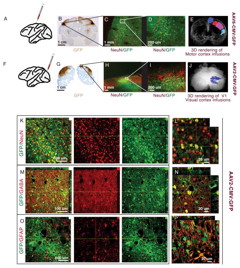
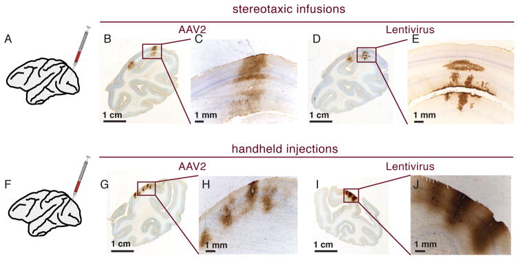
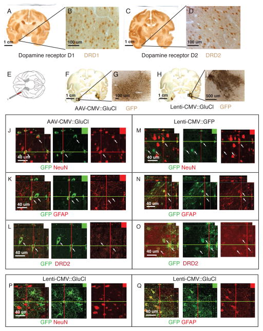
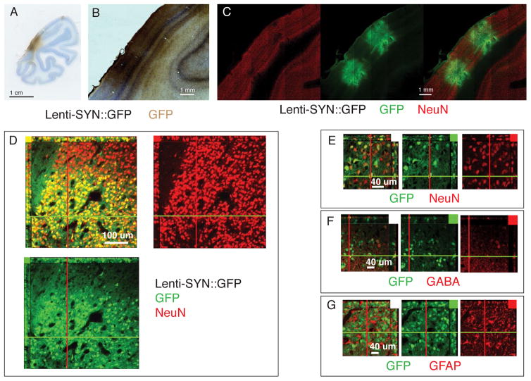
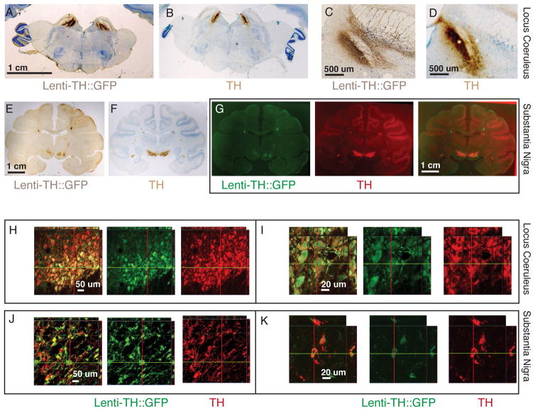
We, like many others, wish to use modern molecular methods to alter neuronal functionality in primates. For us, this requires expression in a large proportion of the targeted cell population. Long generation times make germline modification of limited use. The size and intricate primate brain anatomy poses additional challenges. We surved methods using lentiviruses and serotypes of adeno-associated viruses (AAVs) to introduce active molecular material into cortical and subcortical regions of old-world monkey brains. Slow injections of AAV2 give well-defined expression of neurons in the cortex surrounding the injection site. Somewhat surprisingly we find that in the monkey the use of cytomegalovirus promoter in lentivirus primarily targets glial cells but few neurons. In contrast, with a synapsin promoter fragment the lentivirus expression is neuron specific at high transduction levels in all cortical layers. We also achieve specific targeting of tyrosine hydroxlase (TH)- rich neurons in the locus coeruleus and substantia nigra with a lentvirus carrying a fragment of the TH promoter. Lentiviruses carrying neuron specific promoters are suitable for both cortical and subcortical injections even when injected quickly.


Conflict of interest statement

CONFLICT OF INTEREST

The authors declare no conflict of interest.

Figures

Figure 1
Figure 1. Comparing AAV serotypes in monkey cortex
A–E. Injections into motor areas. A. Schematic representation of injection site. B. Coronal section from monkey injected with AAV6 on the left and AA1 on the right – visualized with DAB-HRP for GFP expression. C,D. Motor cortex of a section adjacent to B, visualized green for GFP and red for NeuN (Alexa 568). Native GFP expressed from AAV6 and NeuN. GFP expression overlaps with cortical layers, but some expression is visible in the white matter. Most GFP expressing cells co-localize with NeuN staining (yellow cells indicate co-expression). E. 3D rendering of HRP stained areas covered by AAV6 injections into the pre-motor cortex. F–J. Injections into visual cortex. F. Schematic representation of injection site. G. Coronal section from monkey injected with AAV-RH10R in the left V1 and AAV2 in the right V1. H, I. Native GFP expressed from AAV2 and NeuN visualized green for GFP and red for NeuN (Alexa 568). Most GFP expressing cells co-localize with NeuN staining (yellow cells) and are concentrated in cortical layers. J. 3D rendering of HRP stained areas covered by AAV2 injections into the visual cortex. K–P. Confocal slices of visual area injected with AAV2, visualized with green for GFP and red for Alexa 568 antibody staining. K, L. GFP expressing cells co-localize with NeuN (Alexa 568). M,N. GFP expression co-localizes with GABA positive (Alexa 568) interneurons. O,P. Only a small percentage of GFP expressing cells co-localize with GFAP positive glia.
Figure 2
Figure 2. Comparing injection parameters for AAV2 and Lentivirus in V1
A. Schematic representation of stereotaxic infusions. B–C. Two overlapping injection of AAV2-GFP at 500nl/min. Injections resulted in contiguous coverage. D–E. Lentivirus-GFP injected at 500nl/min. Staining only covers a small area in the cortical layers. F. Schematic representation for handheld injection. G–H. Handheld injections with AAV2-GFP. Only a limited area around the injection sites express GFP. I–J. Handheld injections with Lentivirus-GFP. Injections result in better cortical coverage around the injection site than slow stereotaxic injections (compare with D,E).
Figure 3
Figure 3. Expression of AAV2 and Lentivirus in the Rhinal Cortex
A,B. Coronal sections stained for DRD1 visualized with DAB-HRP. Cellular staining is visible in areas consistent with known DRD1 expression . C,D. Coronal section stained for DRD2 visualized with DAB-HRP. Cellular staining is visible in areas consistent with known DRD2 expression . E. Schematic of ventral view of macaque brain with virus injections into rhinal cortex. F–I. Coronal sections stained for GFP. Left rhinal injected with AAV2-CMV::GluCl. Right rhinal injected with Lenti-CMV::shRNAi-GFP and Lenti-CMV::GluCl. J–Q. Confocal slices of AAV2-CMV::GluCl injected areas GFP is visualized in green (DyLight 488), other markers in red (Alexa 568). J. AAV expressed GFP co-localizes with NeuN neuron marker (red - cells indicated with white arrows). K. AAV2 expressed GFP does not co-localize with glia marker GFAP (red). L. AAV2 expressed GFP does co-localize with DRD2 antibody staining (red). M. Only a very small percentage of lentivirus GFP expressing cells overlap with NeuN expressing neurons (red). N. Lentivirus GFP is expressed in GFAP positive glia (red). O. Lentivirus GFP is expressed in cells stained with DRD2 antibody (red). P. Only few Lenti-GluCla-CFP expressing cells co-localize with NeuN expressing neurons (red). Q. The majority of Lenti-GluCla-CFP expressing cells overlap with GFAP positive glia (red).
Figure 4
Figure 4. Lentivirus synapsin promoter expression in visual cortex
A–C. Coronal sections of V1 injected with Lenti-SYN::GFP. A,B. GFP visualized with HRP-DAB staining. C. Tiled confocal scan of section stained for GFP and NeuN. Same section in all images with GFP visualized in green (DyLight 488) and NeuN in red (Alexa 568). D–E. Confocal slice to show dense expression of GFP in NeuN positive neurons. Note the sharp transition border of viral GFP expression. F. GFP does co-localized with GABA positive interneurons (red) G. GFP does not co-localized with GFAP positive glia (red).
Figure 5
Figure 5. Lentivirus tyrosine hydroxylase promoter expression in locus coeruleus and substantia nigra
A–D. Coronal section of monkey bilaterally injected into locus coeruleus. A,C. GFP expression detected with HRP-DAB covers most of LC. B,D. Adjacent sections of those shown in A,C but stained for TH expression (note staining in the same area as in A,C). E–I. Coronal sections of monkey bilaterally injected into substantia nigra. E,F. GFP expression detected with HRP-DAB is mostly restricted to SN. G. Scans pseudocolored with green for GFP and red for TH stain. Overlay of both sections in photoshop shows that expression overlaps. H–K. Confocal slices of injected areas. H,I. GFP and TH expression occurs in the same cell population. J,K. Most GFP expressing cells co-localize with TH positive cells.

Similar articles

Cited by

References

    1. Byrne BJ, Falk DJ, Clement N, Mah CS. Gene therapy approaches for lysosomal storage disease: next-generation treatment. Human gene therapy. 2012;23(8):808–15. - PMC - PubMed
    1. Prickett M, Jain M. Gene therapy in cystic fibrosis. Translational research : the journal of laboratory and clinical medicine. 2012 - PubMed
    1. Gavrilov K, Saltzman WM. Therapeutic siRNA: principles, challenges, and strategies. The Yale journal of biology and medicine. 2012;85(2):187–200. - PMC - PubMed
    1. Zhou J, Rossi JJ. Current progress in the development of RNAi-based therapeutics for HIV–1. Gene therapy. 2011;18(12):1134–8. - PMC - PubMed
    1. Poewe W, Mahlknecht P, Jankovic J. Emerging therapies for Parkinson’s disease. Current opinion in neurology. 2012;25(4):448–59. - PubMed

Publication types

MeSH terms