

Rapid target gene validation in complex cancer mouse models using re-derived embryonic stem cells
- PMID: 24401838
- PMCID: PMC3927956
- DOI: 10.1002/emmm.201303297
Rapid target gene validation in complex cancer mouse models using re-derived embryonic stem cells
Abstract
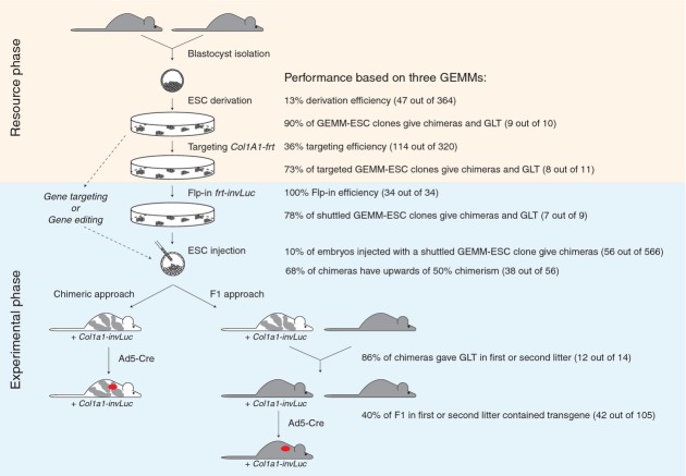
Human cancers modeled in Genetically Engineered Mouse Models (GEMMs) can provide important mechanistic insights into the molecular basis of tumor development and enable testing of new intervention strategies. The inherent complexity of these models, with often multiple modified tumor suppressor genes and oncogenes, has hampered their use as preclinical models for validating cancer genes and drug targets. In our newly developed approach for the fast generation of tumor cohorts we have overcome this obstacle, as exemplified for three GEMMs; two lung cancer models and one mesothelioma model. Three elements are central for this system; (i) The efficient derivation of authentic Embryonic Stem Cells (ESCs) from established GEMMs, (ii) the routine introduction of transgenes of choice in these GEMM-ESCs by Flp recombinase-mediated integration and (iii) the direct use of the chimeric animals in tumor cohorts. By applying stringent quality controls, the GEMM-ESC approach proofs to be a reliable and effective method to speed up cancer gene assessment and target validation. As proof-of-principle, we demonstrate that MycL1 is a key driver gene in Small Cell Lung Cancer.
Figures
 male,
male,  female,
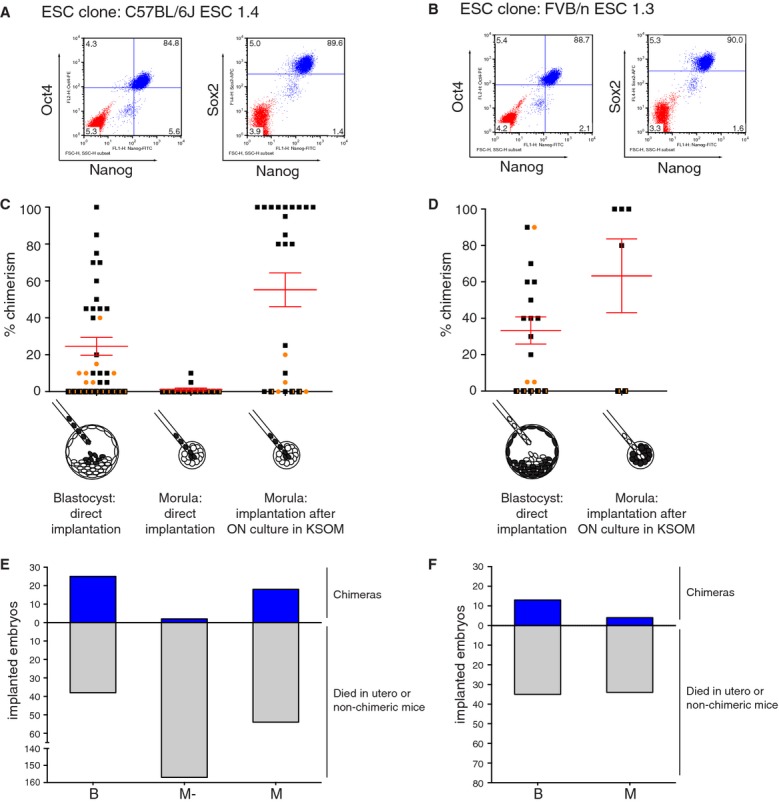
  n.d. D Two ESC injection procedures for FVB/n ESC clone 1.3 were evaluated on basis of chimeric contribution. Blastocyst injections resulted in reasonable chimeras whereas ESC injections into morulae in combination with overnight culture improved chimerism with three out of four live borns showing 100% chimerism (entirely white). The 80% chimera was a runt and died before weaning. male, female, n.d. E,F Efficiency of ESC injection procedures shown in (C) and (D) based on number of viable chimeras born compared to the total number of implanted embryos for C57BL/6J ESC clone 1.4 (E) and FVB/n ESC clone 1.3 (F). Note, for both ESC clones fewer chimeras were observed relative to the total number of implanted embryos when comparing ESC injected morulae to ESC injected blastocysts.
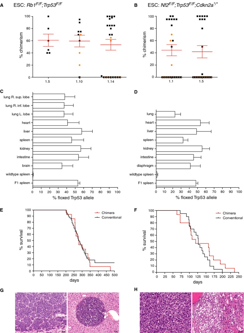 male, female, n.d. C,D Comparison between chimeric contribution estimated on basis of coat-color versus genetic chimerism, tested in various tissues. Southern blot analysis was performed with a probe that distinguishes between a wild-type Trp53 allele or the floxed Trp53 allele reflecting the contribution by the host ESCs or cultured ESCs, respectively (example in supplementary Fig S3). Controls are wild-type spleen (0% chimerism expected) and F1 offspring of chimeras (50% chimerism expected). (C) Genetic chimerism of Rb1F/F
;Trp53F/F chimeras with coat color chimerism ranging from 70 to 100% (average 84%, n = 7). (D) Genetic chimerism of Nf2F/F
;Trp53F/F
;Cdkn2a*/* chimeras with coat color chimerism ranging from 85 to 100% (average 95%, n = 4). E Survival curves of Rb1F/F
;Trp53F/F mice intratracheally injected with Ad5-Cre. Black line, conventional mice; red line, chimeras. F Survival curves of Nf2F/F
;Trp53F/F
;Cdkn2a*/* mice intrathoracically injected with Ad5-Cre. Black line: conventional mice; Red line: chimeras. G Typical example of a neuroendocrine carcinoma (Small Cell Lung Cancer) in the lung (left panel) and a metastatic lesion in the liver (right panel). H Typical example of a mesotheliomatous lesion in the thoracic cavity. Tumor cells are either spindle sarcomatoid cells (left panel) or vacuolated epithelioid cells (right panel).
male, female, n.d. C,D Comparison between chimeric contribution estimated on basis of coat-color versus genetic chimerism, tested in various tissues. Southern blot analysis was performed with a probe that distinguishes between a wild-type Trp53 allele or the floxed Trp53 allele reflecting the contribution by the host ESCs or cultured ESCs, respectively (example in supplementary Fig S3). Controls are wild-type spleen (0% chimerism expected) and F1 offspring of chimeras (50% chimerism expected). (C) Genetic chimerism of Rb1F/F
;Trp53F/F chimeras with coat color chimerism ranging from 70 to 100% (average 84%, n = 7). (D) Genetic chimerism of Nf2F/F
;Trp53F/F
;Cdkn2a*/* chimeras with coat color chimerism ranging from 85 to 100% (average 95%, n = 4). E Survival curves of Rb1F/F
;Trp53F/F mice intratracheally injected with Ad5-Cre. Black line, conventional mice; red line, chimeras. F Survival curves of Nf2F/F
;Trp53F/F
;Cdkn2a*/* mice intrathoracically injected with Ad5-Cre. Black line: conventional mice; Red line: chimeras. G Typical example of a neuroendocrine carcinoma (Small Cell Lung Cancer) in the lung (left panel) and a metastatic lesion in the liver (right panel). H Typical example of a mesotheliomatous lesion in the thoracic cavity. Tumor cells are either spindle sarcomatoid cells (left panel) or vacuolated epithelioid cells (right panel).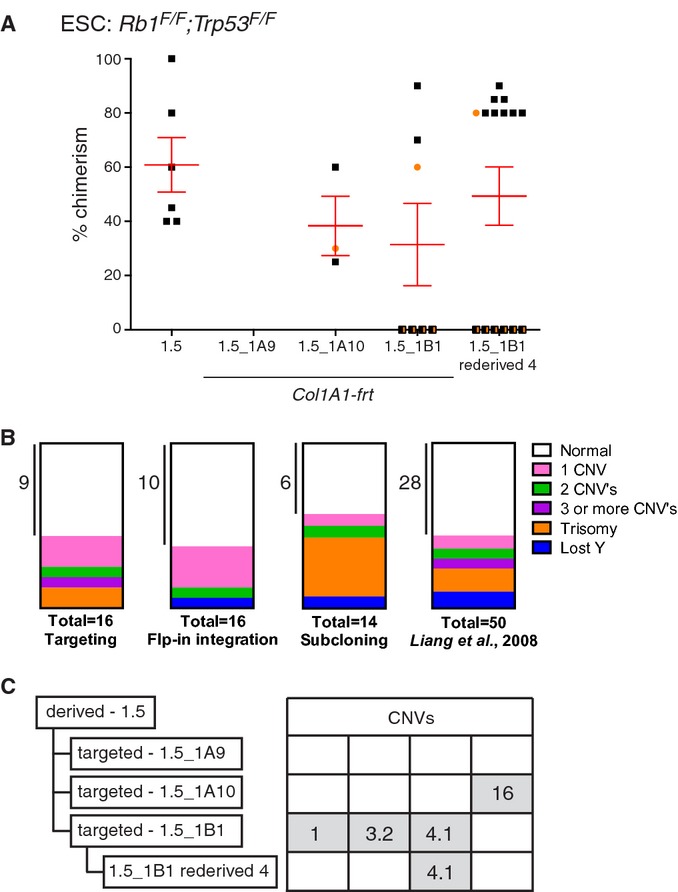
Comparison of chimeric contribution between the parental Rb1F/F ;Trp53F/F ESC clone 1.5 and three Col1a1-frt targeted derivatives. Correct targeting was confirmed by Southern blot analysis using a 3′ probe in the Col1a1 locus (supplementary Fig S4A and B). Two Col1a1-frt targeted clones, i.e. 1.5_1A10 and 1.5_1B1, provided good and germline-competent chimeras (supplementary Table S1). One chimera from the Rb1F/F ;Trp53F/F ESC clone 1.5_1B1 was backcrossed twice to the original strain and ESC were re-derived, i.e. clone 1.5_1B1 re-derived 4 (Table 1). This ESC clone resulted in improved chimeras compared to the parental clones.
male, female, n.d.Parts of whole representation of genetic aberrations observed in GEMM-ESCs cultured in 2i medium and subjected to either gene targeting, Flp-in integration and subcloning (supplementary Table S4). Last box represent the genetic aberrations observed in ESCs cultured under classic culture conditions as reported by Liang et al, .
Summary of CNVs observed in Rb1F/F ;Trp53F/F ESC clones as detected by aCGH. Two Col1a1-frt targeted clones acquired four independent CNVs. Some CNVs can be transmitted via the germ line as CNV-4.1 was maintained after backcrossing twice to the original strain, see ESC clone 1.5_1B1 re-derived 4. A detailed description of all CNVs is provided in supplementary Table S3.
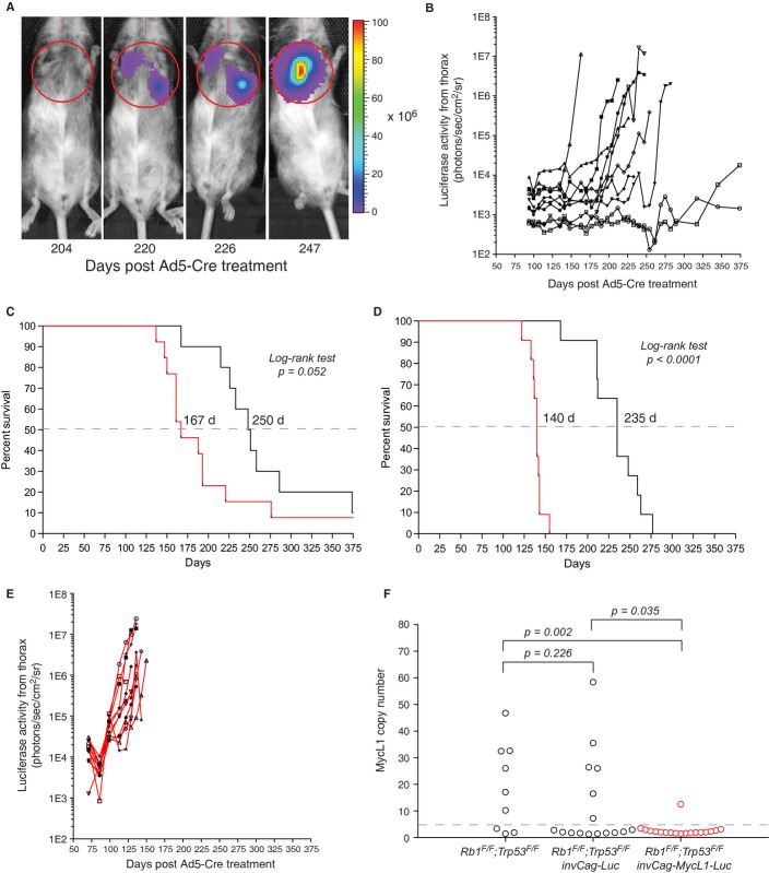
In vivo imaging of a invCAG-Luc;Rb1F/F ;Trp53F/F chimeric mouse injected intrathoracically with Ad5-Cre. Tumor growth was monitored weekly by bioluminescence imaging.
Luciferase activity emitted from the thorax of 10 chimeric invCAG-Luc;Rb1F/F ;Trp53F/F mice. Each line represents measurements of an individual mouse. The chimeric mouse with the lowest coat-color chimerism (○, 20%) did not develop a tumor, while the second lowest chimera (□, 35%) did develop SCLC though with a long latency. One chimera (♦, 962975) failed to show any Luciferase activity but did develop SCLC. Analysis of the tumor revealed a lack of Cre-mediated switching of the invCag-Luc transgene (supplementary Fig S7).
Survival curves of chimeric Rb1F/F ;Trp53F/F mice containing either the invCag-Luc (black line) or the invCag-MycL1-Luc (red line) transgene, intratracheally injected with Ad5-Cre. Median survival indicated by the dotted line was 250 and 167 days, respectively.
Survival curves of F1 Rb1F/F ;Trp53F/F mice containing either the invCag-Luc (black line) or the invCag-MycL1-Luc (red line) transgene, intratracheally injected with Ad5-Cre. Median survival indicated by the dotted line was 235 and 140 days, respectively.
Luciferase activity emitted from the thorax of 11 F1 invCAG-MycL1-Luc;Rb1F/F ;Trp53F/F mice. Each line represents measurements of an individual mouse.
MycL1 copy number in SCLC tumors from three different genotypes determined by real-time PCR and aCGH. Each circle represents a primary SCLC tumor. All tumors with more than four copies (dotted line) were considered positive for MycL1 amplification. Note that overexpression of MycL1 by the transgene significantly reduces the frequency of genomic MycL1 amplifications in tumors as compared to the Rb1F/F ;Trp53F/F control ( P = 0.002 Fisher's Exact Test) and the invCAG-Luc;Rb1F/F ;Trp53F/F control ( P = 0.035 Fischer's Exact Test).
Similar articles
-
Rapid validation of cancer genes in chimeras derived from established genetically engineered mouse models.Bioessays. 2011 Sep;33(9):701-10. doi: 10.1002/bies.201100018. Epub 2011 Jul 7. Bioessays. 2011. PMID: 21735458 Free PMC article.
-
Using the GEMM-ESC strategy to study gene function in mouse models.Nat Protoc. 2015 Nov;10(11):1755-85. doi: 10.1038/nprot.2015.114. Epub 2015 Oct 15. Nat Protoc. 2015. PMID: 26492136
-
The perfect host: a mouse host embryo facilitating more efficient germ line transmission of genetically modified embryonic stem cells.PLoS One. 2013 Jul 2;8(7):e67826. doi: 10.1371/journal.pone.0067826. Print 2013. PLoS One. 2013. PMID: 23844102 Free PMC article.
-
Stem cell potency and the ability to contribute to chimeric organisms.Reproduction. 2013 Mar 7;145(3):R81-8. doi: 10.1530/REP-12-0396. Print 2013 Mar 1. Reproduction. 2013. PMID: 23221011 Free PMC article. Review.
-
Haploid mouse embryonic stem cells: rapid genetic screening and germline transmission.Annu Rev Cell Dev Biol. 2014;30:705-22. doi: 10.1146/annurev-cellbio-100913-012920. Annu Rev Cell Dev Biol. 2014. PMID: 25288120 Review.
Cited by
-
YAP drives fate conversion and chemoresistance of small cell lung cancer.Sci Adv. 2021 Oct;7(40):eabg1850. doi: 10.1126/sciadv.abg1850. Epub 2021 Oct 1. Sci Adv. 2021. PMID: 34597132 Free PMC article.
-
Complexity galore: 3D cultures, biomechanics and systems medicine at the eighth ENBDC workshop "Methods in Mammary Gland Development and Cancer".Breast Cancer Res. 2016 Nov 25;18(1):115. doi: 10.1186/s13058-016-0777-2. Breast Cancer Res. 2016. PMID: 27887657 Free PMC article.
-
MYCN drives chemoresistance in small cell lung cancer while USP7 inhibition can restore chemosensitivity.Genes Dev. 2020 Sep 1;34(17-18):1210-1226. doi: 10.1101/gad.340133.120. Epub 2020 Aug 20. Genes Dev. 2020. PMID: 32820040 Free PMC article.
-
Tumor Heterogeneity Underlies Differential Cisplatin Sensitivity in Mouse Models of Small-Cell Lung Cancer.Cell Rep. 2019 Jun 11;27(11):3345-3358.e4. doi: 10.1016/j.celrep.2019.05.057. Cell Rep. 2019. PMID: 31189116 Free PMC article.
-
Targeting transcriptional addictions in small cell lung cancer with a covalent CDK7 inhibitor.Cancer Cell. 2014 Dec 8;26(6):909-922. doi: 10.1016/j.ccell.2014.10.019. Cancer Cell. 2014. PMID: 25490451 Free PMC article.
References
-
- Barakat TS, Gribnau J. X chromosome inactivation and embryonic stem cells. Adv Exp Med Biol. 2010;695:132–154. - PubMed
-
- Beard C, Hochedlinger K, Plath K, Wutz A, Jaenisch R. Efficient method to generate single-copy transgenic mice by site-specific integration in embryonic stem cells. Genesis. 2006;44:23–28. - PubMed
-
- Belteki G, Gertsenstein M, Ow DW, Nagy A. Site-specific cassette exchange and germline transmission with mouse ES cells expressing phiC31 integrase. Nat Biotechnol. 2003;21:321–324. - PubMed
-
- Calbo J, Meuwissen R, van Montfort E, van Tellingen O, Berns A. Genotype-phenotype relationships in a mouse model for human small-cell lung cancer. Cold Spring Harb Symp Quant Biol. 2005;70:225–232. - PubMed
Publication types
MeSH terms
Substances
LinkOut - more resources
Full Text Sources
Other Literature Sources
Medical
Molecular Biology Databases
Research Materials
