. 2014 Jan 8;6(218):218ra6.
doi: 10.1126/scitranslmed.3006557.

A blood-resistant surgical glue for minimally invasive repair of vessels and heart defects

Affiliations

A blood-resistant surgical glue for minimally invasive repair of vessels and heart defects

Nora Lang et al. Sci Transl Med. .

Abstract

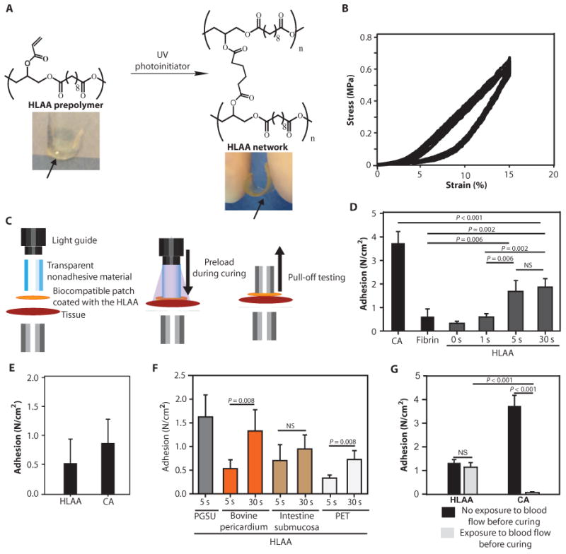
Currently, there are no clinically approved surgical glues that are nontoxic, bind strongly to tissue, and work well within wet and highly dynamic environments within the body. This is especially relevant to minimally invasive surgery that is increasingly performed to reduce postoperative complications, recovery times, and patient discomfort. We describe the engineering of a bioinspired elastic and biocompatible hydrophobic light-activated adhesive (HLAA) that achieves a strong level of adhesion to wet tissue and is not compromised by preexposure to blood. The HLAA provided an on-demand hemostatic seal, within seconds of light application, when applied to high-pressure large blood vessels and cardiac wall defects in pigs. HLAA-coated patches attached to the interventricular septum in a beating porcine heart and resisted supraphysiologic pressures by remaining attached for 24 hours, which is relevant to intracardiac interventions in humans. The HLAA could be used for many cardiovascular and surgical applications, with immediate application in repair of vascular defects and surgical hemostasis.


Figures

Fig. 1
Fig. 1. Elastic and adhesive properties of the HLAA on wet biological tissues
(A) Chemical structure of the HLAA before and after exposure to UV light. (B) HLAA properties under cyclical loading. (C) Pull-off method for testing adhesive strength of the HLAA. First, the tissue was glued onto a flat metal substrate, and the patch coated with the HLAA was pressed against the tissue using a transparent nonadhesive material and the UV light guide with a preload of −3 N. UV light was applied to activate the HLAA and the transparent nonadhesive material, and the UV light guide was then removed. A second flat metal substrate was glued to the patch for pull-off testing. (D) Pull-off adhesion strength of fibrin sealant, CA, and HLAA exposed to UV light for different periods of time (0, 1, 5, or 30 s) against epicardial tissue (fibrin: n = 4, CA: n = 3, HLAA: n = 4). PGSU patch material was used for all the experiments. P values were determined by one-way analysis of variance (ANOVA) with the Tukey post hoc test. (E) In vivo adhesion strength of HLAA-and CA-coated patches after 2 days of implantation. Strengths were determined using a pull-off procedure on explanted rat epicardium (n = 3). (F) Adhesion strength using PGSU and clinically used patch materials against fresh cardiac tissue (n = 5). P values were determined by Mann-Whitney rank sum test. PET, polyethylene terephthalate. (G) HLAA and CA adhesion with or without exposure to blood before contact with cardiac tissue (n = 3). P values were determined by one-way ANOVA with Tukey post hoc testing. Data in (D) to (G) are means ± SD. NS, not significant.
Fig. 2
Fig. 2. Interaction between the HLAA and collagen substrates
(A) The HLAA-coated patch was applied ex vivo to porcine epicardium, and the interface was examined using MT staining. M, myocardium; C, collagen; A, HLAA; P, PGSU patch. The HLAA appeared to entangle with epicardium collagen fibers (arrows). Scale bars, 500 μm (left) and 20 μm (right). (B) Freeze-fractured HLAA-tissue interface observed with SEM for endocardium (left) and epicardium (right) tissues. Scale bars, 30 and 2 μm, respectively. (C) Controlled adhesion tests were performed on glass slides coated with collagen type I. Data are means ± SD (n = 3). (D) HLAA pull-off adhesion strength against the adventitia of pig carotid artery and epicardium tissue. Data are means ± SD (n = 5). P values in (C) and (D) were determined by unpaired t test.
Fig. 3
Fig. 3. In vivo biocompatibility and adhesive strength testing of HLAA- and CA-coated elastomeric patches
(A) Experimental methodology and time points selected to evaluate the biocompatibility of the HLAA. CA was used as the control. (B) Explanted hearts 14 days after implantation and corresponding hematoxylin and eosin (H&E) staining of the cardiac tissue in contact with the adhesives. A thicker layer of inflammatory cells was observed for CA, as indicated by arrowheads. Scale bars, 500 μm. (C) The degree of tissue necrosis and inflammation for the HLAA and CA (0, negligible; 1, reduced; 2, moderate; 3, severe) was scored by a blinded pathologist. Data are means ± SD (n = 3). P values were determined by one-way ANOVA with Tukey post hoc testing. (D) Experimental methodology and selected time points for closure of LV free wall cardiac defects in the rat. A 2-mm defect was created and closed with a patch (diameter, 6 mm) coated with the HLAA. Closure of the defect with sutures was used as a control. (E) Representative H&E- and MT-stained sections at 1 and 6 months after defect closure for the suture and HLAA groups (n = 3). Scale bars, 1 mm.
Fig. 4
Fig. 4. HLAA-coated patches attach to beating hearts in vivo in a large animal model
(A) Device and technique for minimally invasive patch delivery and attachment to the pig heart through a single LV incision. (B) Echocardiographic evaluation of patch location 0 hours (n = 4) and 24 hours (n = 2) after patch attachment. LV, left ventricle; RV, right ventricle. (C) Location of the deployed patch 24 hours after initial application (A, apex; P, patch; PM, papillary muscles; LV, left ventricle). (D) H&E stain of the tissue surrounding the patch material after 4 hours (n = 2) and 24 hours (n = 2) of implantation. M, myocardium; P, patch; C, capsule. Scale bars, 1 mm (left) and 200 μm (right).
Fig. 5
Fig. 5. HLAA alone can close vascular defects
(A) A 2-mm incision on ex vivo porcine carotid arteries was closed with the HLAA, without a patch. (B) Carotid artery after defect creation and 24 hours after closure with the HLAA. (C) Doppler images demonstrating vessel flow before defect creation and 24 hours after closure. (D) H&E-stained sections from the HLAA-treated vessels (left) and a noninjured carotid artery (right). Arrow points to the defect created. Images are representative of n = 4 animals. Scale bars, 1 mm (left) and 50 μm (right). (E) Thrombogenic response of HLAA. The lactate dehydrogenase colorimetric signal was determined after incubating glass, HLAA, and PGSU with porcine blood for 1 hour. Data are means ± SD (n = 5). P values were determined by one-way ANOVA with Tukey post hoc test.

References

    1. Spotnitz WD. Hemostats, sealants, and adhesives: A practical guide for the surgeon. Am Surg. 2012;78:1305–1321. - PubMed
    1. Dolgin E. The sticking point. Nat Med. 2013;19:124–125. - PubMed
    1. Chang EI, Galvez MG, Glotzbach JP, Hamou CD, El-ftesi S, Rappleye CT, Sommer KM, Rajadas J, Abilez OJ, Fuller GG, Longaker MT, Gurtner GC. Vascular anastomosis using controlled phase transitions in poloxamer gels. Nat Med. 2011;17:1147–1152. - PubMed
    1. Cheema FH, Younus MJ, Roberts HG., Jr Repairing the posterior postinfarction ventricular septal defect: A left ventricular approach with a sealant reinforced multipatch technique. Semin Thorac Cardiovasc Surg. 2012;24:63–66. - PubMed
    1. Predescu D, Chaturvedi RR, Friedberg MK, Benson LN, Ozawa A, Lee KJ. Complete heart block associated with device closure of perimembranous ventricular septal defects. J Thorac Cardiovasc Surg. 2008;136:1223–1228. - PubMed

Publication types

MeSH terms