. 2014 Jan 16;505(7483):372-7.
doi: 10.1038/nature12928. Epub 2014 Jan 8.

UvrD facilitates DNA repair by pulling RNA polymerase backwards

Affiliations

UvrD facilitates DNA repair by pulling RNA polymerase backwards

Vitaly Epshtein et al. Nature. .

Abstract

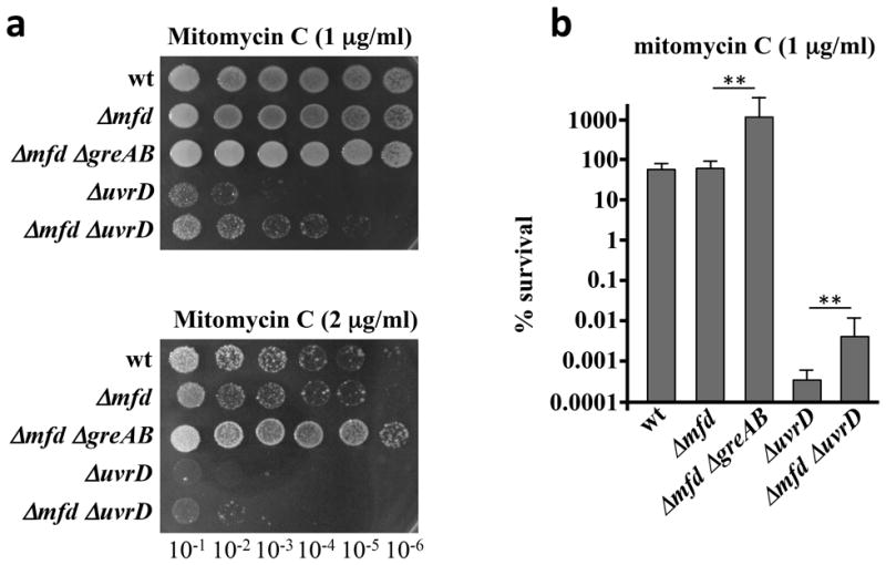
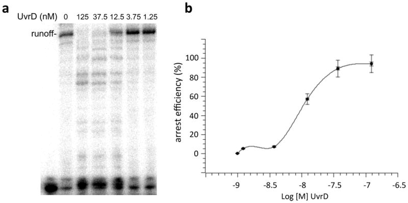
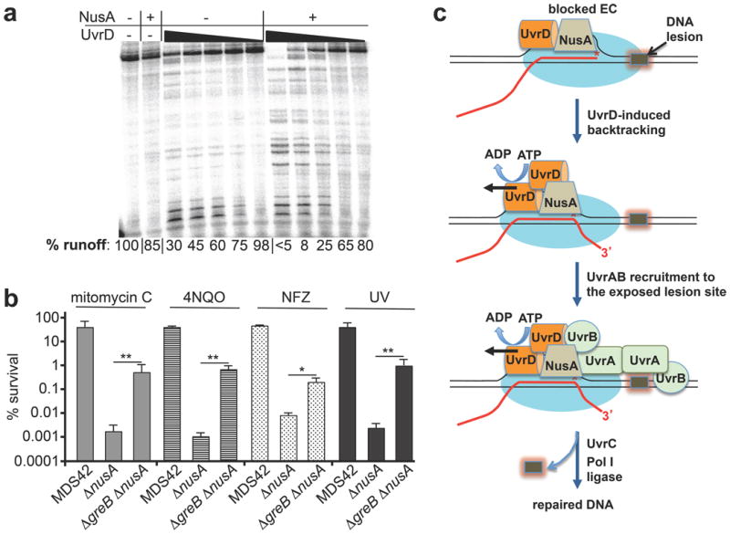
UvrD helicase is required for nucleotide excision repair, although its role in this process is not well defined. Here we show that Escherichia coli UvrD binds RNA polymerase during transcription elongation and, using its helicase/translocase activity, forces RNA polymerase to slide backward along DNA. By inducing backtracking, UvrD exposes DNA lesions shielded by blocked RNA polymerase, allowing nucleotide excision repair enzymes to gain access to sites of damage. Our results establish UvrD as a bona fide transcription elongation factor that contributes to genomic integrity by resolving conflicts between transcription and DNA repair complexes. Furthermore, we show that the elongation factor NusA cooperates with UvrD in coupling transcription to DNA repair by promoting backtracking and recruiting nucleotide excision repair enzymes to exposed lesions. Because backtracking is a shared feature of all cellular RNA polymerases, we propose that this mechanism enables RNA polymerases to function as global DNA damage scanners in bacteria and eukaryotes.


Figures

Extended Data Figure 1
Extended Data Figure 1. UvrD binds RNAP
a, A sample list of RNAP-bound proteins from exponentially grown E. coli. Abundance is based on emPAI score. b, UvrD + 6His–RNAP, UvrD, or 6His–RNAP were affinity-purified on nickel beads and electrophoresed alongside pure UvrD, for reference; The asterisk indicates UvrD. c, RNAP-UvrD complex isolated by size-exclusion chromatography. UvrD–RNAP were mixed 3:1, and run over a Superdex 200 10/300GL column. RNAP core (red) and UvrD (green) chromatograms are overlaid for comparison. The inlaid polyacrylamide gel displays fractions taken from each chromatographic peak.
Extended Data Figure 2
Extended Data Figure 2. UvrD promotes long-range backtracking
Biotinylated RNAP was used to prepare the startup EC11 immobilizing on beads. It was walked to position 39, followed by incubation with UvrD, washing (to remove UvrD and NTPs), and then treatment with GreB for indicated times. Numbers on the right indicate the size of 5′-labelled RNAs.
Extended Data Figure 3
Extended Data Figure 3. Thymine dimer in the template strand blocks the elongation complex
Schematic diagram of the T7A1 promoter template shows the position of CPD (red). Both control (no TT) and CPD-bearing template strands were radiolabelled at their 5′ ends (top band). The sequencing gel also shows the radiolabelled RNA products before and after chase of EC11. The EC was completely halted by CPD (indicated by red TT). On the control template it formed the runoff products.
Extended Data Figure 4
Extended Data Figure 4. Effect of greAB, mfd and ribosome inactivation on uvrD sensitivity to DNA damaging agents and UV
a, Representative efficiencies of colony formation of wild-type (MG1655) and mutant E. coli cells in the presence of the indicated amounts of mitomycin C, 4NQO and cisplatin. Cells were spotted on LB agar plates in serial tenfold dilutions and incubated at 30 °C for 24 h. b, Representative efficiencies of colony formation of wild-type and ΔuvrD cells in the presence of the indicated amounts of mitomycin, 4NQO and chloramphenicol. Cells were spotted on LB agar plates in serial tenfold dilutions and incubated at 30 °C for 24 h. c, Representative efficiencies of colony formation of wild-type (MG1655) and mutant E. coli cells after UV irradiation. Cells were spotted on LB agar plates in serial tenfold dilutions and incubated at 30 °C for 24 h. d, Representative efficiencies of colony formation of wild-type and mutant E. coli cells after UV irradiation in the presence of a sublethal dose of chloramphenicol. Cells were spotted on LB agar plates in serial tenfold dilutions and incubated at 30 °C for 24 h.
Extended Data Figure 5
Extended Data Figure 5. UvrD inactivation suppresses temperature sensitivity of greAB cells
E. coli strains were streaked on LB agar plates and incubated at the indicated temperatures for 24 h.
Extended Data Figure 6
Extended Data Figure 6. UvrD–RNAP crosslinks
a, Three inter-protein crosslinks indicate that UvrD binds near the β flap on RNAP. UvrD (grey, PDB accession code 2IS4) cross-links to RNAP (PDB accession code 4IGC) at three distinct positions that span the β (pale yellow) and β′ (light blue) subunits of RNAP. The non-template strand (blue, PDB accession code 4G7O) is indicated for reference. Crosslinked lysines are colored magenta and pairs are connected with a black line. b, MS2 spectrum of a representative crosslinked pair (β′ K40)–(UvrD K448). The peptide sequences with cross-linked lysine residues are shown (top right). Observed peptide backbone cleavage is indicated and b- and y-type fragment ions are labelled in the spectrum. The m/z tolerances of fragment ions are presented in the inset below the spectra. Spectra were annotated using pLabel.
Extended Data Figure 7
Extended Data Figure 7. GreB inactivation suppresses nusA sensitivity to genotoxic chemicals and UV
a, Representative efficiencies of colony formation of wild-type (MG1655) and mutant E. coli cells in the presence of indicated amounts of NFZ and 4NQO. Cells were spotted on LB agar plates in serial tenfold dilutions and incubated at 30 °C for 24 h. b, Data from three independent experiments are presented as the mean ± s.e.m.; **P < 0.01. c, Representative efficiencies of colony formation of MDS42 and mutant E. coli cells in the presence of indicated amounts of mitomycin, 4NQO, NFZ and after UV irradiation. Cells were spotted on LB agar plates in serial tenfold dilutions and incubated at 30 °C for 24 h.
Extended Data Figure 8
Extended Data Figure 8. Deletion of mfd partially suppresses uvrD sensitivity to mitomycin C
a, Representative efficiencies of colony formation of wild-type (MG1655) and mutant E. coli cells in the presence of indicated amounts of mitomycin C. Cells were spotted on LB agar plates in serial tenfold dilutions and incubated at 30 °C for 24 h. b, Data from three independent experiments are presented as the mean ±s.e.m.; **P < 0.01.
Extended Data Figure 9
Extended Data Figure 9. Transcriptional arrest as a function of UvrD concentration
a, A representative chase experiment demonstrating multiple transcriptional arrests as a function of UvrD concentration. b, Data from three independent experiments are plotted as the mean ± s.e.m. Arrest efficiency (%) was calculated as a fraction of all arrested complexes in relation to the full-length runoff. Extended Data Table 1 Escherichia coli strains used in this study.
Figure 1
Figure 1. UvrD promotes RNAP backtracking
a, EC20 was combined with wild-type UvrD (lanes 2–5), catalytically inactive UvrDE221Q, (lanes 6, 7), or no UvrD (lane 1) before NTP chase (lanes 1, 2, 6) and wash (lanes 3, 7). Red lines connect corresponding RNAs from arrested elongation complexes before and after GreB cleavage (lanes 4, 5). b, EC11 was walked to positions 36, 38 and 39. UvrD was added ± ATP and chased. Inactivated EC36–39 (%) is indicated. c, The p1EC constructs (left) and primer extension analyses (right). CAA modifications on the non-template strand of p1EC + pVector (lanes 2, 3) or p1EC + pUvrD (lanes 4, 5). The lac operator (Lac) and transcription bubble are indicated. Red lines show elongation complex position.
Figure 2
Figure 2. RNAP backtracking facilitates NER
a, UvrD pulls RNAP from thymine dimers (TT). The T7A1 promoter template with TT (red) and schematic overview (top); EC11 was chased to TT (lanes 9–16) (bottom). Where indicated, UvrD was added for 5 min. Red boxes show UvrC-mediated DNA cleavage products (19 nucleotides); per cent cleavage is averaged ( ± s.e.m.) from four independent experiments P < 0.05. b, Left, the primer extension experimental overview. Right, primer extension products reflect the location of lesions. Orange lines indicate repair zones for percentage repair. Asterisks indicate representative lesions repaired faster in greAB than in wild-type cells (blue), at the same rate (green) or ‘non-disappearing’ bands for normalization (orange).
Figure 3
Figure 3. Anti-backtracking factors obstruct UvrD activity in NER
a, Inactivating greAB or slowing ribosomal translocation (with 1 μg ml−1 chloramphenicol) suppresses uvrD sensitivity to mitomycin C (1 μg ml−1; filled bars) and 4NQO (1 μM; striped bars). Data from three independent experiments are presented as the mean ± s.e.m.; *P < 0.05, **P < 0.01. Cm, chloramphenicol; wt, wild type. b, Inactivating greAB, mfd or slowing ribosomal translocation suppresses uvrD sensitivity to UV irradiation (5 J per m2 at 30 °C). Data from three independent experiments are presented as the mean ± s.e.m.; **P < 0.01. c, Cartoon summarizing interference with NER.
Figure 4
Figure 4. Mapping UvrD interactions with the elongation complex
a, The 5′-radiolabelled scaffold carries a single photo-inducible 4-thio-dU (red) in the template strand (top). Protein crosslinking adducts corresponding to the β and β′ subunits of RNAoP and UvrD (red asterisk) (middle). A model of UvrD binding (bottom). b, UvrD (top, Protein Data Bank (PDB) accession number 2IS4) is cross-linked to RNAP (middle, PDB accession number 4IGC) at three positions (magenta) that span the β (yellow) and β′ (light blue) subunits, proximal to the non-template strand (blue, PDB accession number 4G7O). The β flap-tip-helix (green) is indicated and the suggested binding interface is circled. Schematic summarizing UvrD–RNAP binding shown below.
Figure 5
Figure 5. UvrD and NusA cooperate in backtracking-mediated NER
a, NusA facilitates UvrD-mediated backtracking. The fraction of full-length transcript (percentage runoff) is indicated. b, GreB inactivation in MDS43 cells suppresses ΔnusA sensitivity to mitomycin C (0.5 μg ml−1), 4NQO (3 μM), nitrofurazone (NFZ, 2 μM), and UV irradiation (15 J per m2). Data from three independent experiments are presented as the mean ± s.e.m.; *P < 0.05, **P < 0.01. c, Model for backtracking-mediated NER.

Comment in

Similar articles

Cited by

References

    1. Reardon JT, Sancar A. Nucleotide excision repair. Prog Nucleic Acid Res Mol Biol. 2005;79:183–235. Medline CrossRef. - PubMed
    1. Van Houxen B, McCullough A. Nucleotide excision repair in E. coli. Ann NY Acad Sci. 1994;726:236–251. Medline CrossRef. - PubMed
    1. Ganesan A, Spivak G, Hanawalt PC. Transcription-coupled DNA repair in prokaryotes. Prog Mol Biol Transl Sci. 2012;110:25–40. Medline CrossRef. - PubMed
    1. Truglio JJ, Croteau DL, Van Houten B, Kisker C. Prokaryotic nucleotide excision repair: the UvrABC system. Chem Rev. 2006;106:233–252. Medline CrossRef. - PubMed
    1. Mellon I, Hanawalt PC. Induction of the Escherichia coli lactose operon selectively increases repair of its transcribed DNA strand. Nature. 1989;342:95–98. Medline CrossRef. - PubMed

Publication types

MeSH terms