. 2014 Jul;26(1):99-113.
doi: 10.1007/s12640-013-9455-7. Epub 2014 Jan 9.

Group III mGlu receptor agonist, ACPT-I, exerts potential neuroprotective effects in vitro and in vivo

Affiliations

Group III mGlu receptor agonist, ACPT-I, exerts potential neuroprotective effects in vitro and in vivo

Helena Domin et al. Neurotox Res. 2014 Jul.

Abstract

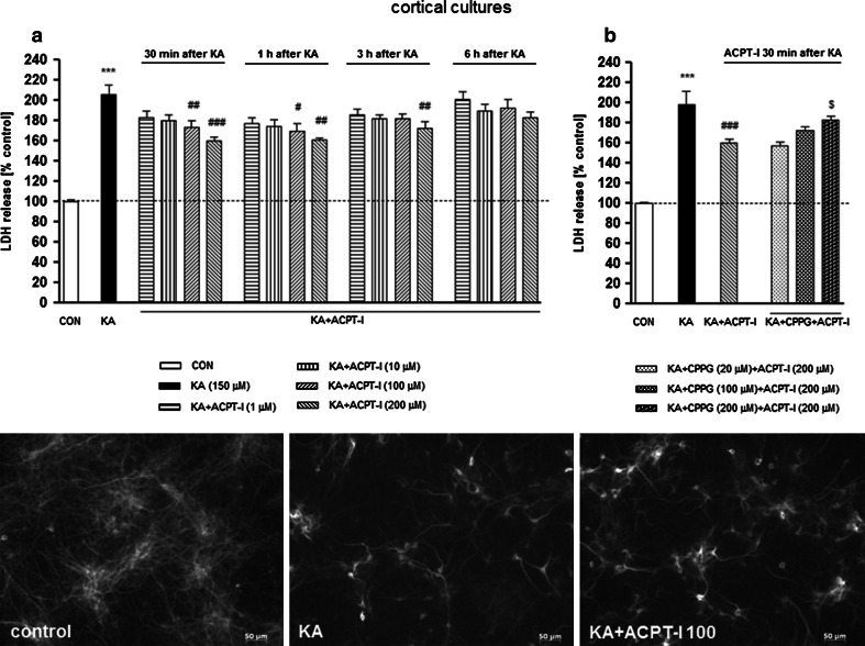
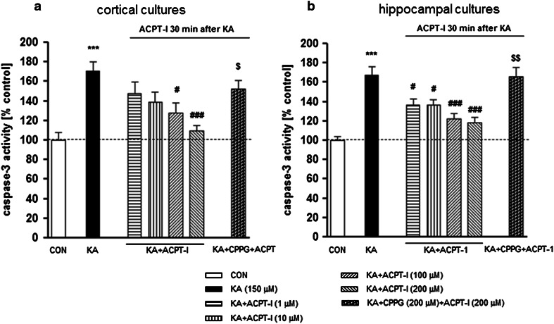
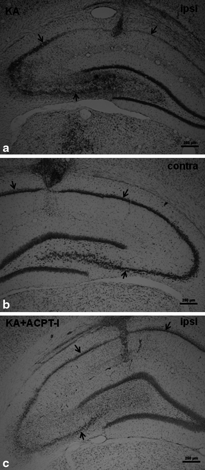
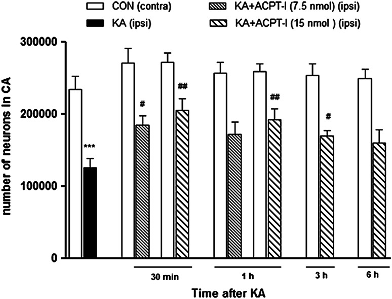
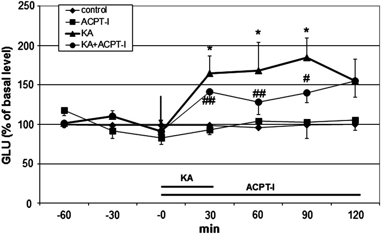
Many evidence suggest that metabotropic glutamate receptors (mGluRs) may modulate glutamatergic transmission, hence, these receptors are regarded as potential targets for neuroprotective drugs. Since group III mGlu receptor agonists are known to reduce glutamatergic transmission by inhibiting glutamate release, we decided to investigate the neuroprotective potential of the group III mGlu receptor agonist, (1S,3R,4S)-1-aminocyclopentane-1,2,4-tricarboxylic acid (ACPT-I) against kainate (KA)-induced excitotoxicity in vitro and in vivo. In primary neuronal cell cultures ACPT-I (1-200 μM), applied 30 min-3 h after starting the exposure to KA (150 μM), significantly attenuated the KA-induced LDH release, increased cell viability, and inhibited caspase-3 activity both in cortical and hippocampal cell cultures. The effects were dose-, time- and structure-dependent. The neuroprotective effects of ACPT-I were reversed by (RS)-alpha-cyclopropyl-4-phosphonophenyl glycine, a group III mGluR antagonist. In the in vivo studies, KA (2.5 nmol/1 μl) was unilaterally injected into the rat dorsal CA1 hippocampal region and the size of degeneration was examined by stereological counting of surviving neurons in the CA pyramidal layer. It was found that ACPT-I (7.5 or 15 nmol/1 μl), injected into the dorsal hippocampus 30 min, 1 or 3 h after KA in dose-dependent manner prevented the KA-induced neuronal damage. Moreover, in vivo microdialysis studies in the rat hippocampus showed that ACPT-I (200 μM) given simultaneously with KA (50 μM) significantly diminished the KA-induced glutamate release in the hippocampus. This mechanism seems to play a role in mediating the neuroprotective effect of ACPT-I.


Figures

Fig. 1
Fig. 1
(upper panel) a The effect of ACPT-I on kainate (KA; 150 μM)-induced LDH release in the primary cultures of a mouse cortical neurons. ACPT-I (1, 10, 100, or 200 μM) was added to the culture medium 30 min, 1, 3, or 6 h after starting the exposure to KA. (upper panel) b The effect of CPPG on changes in LDH release induced by KA and ACPT-I in primary cultures of a mouse cortical neurons. ACPT-I (200 μM) was added to the culture medium 30 min after KA; CPPG (20, 100, or 200 μM) was applied 10 min before the ACPT-I. LDH was measured 48 h after KA administration. The data were normalized as a percentage of control value and expressed as the mean of n ≥ 6 platings ± SEM from 3 to 4 independent experiments. *** P < 0.001 (vs. control cultures); # P < 0.05, ## P < 0.01, ### P < 0.001 (vs. KA-treated cultures); and $ P < 0.05 (vs. KA + ACPT-I-treated cultures). (Bottom panel) Microphotographs from MAP-2 immunofluorescence of cortical neurons. Numerous clusters of neurons are seen in control cultures. The density of immunostained neurons visibly decreased after 48 h incubation with KA. ACPT-I (100 μM; 1 h after KA) partially prevents the reduction in the number of MAP-2 positive cells in KA-treated cortical neurons. Calibration bars 50 μm
Fig. 2
Fig. 2
(upper panel) a The effect of ACPT-I on kainate (KA; 150 μM)-induced LDH release in the primary cultures of a mouse hippocampal neurons. ACPT-I (1, 10, 100, or 200 μM) was added to the culture medium 30 min, 1, 3, or 6 h after starting the exposure to KA. (upper panel) b The effect of CPPG on changes in LDH release induced by KA and ACPT-I in primary cultures of a mouse hippocampal neurons. ACPT-I (200 μM) was added to the culture medium 30 min after KA; CPPG (20, 100 or 200 μM) was applied 10 min before the ACPT-I. LDH was measured 24 h after KA administration. The data were normalized as a percentage of control value and expressed as the mean of n ≥ 6 platings ± SEM from 3 to 4 independent experiments. *** P < 0.001 (vs. control cultures); # P < 0.05, ## P < 0.01, ### P < 0.001 (vs. KA-treated cultures); and P < 0.01 (vs. KA + ACPT-I-treated cultures). (Bottom panel) Microphotographs from MAP-2 immunofluorescence of hippocampal neurons. Numerous neurons with processes are seen in control cultures. The decrease in neurons density and diminution of their processes are found after 24 h incubation with KA. ACPT-I (100 μM; 1 h after KA) partially prevents the reduction in the number of MAP-2 positive cells in KA-treated hippocampal neurons. Calibration bars 50 μm
Fig. 3
Fig. 3
The effect of ACPT-I and CPPG on the KA-induced increase in caspase-3 activity in a mouse’s primary cortical (a) and hippocampal (b) cultures. Caspase-3 was measured 6 h after starting the exposure to KA. ACPT-I (1, 10, 100, or 200 μM) was added to the culture medium 30 min after KA; CPPG (200 μM) was applied 10 min before the ACPT-I. The data were normalized as a percentage of control value and expressed as the mean of n ≥ 6 platings ± SEM from 3 to 4 independent experiments. *** P < 0.001 (vs. control cultures); # P < 0.05, ### P < 0.001 (vs. KA-treated cultures); and $ P < 0.05, P < 0.01 (vs. KA + ACPT-I-treated cultures)
Fig. 4
Fig. 4
Microphotographs of coronal sections of rat brain hippocampi stained with cresyl violet. Arrows indicate a CA pyramidal layer where the neurons were counted. Calibration bars 250 μm. a Loss of neurons and extensive gliosis can be seen in CA after KA microinjection (2.5 nmol/1 μl) in comparison with the non-degenerated contralateral side (b). c Neuroprotective effect of ACPT-I (15 nmol/rat) injected into the hippocampus 3 h after KA. The lesion is much smaller than after KA alone
Fig. 5
Fig. 5
The effect of intrahippocampal injections of KA (2.5 nmol/1 μl) and KA followed by ACPT-I on the number of neurons in the pyramidal layer of CA regions. The results of stereological counting showed neurodegeneration after KA (50 % loss) and neuroprotection induced by ACPT-I given 30 min, 1, or 3 h after KA. No protection was seen when ACPT-I was given 6 h after KA. Each bar represents the mean ± SEM of n = 6 per group. *** P < 0.001 KA (ipsilateral) versus contralateral side, # P < 0.05, ## P < 0.01 KA +ACPT-I (ipsilateral) versus KA-lesioned (ipsilateral) hippocampi
Fig. 6
Fig. 6
The effect of ACPT-I (200 μM) on the extracellular GLU level induced by kainic acid (KA, 50 μM) in the rat hippocampus. Drug administration is indicated with an arrow, while the horizontal bar shows the duration of the treatment. The basal extracellular GLU levels (μM) were 0.78 ± 0.08, 0.55 ± 0.06, 1.01 ± 0.06 and 0.84 ± 0.09 in control, ACPT-I, KA and KA + ACPT-I group, respectively. Data are mean ± SEM (n = 4–6). Repeated measures of ANOVA and Tukey’s post hoc test. * P < 0.05 versus control; # P < 0.05, ## P < 0.01 versus KA-treated group

References

    1. Acher FC, Tellier FJ, Azerad R, Brabet IN, Fagni L, Pin JP. Synthesis and pharmacological characterization of aminocyclopentanetricarboxylic acids: new tools to discriminate between metabotropic glutamate receptor subtypes. J Med Chem. 1997;40:3119–3129. doi: 10.1021/jm970207b. - DOI - PubMed
    1. Arundine M, Tymianski M. Molecular mechanisms of calcium-dependent neurodegeneration in excitotoxicity. Cell Calcium. 2003;34:325–337. doi: 10.1016/S0143-4160(03)00141-6. - DOI - PubMed
    1. Baskys A, Malenka RC. Agonists at metabotropic glutamate receptors presynaptically inhibit EPSCs in neonatal rat hippocampus. J Physiol. 1991;444:687–701. - PMC - PubMed
    1. Bradley SR, Levey AI, Hersch SM, Conn PJ. Immunocytochemical localization of group III metabotropic glutamate receptors in the hippocampus with subtype-specific antibodies. J Neurosci. 1996;16:2044–2056. - PMC - PubMed
    1. Brewer GJ. Serum-free B27/neurobasal medium supports differentiated growth of neurones from the striatum, substantia nigra, septum, cerebral cortex, cerebellum and dentate gyrus. J Neurosci Res. 1995;42:674–683. doi: 10.1002/jnr.490420510. - DOI - PubMed

Publication types

MeSH terms

LinkOut - more resources