Review
. 2014 Jan 8;11(92):20130918.
doi: 10.1098/rsif.2013.0918. Print 2014 Mar 6.

A linear-encoding model explains the variability of the target morphology in regeneration

Affiliations
Review

A linear-encoding model explains the variability of the target morphology in regeneration

Daniel Lobo et al. J R Soc Interface. .

Abstract

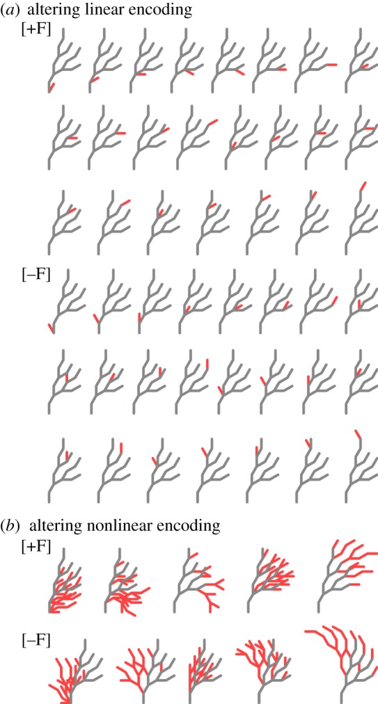
A fundamental assumption of today's molecular genetics paradigm is that complex morphology emerges from the combined activity of low-level processes involving proteins and nucleic acids. An inherent characteristic of such nonlinear encodings is the difficulty of creating the genetic and epigenetic information that will produce a given self-assembling complex morphology. This 'inverse problem' is vital not only for understanding the evolution, development and regeneration of bodyplans, but also for synthetic biology efforts that seek to engineer biological shapes. Importantly, the regenerative mechanisms in deer antlers, planarian worms and fiddler crabs can solve an inverse problem: their target morphology can be altered specifically and stably by injuries in particular locations. Here, we discuss the class of models that use pre-specified morphological goal states and propose the existence of a linear encoding of the target morphology, making the inverse problem easy for these organisms to solve. Indeed, many model organisms such as Drosophila, hydra and Xenopus also develop according to nonlinear encodings producing linear encodings of their final morphologies. We propose the development of testable models of regeneration regulation that combine emergence with a top-down specification of shape by linear encodings of target morphology, driving transformative applications in biomedicine and synthetic bioengineering.

Keywords: deer antler; fiddler crab; in silico modelling; morphology encoding; planaria; regeneration.


Figures

Figure 1.
Figure 1.
Deer antler variable regenerative morphology. (a) Using computed tomography scans, we reconstructed in three dimensions the shed antlers from a white-tailed buck from years 5 to 8. In year 6, the left antler suffered an injury during the early developmental stages of antlerogenesis, producing a ‘royal’ instead of the usual single tine (red arrow). This injury caused the alteration of the regenerative target morphology: in the following years, the left antlers regenerated the ectopic royal in the same location as the original injury (green arrows), and the right antlers (which were never injured) developed a less developed royal in the reciprocal location (blue arrows). (b) On a Siberian wapiti, a cut off the dorsal portion of the germinative bud when the antler had reached nearly 40% of its normal length produced a slight cicatrize in that year (red arrow). The injury altered the target morphology, producing during the following 2 years a new tine (green arrows). Diagrams in (b) modified after [65].
Figure 2.
Figure 2.
Planaria variable regenerative morphology. (a) The planarian wild-type morphology can be divided into three regions (head–trunk–tail), a pattern that is regenerated after almost any kind of amputation. (b) However, certain cuts under the influence of octanol in the media can produce worms with double, triple, and even quadruple heads. (c) A multi-headed worm not only presents an altered morphology, but also suffers a permanent alteration in the regenerative target morphology. (d,e) Subsequent cut fragments, even without the drug that induced the alteration, regenerate the same altered morphology. Worm experiment diagrams extracted from Planform [151].
Figure 3.
Figure 3.
Fiddler crab variable regenerative morphology. (a) Fiddler crabs do not possess an innate handedness, developing two similar chelipeds during development. (b) During growth, one of their chelipeds, with equal probability, is lost. (c) This event establishes the location of the giant cheliped and the regenerative target morphology in the crab—left- or right-handed. (d,e) Further amputations of any or both chelipeds result in the regeneration of the same morphology, that is, the same handedness established with the first amputation.
Figure 4.
Figure 4.
Different types of encodings for an artificial branching morphology. (a) A one-to-one encoding uses a blueprint (blue) to encode the morphology (green). (b) A linear encoding uses a simple algorithm (turtle geometry in this example) to transform a string of instructions (blue) into the morphology (green): every symbol in the string represents a movement for a ‘turtle’ leaving a trace. (c) A nonlinear encoding uses a complex algorithm (L-system rewriting grammar in this example) to create from rules (blue) the morphology (green): a final string of turtle-geometry symbols is emergently created by iteratively applying the rule three times: in each iteration, every ‘F’ symbol in the string is replaced by the string ‘F[+F[−F]]’.
Figure 5.
Figure 5.
Slightly altering the code causes a small local change in a linear-encoded morphology, but a large global change in a nonlinear-encoded morphology. (a) In a linear encoding, a single substring insertion (‘[+F]’ or ‘[−F]’) changes the morphology locally, adding an extra tine (red) to the original morphology (grey). Any possible morphology with an extra tine can be generated with a single simple substring insertion in the linear encoding. (b) In a nonlinear encoding, the effect of a substring insertion is amplified due the emergent nature of the encoding, producing a generalized large change in the generated morphologies. Only a limited set of globally changed morphologies can be generated with a single simple substring insertion in the nonlinear encoding.
Figure 6.
Figure 6.
Many organisms develop according to a nonlinear encoding producing a one-to-one linear encoding of the final morphology. (a) In Drosophila, the maternal gene factors and a complex gene network including gap, pair-rule, and segment polarity genes form together a nonlinear encoding of the homeotic gene expression pattern that is produced in the larva embryo. This expression pattern is a blueprint, a one-to-one encoding, of the final morphology of the fly: each part of the fly morphology is determined by the expression of a homeotic selector gene. (b) In hydra, a reaction–diffusion mechanism (nonlinear encoding) produces a concentration prepattern (one-to-one encoding) of the HyAlx gene, which establishes the location where the hydra tentacles will grow. (c) In Xenopus, a bioelectric network (nonlinear encoding) produces an electrical prepattern (one-to-one encoding) of the tadpole face morphology: ectoderm regions with hyperpolarized cells (brighter) establish the developmental location of the eyes (blue marks) and mouth (red mark). Embryo and adult Drosophila cartoons adapted from [128]. Hydra pictures adapted from [129]. Electric frog embryo picture adapted from [130].
Figure 7.
Figure 7.
A linear encoding can explain the variable target morphology in regeneration. (a) During development, a nonlinear encoding mechanism (such as a genetic network) produces a linear encoding that establishes the target morphology of development and regeneration (antler structure or planaria bodyplan). (b) Injuries or drugs can alter locally the linear encoding of the target morphology, which will cause a corresponding local change in the regenerated morphology. An injury in an antler tine (red arrow) can alter the encoding of that tine in the code, which will cause the regeneration of the antler with a modified morphology (a royal, green arrow) exactly at the location of the tine—a phenomenon hardly possible with a nonlinear encoding. Similarly in planaria, injuries combined with octanol cause the modification of the linear encoding of the body pattern (represented by the patterned circle in the figure), causing the subsequent regeneration of the same altered morphology. In the fiddler crab, the first amputation of a cheliped establishes a linear encoding (represented with a coloured body in the figure) of the crab handedness, which dictates the location of the giant cheliped in subsequent amputations.

References

    1. Crick F. 1970. Diffusion in embryogenesis. Nature 225, 420–422. (10.1038/225420a0) - DOI - PubMed
    1. Meinhardt H, Gierer A. 2000. Pattern formation by local self-activation and lateral inhibition. Bioessays 22, 753–760. (10.1002/1521-1878(200008)22:8<753::AID-BIES9>3.0.CO;2-Z) - DOI - PubMed
    1. Lewis J. 2008. From signals to patterns: space, time, and mathematics in developmental biology. Science 322, 399–403. (10.1126/science.1166154) - DOI - PubMed
    1. Cui M-L, Copsey L, Green A, Bangham A, Coen E. 2010. Quantitative control of organ shape by combinatorial gene activity. PLoS Biol. 8, e1000538 (10.1371/journal.pbio.1000538) - DOI - PMC - PubMed
    1. Lecuit T, Le Goff L. 2007. Orchestrating size and shape during morphogenesis. Nature 450, 189–192. (10.1038/nature06304) - DOI - PubMed

Publication types