. 2014 Jan 8;11(92):20131043.
doi: 10.1098/rsif.2013.1043. Print 2014 Mar 6.

The energy components of stacked chromatin layers explain the morphology, dimensions and mechanical properties of metaphase chromosomes

Affiliations

The energy components of stacked chromatin layers explain the morphology, dimensions and mechanical properties of metaphase chromosomes

Joan-Ramon Daban. J R Soc Interface. .

Abstract

The measurement of the dimensions of metaphase chromosomes in different animal and plant karyotypes prepared in different laboratories indicates that chromatids have a great variety of sizes which are dependent on the amount of DNA that they contain. However, all chromatids are elongated cylinders that have relatively similar shape proportions (length to diameter ratio approx. 13). To explain this geometry, it is considered that chromosomes are self-organizing structures formed by stacked layers of planar chromatin and that the energy of nucleosome-nucleosome interactions between chromatin layers inside the chromatid is approximately 3.6 × 10(-20) J per nucleosome, which is the value reported by other authors for internucleosome interactions in chromatin fibres. Nucleosomes in the periphery of the chromatid are in contact with the medium; they cannot fully interact with bulk chromatin within layers and this generates a surface potential that destabilizes the structure. Chromatids are smooth cylinders because this morphology has a lower surface energy than structures having irregular surfaces. The elongated shape of chromatids can be explained if the destabilizing surface potential is higher in the telomeres (approx. 0.16 mJ m(-2)) than in the lateral surface (approx. 0.012 mJ m(-2)). The results obtained by other authors in experimental studies of chromosome mechanics have been used to test the proposed supramolecular structure. It is demonstrated quantitatively that internucleosome interactions between chromatin layers can justify the work required for elastic chromosome stretching (approx. 0.1 pJ for large chromosomes). The high amount of work (up to approx. 10 pJ) required for large chromosome extensions is probably absorbed by chromatin layers through a mechanism involving nucleosome unwrapping.

Keywords: biomechanics; bionanoscience; chromatin higher order structure; metaphase chromosome structure; supramolecular structures.


Figures

Figure 1.
Figure 1.
Chromosome dimensions and structural models for metaphase chromatin folding. (a) Schematic illustration of different models for chromatin structure in metaphase chromosomes: (a) chromatin fibres (grey lines) form loops that are bound to a central protein scaffold (brown) [–10], (b) chromatin fibres are irregularly folded [–13], and (c) chromatin has a laminar structure with many staked layers oriented perpendicular to the chromatid axis [,–18]; parallel lines represent the side view of the stacked layers. (b) Examples showing the differences in metaphase chromosome sizes that exist among animal species: crested newt (T. cristatus) (a), human (b), and D. melanogaster (c); the three chromosome sets are represented at the same magnification; figure reproduced from [19] with permission from the Royal Society.
Figure 2.
Figure 2.
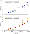
Dimensions of chromosomes calculated using equation (2.2). The calculated diameters (a) and lengths (b) of chromatids containing different amounts of DNA are indicated with diamonds and the corresponding experimental values with squares.
Figure 3.
Figure 3.
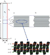
Idealized representation of the different energy regions in chromosomes formed by stacked chromatin layers and description of different variables considered in the text. The schematized chromatid has a length L and a radius R (=D/2) and contains a molecule of DNA of NMb megabases coiled in Nn nucleosomes. The number of nucleosomes in the surface of the telomeres and in the lateral surface are NT and NL, respectively. (a) The energy per nucleosome in bulk metaphase chromatin (ɛ0) is lower than that of a nucleosome in the chromatid surface. Nucleosomes in any internal layer (i) are stabilized by interactions within the layer and by interactions with two adjacent layers (i − 1 and i + 1). In the two telomere surfaces (layers 1 and n, indicated in black), nucleosomes have an extra energy (ɛT) because each one of these layers can interact only with one adjacent layer (2 and n − 1, respectively); this generates a surface potential μT (the surface area of telomeres is ST). Nucleosomes in the chromatid lateral surface (red) have also an extra energy (ɛL) because they are more exposed to the medium and are less stabilized by the attractive interactions within the layer; the corresponding surface potential is μL (the lateral surface area is SL). (b) Irregular stacking of chromatin layers destabilizes the structure because there are more nucleosomes exposed to the medium, increasing the lateral surface area. (c) Schematic representation of nucleosome organization in successive chromatin layers according to [16] (reproduced with permission from the American Chemical Society); nucleosomes are represented by two turns of DNA (black) wrapped around the core histone octamer (grey); layers are indicated by discontinuous red and green lines; the consecutive nucleosomes in each layer are connected with linker DNA (not shown in the drawing). The energy per nucleosome in bulk chromatin ɛ0 has two components: the energy ɛwl due to all interactions within each layer and the energy ɛnn due to the interactions with the interdigitated nucleosomes of the two adjacent layers.
Figure 4.
Figure 4.
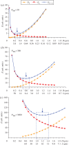
Surface energy of metaphase chromosomes as a function of their diameter and length. The plots correspond to chromatids containing different amounts of DNA: (a) 30, (b) 300 and (c) 3000 Mb. The three chromatids have the same DNA density ρ = 166 Mb µm−3 [51], and the corresponding volume V = NMb/ρ is maintained constant in each plot (i.e. R and L satisfy the equation πR2L = NMb/ρ). The relative energies of nucleosomes in the surface of the telomeres (ET = 2πR2μT) and in the lateral surface of the chromatid (EL = 2πRLμL) were calculated considering that μT/μL = L/D = 13 (see text); according to the approximate value of the surface potentials μT and μL given in the text, 1 arbitrary unit ≈ 1.2 × 10⁻¹⁷ J. The total surface energy (ET+L = ET + EL) show minimum values (indicated by blue arrows) corresponding to diameters of approximately 0.3 (a), approximately 0.6 (b) and approximately 1.2 (c) µm. In each plot, there is a range of chromatid dimensions (indicated by blue rectangles) that have energies closely similar to the minimum value.
Figure 5.
Figure 5.
Simplified solid model for the discussion of some basic aspects of chromosome mechanics. The blue plastic discs represent part of a multilayered unstretched chromatid (a) and after extension up to 5 (b) and 10 (c) times its initial length. It is represented (using staples placed at random locations) that part of the interactions between adjacent layers is not broken even in large extensions. This facilitates the complete recovery (five times extension) or partial recovery (10 times extension) of the native length through a two-dimensional re-zipping mechanism based on the restoration of the initial interactions between layers. (d) It is shown that chromatid bending produces the breakage (concentrated in a lateral region) of the same interactions between layers considered in (b). Reversible and irreversible transformations are indicated by parallel arrows and a single arrow, respectively.
Figure 6.
Figure 6.
Structural possibilities for the maintenance of the covalent continuity of the single chromatin filament contained in a chromatid. (a) Three-dimensional scheme corresponding to part of a chromatid formed by stacked planar layers; the connections between layers introduce local structural heterogeneities that could be located in internal regions or in the periphery of the chromatid (some peripheral connections are schematically indicated by short vertical lines between layers). (b) A helicoid could facilitate a continuous folding of the chromatin filament without any structural heterogeneity. Note that the side view of a chromatid with N stacked circular layers of radius R formed with thin sheets of planar chromatin in close contact (i.e. having a thickness approximately equal to the distance h between layers) is essentially equivalent to the side view of a helicoid of N turns constructed with a single sheet of chromatin having the same thickness than the planar layers. This right-handed helicoid of pitch h can be defined by the parametric equations: x = r cosφ, y = r sinφ, z = /2π, with 0 ≤ rR and 0 ≤ φN2π. Equivalent equations define a left-handed helicoid, but in this case z = −hφ/2π. According to previous electron microscopy, AFM and electron tomography measurements of layer thickness [15,16], h ≈ 5–6 nm.

Similar articles

Cited by

References

    1. Luger K, Mäder AW, Richmond RK, Sargent DF, Richmond TJ. 1997. Crystal structure of the nucleosome core particle at 2.8 Å resolution. Nature 389, 251–260. (10.1038/38444) - DOI - PubMed
    1. Daban JR. 2011. Electron microscopy and atomic force microscopy studies of chromatin and metaphase chromosome structure. Micron 42, 733–750. (10.1016/j.micron.2011.05.002) - DOI - PubMed
    1. Grigoryev SA, Woodcock CL. 2012. Chromatin organization: the 30 nm fiber. Exp. Cell Res. 318, 1448–1455. (10.1016/j.yexcr.2012.02.014) - DOI - PubMed
    1. Luger K, Dechassa ML, Tremethick DJ. 2012. New insight into nucleosome and chromatin structure: an ordered state or a disordered affair? Nat. Rev. Mol. Cell Biol. 13, 436–447. (10.1038/nrm3382) - DOI - PMC - PubMed
    1. Rippe K. 2012. The folding of nucleosome chains. In Genome organization and function in the cell nucleus (ed. Rippe K.), pp. 139–167. Weinheim, Germany: Wiley-VCH.

Publication types

LinkOut - more resources