. 2014 Feb 21;289(8):4600-25.
doi: 10.1074/jbc.M113.538215. Epub 2014 Jan 8.

Differential loss of prolyl isomerase or chaperone activity of Ran-binding protein 2 (Ranbp2) unveils distinct physiological roles of its cyclophilin domain in proteostasis

Affiliations

Differential loss of prolyl isomerase or chaperone activity of Ran-binding protein 2 (Ranbp2) unveils distinct physiological roles of its cyclophilin domain in proteostasis

Kyoung-in Cho et al. J Biol Chem. .

Abstract

The immunophilins, cyclophilins, catalyze peptidyl cis-trans prolyl-isomerization (PPIase), a rate-limiting step in protein folding and a conformational switch in protein function. Cyclophilins are also chaperones. Noncatalytic mutations affecting the only cyclophilins with known but distinct physiological substrates, the Drosophila NinaA and its mammalian homolog, cyclophilin-B, impair opsin biogenesis and cause osteogenesis imperfecta, respectively. However, the physiological roles and substrates of most cyclophilins remain unknown. It is also unclear if PPIase and chaperone activities reflect distinct cyclophilin properties. To elucidate the physiological idiosyncrasy stemming from potential cyclophilin functions, we generated mice lacking endogenous Ran-binding protein-2 (Ranbp2) and expressing bacterial artificial chromosomes of Ranbp2 with impaired C-terminal chaperone and with (Tg-Ranbp2(WT-HA)) or without PPIase activities (Tg-Ranbp2(R2944A-HA)). The transgenic lines exhibit unique effects in proteostasis. Either line presents selective deficits in M-opsin biogenesis with its accumulation and aggregation in cone photoreceptors but without proteostatic impairment of two novel Ranbp2 cyclophilin partners, the cytokine-responsive effectors, STAT3/STAT5. Stress-induced STAT3 activation is also unaffected in Tg-Ranbp2(R2944A-HA)::Ranbp2(-/-). Conversely, proteomic analyses found that the multisystem proteinopathy/amyotrophic lateral sclerosis proteins, heterogeneous nuclear ribonucleoproteins A2/B1, are down-regulated post-transcriptionally only in Tg-Ranbp2(R2944A-HA)::Ranbp2(-/-). This is accompanied by the age- and tissue-dependent reductions of diubiquitin and ubiquitylated proteins, increased deubiquitylation activity, and accumulation of the 26 S proteasome subunits S1 and S5b. These manifestations are absent in another line, Tg-Ranbp2(CLDm-HA)::Ranbp2(-/-), harboring SUMO-1 and S1-binding mutations in the Ranbp2 cyclophilin-like domain. These results unveil distinct mechanistic and biological links between PPIase and chaperone activities of Ranbp2 cyclophilin toward proteostasis of selective substrates and with novel therapeutic potential.

Keywords: Chaperone Chaperonin; Enzymes; Proteasome; Protein Misfolding; Ubiquitination.


Figures

FIGURE 1.
FIGURE 1.
Mutations impairing the PPIase activity of CY domain of Ranbp2. A, primary structure and domains of Ranbp2. Domains are not drawn to scale. CY of Ranbp2 is circled in red. LD, leucine-rich domain; RBDn = 1–4, Ran GTPase-binding domains, n = 1–4; ZnFn = 7, zinc finger-rich domains; KBD, kinesin-1-binding domain; CLD, cyclophilin-like domain; IR, internal repeat; CY, cyclophilin domain. B, structural superposition of the CY of Ranbp2 model (green ribbon), CyPA/PPIA (red ribbon), and CyPB/PPIB (gray ribbon). The residues of the PPIase active site are shown in stick representation. Predicted changes in loops are depicted. C, ribbon representation of the CY of Ranbp2 modeled to the template structure of CyPA/PPIA. The residue, Ser-3036, is a substrate for phosphorylation, whereas Arg-2944 is a critical residue for catalysis of peptidyl cis-trans-prolyl isomerization. Residues Ser-3036 and Arg-2944 are shown in orange stick and other residues of the catalytic PPIase site of CY are shown in green stick. D, immunoquantitation of phosphorylation of serine/threonine residues with an anti-Ser(P) antibody of immunoprecipitated wild-type mRFP-CY and mutant mRFP-CYR2944A and mRFP-CYS3036E constructs, upon ectopic expression in HeLa cells. The phosphomimetic substitution, mRFP-CYS3036E, results in a significant reduction of immunodetection of phosphorylation, whereas the mRFP-CYR2944A mutation had no effect on the phosphorylation status of CY (n = 4). Quantitation analysis was normalized against mRFP. Representative immunoblots of immunoquantitation assays are shown below the graph. A.U., arbitrary units. E, PPIase catalytic activity (kcat/Km) of wild-type CY, mutant CYR2944A and CYS3036E, and RBD4-CY in the presence of various peptidyl-prolyl substrates. Note that the CYS3036E has reduced PPIase activity across all peptidyl-prolyl substrates, whereas CYR2944A completely abolished the PPIase activity toward all prolyl substrates. The RBD4-CY construct presents reduced activity toward selective prolyl substrates, such as cis-Suc-Ala-Ile-Pro-Phe-p-nitroanilide (AIPF). The CY of Ranbp2 also exhibits distinct catalytic efficiencies against peptidyl-prolyl substrates with the lowest recorded for cis-Suc-Ala-Gly-Pro-Phe-p-nitroanilide (AGPF). Data shown represent the mean ± S.D., n = 3–4.
FIGURE 2.
FIGURE 2.
Expression of Tg-Ranbp2WT-HA::Ranbp2−/− and Tg-Ranbp2R2944A-HA::Ranbp2−/−. A, representation of the constitutively disrupted Ranbp2 allele (Ranbp2+/Gt(pGT0pfs)630Wcs) by the promoterless bicistronic insertion (fused neo/lacZ and placental alkaline phosphatase (PLAP)) of a cassette with a splicing acceptor (SA) site between exons 1 and 2 of Ranbp2 (top). Splicing of the insertion cassette with exon 1 causes the fusion and terminal translation of exon 1 with the neo/lacZ cassette. B, diagram of the transgenic BAC recombineering constructs, Tg-Ranbp2WT-HA and Tg-Ranbp2R2944A-HA, employed in this study. Tg-Ranbp2WT-HA contains wild-type Ranbp2 with a C-terminal hemagglutinin (HA) tag insertion at the end of the terminal exon encoding CY of Ranbp2, whereas Tg-Ranbp2R2944A-HA contains this HA tag modification and the loss-of-function PPIase mutation, Ranbp2R2944A, in CY of Ranbp2. Note drawing is not to scale. C, transcriptional expression of 4–6-week-old nontransgenic wild-type Ranbp2 and transgenic Tg-Ranbp2WT-HA and Tg-Ranbp2R2944A-HA by qRT-PCR. Nontransgenic wild-type Ranbp2 and Tg-Ranbp2R2944A-HA are expressed at comparable levels, whereas Tg-Ranbp2WT-HA are ∼10- and 4-fold higher in the liver and retina, respectively, than wild-type Ranbp2 and Tg-Ranbp2R2944A-HA. Primers toward the 5′ and 3′ end of Ranbp2 were employed for qRT-PCR. D, quantitation of protein expression levels of Ranbp2 and Tg-Ranbp2R2944A-HA were comparable between 4- and 6-week-old wild-type and Tg-Ranbp2R2944A-HA::Ranbp2−/− mice (top graph), respectively, whereas the Tg-Ranbp2WT-HA levels in Ranbp2WT-HA::Ranbp2−/− were ∼4.5-fold higher than Ranbp2 in wild-type mice (lower graph). Representative immunoblots of Ranbp2 and loading controls are shown below the graphs. Nup153 and Hsc70 are the nucleoporin 153 and the cytosolic heat shock protein 70, respectively, used as loading controls. Data shown represent the mean ± S.D., n = 4; n.s., nonsignificant; −/−, Ranbp2−/−; Tg-R2944A-HA, Tg-Ranbp2R2944A-HA; Tg-WT-HA, Tg-Ranbp2WT-HA.
FIGURE 3.
FIGURE 3.
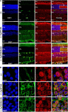
Immunolocalization of native Ranbp2, Tg-Ranbp2R2944A-HA, and Tg-Ranbp2WT-HA proteins in radial retinal sections (A) and retinal flat mounts of cell bodies of ganglion neurons (B). Endogenous and transgenic Ranbp2 proteins of 4–6-week-old mice were detected with antibodies against the IR domain of Ranbp2 and HA tag, respectively. The native localization of Ranbp2 (A, 1st row panel) was distributed throughout the cell bodies of retinal neurons with prominent expressions in the ciliary region of photoreceptors and cell bodies of ganglion neurons. A similar subcellular distribution of transgenic Ranbp2 was observed with Tg-Ranbp2R2944A-HA and Tg-Ranbp2WT-HA, but there is an accumulation of Tg-Ranbp2WT-HA in the ciliary region of photoreceptors (A, 2nd row panel). Insets are higher magnifications of the boxed ciliary regions. B, high magnifications of ganglion neurons captured from retinal flat mounts and depicting the localization of native and transgenic Ranbp2 at the nuclear pores of the nuclear rim. No discernible differences were observed between nontransgenic and transgenic genotypes. −/−, Ranbp2−/−; Tg-R2944A-HA, Tg-Ranbp2R2944A-HA; Tg-WT-HA, Tg-Ranbp2WT-HA; OS, outer segments of photoreceptors; IS, inner segments of photoreceptors; ONL, outer nuclear layer (cell bodies of photoreceptors); INL, inner nuclear layer (cell bodies of second-order neurons); GC, ganglion cells. Scale bars, 50 μm (A), 20 μm (A, inset), and 10 μm (B).
FIGURE 4.
FIGURE 4.
Topographic distribution of M- and S-cone photoreceptors in wild-type, Tg-Ranbp2R2944A-HA::Ranbp2−/−, and Tg-Ranbp2WT-HA::Ranbp2−/−. No changes in the distribution of M-cone (left) and S-cone photoreceptors (right) are observed between all genotypes across all regions of the retina of 10–12-week-old mice. Representative confocal Z-stack images of dorsal (D), dorsal-central (DC), ventral-central (VC), and ventral (V) regions of the retinal flat mounts are shown (top panels). Quantitative distribution of M-cone (left) and S-cone (right) photoreceptors between genotypes are shown below image panels. No significant differences were found between genotypes (p > 0.05). Data shown represent the mean ± S.D., n = 3–4; n.s., nonsignificant; −/−, Ranbp2−/−; Tg-R2944A-HA, Tg-Ranbp2R2944A-HA; Tg-WT-HA, Tg-Ranbp2WT-HA. Scale bars, 25 μm.
FIGURE 5.
FIGURE 5.
Impairment of M-opsin biogenesis in Tg-Ranbp2R2944A-HA::Ranbp2−/− and Tg-Ranbp2WT-HA::Ranbp2−/−. A, three-dimensional subcellular localization of M- and S-opsin and PNA in the outer segments of cone photoreceptors. Accumulation and aggregation foci of M-opsin are visible in 10–12-week-old Tg-Ranbp2R2944A-HA::Ranbp2−/− and Tg-Ranbp2WT-HA::Ranbp2−/− at the base and throughout the outer segment of cone photoreceptors singly expressing M-opsin and co-expressing M/S opsins. Distribution of S-opsin is normal between nontransgenic and transgenic mice. B, magnified images of inset regions of A. C, immunoblots of retinal homogenates showing the selective accumulation of M-opsin (∼3-fold) in 10–12-week-old Tg-Ranbp2R2944A-HA::Ranbp2−/− and Tg-Ranbp2WT-HA::Ranbp2−/−. The expression level of S-opsin remains unchanged between nontransgenic and transgenic mice. Graphs below the blots represent the quantitative analysis of immunoblots. Hsc70 is the cytosolic heat shock protein 70, which is used as loading control. D, transcriptional levels of M-opsin (Opn1mw) and S-opsin (Opn1sw) and rhodopsin (Rho) by qRT-PCR. There is a decrease by ∼20% of M-opsin selectively in Tg-Ranbp2R2944A-HA::Ranbp2−/−, whereas S-opsin is decreased by ∼50% in Tg-Ranbp2R2944A-HA::Ranbp2−/− and Tg-Ranbp2WT:Ranbp2−/−. Rhodopsin (Rho) levels remain unchanged across all genotypes. Data shown represent the mean ± S.D., n = 4; n.s., nonsignificant; −/−, Ranbp2−/−; Tg-R2944A-HA, Tg-Ranbp2R2944A-HA; Tg-WT-HA, Tg-Ranbp2WT-HA. Scale bars, 25 μm (A) and 5 μm (B).
FIGURE 6.
FIGURE 6.
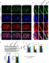
STAT3 and STAT5 are partners of CY of Ranbp2 and their proteostasis are unimpaired in Tg-Ranbp2R2944A-HA::Ranbp2−/− and Tg-Ranbp2WT-HA::Ranbp2−/−. A, ectopically expressed CY alone of Ranbp2 associates with native STAT3/STAT5 in cultured cells. Immunoblots of co-immunoprecipitates of HeLa cell extracts transfected with wild-type mRFP-CY and mutant mRFP-CYR2944A and mRFP-CYS3036E constructs. Graphs represent quantitation of immunoblots shown on left. The association of STAT3 (STAT3β) and STAT5 are selectively and significantly decreased in mRFP-CYR2944A. Co-immunoprecipitated STAT3 and STAT5 were normalized for the amount of immunoprecipitated mRFP-fused constructs. B, levels of STAT3 (STAT3β) and STAT5 expressions are unaffected in retinal homogenates of Tg-Ranbp2R2944A-HA::Ranbp2−/− and Tg-Ranbp2WT-HA::Ranbp2−/−. C, relative levels of STAT3 (STAT3β) between the retina and midbrain homogenates. In comparison with the retina, the basal expression level of STAT3 is ∼25-fold higher in the midbrain of 10–12-week-old mice. Quantitative analysis (right) of the representative immunoblot (left) is shown. D, quiescent and activated (phosphorylated) STAT3 (STAT3β and STAT3α) and STAT5 associate in vivo with Tg-Ranbp2R2944A-HA and Tg-Ranbp2WT-HA in midbrain extracts of Tg-Ranbp2R2944A-HA::Ranbp2−/− and Tg-Ranbp2WT-HA::Ranbp2−/− mice. Immunoblots are co-immunoprecipitates of transgenic Ranbp2 proteins immunoprecipitated with anti-HA antibody. Quantitative analysis (left) of the representative immunoblot (right) is shown. There are no significant differences in the amount of STAT3β and STAT5 co-immunoprecipitated by Tg-Ranbp2R2944A-HA and Tg-Ranbp2WT-HA. E, immunoquantitation of activation of STAT3 (P-STAT3β) upon light treatment (LT) between 10- and 12-week-old nontransgenic wild-type and Tg-Ranbp2R2944A-HA::Ranbp2−/− mice. There is a 30-fold activation of P-STAT3 upon light treatment in the retina, but there is no significant difference in such activation between nontransgenic wild-type and Tg-Ranbp2R2944A-HA::Ranbp2−/− mice. Image panels on the right show the immunolocalization of P-STAT3 in cell bodies and fibers of photoreceptors. F, TUNEL+ (apoptotic) photoreceptors between nontransgenic wild-type and Tg-Ranbp2R2944A-HA::Ranbp2−/− mice under cyclic (NT, nontreated) and chronic light treatment (LT, light-treated). No significant differences were observed between and within the cyclic and light-treated groups (n = 9, p >0.05). Representative images (left) and quantitation of TUNEL+ photoreceptors by dot-box plot analyses (right) in central and peripheral regions of the retina are shown (n = 9). Statistical analysis was done using Mann-Whitney U test at significance level 0.05. Data shown represent the mean ± S.D., n = 4 (unless otherwise noted); n.s., nonsignificant; −/−, Ranbp2−/−; Tg-R2944A-HA, Tg-Ranbp2R2944A-HA; Tg-WT-HA, Tg-Ranbp2WT-HA. Scale bar, 10 μm (E) and 25 μm (F). A.U., arbitrary units.
FIGURE 7.
FIGURE 7.
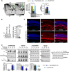
Post-transcriptional down-regulation of hnRNPA2/B1 and impairment of the subcellular partitioning of HDAC4 selectively in Tg-Ranbp2R2944A-HA::Ranbp2−/−. A, 2D-DIGE of retinal homogenates of 4–6-week-old wild-type (+/+) and Tg-Ranbp2R2944A-HA::Ranbp2−/− mice. Insets are volumetric analyses of spot intensity marked in yellow; there was an ∼3-fold difference between wild-type (+/+) and Tg-Ranbp2R2944A-HA::Ranbp2−/−. Spot identification (marked in yellow) by mass spectrometry analysis showed that it was hnRNPA2/B1. Validation and comparison of changes in hnRNPA2/B1 expression between genotypes show selective and about 60% reduction of hnRNPA2/B1 expression between 4- and 6-week-old Tg-Ranbp2R2944A-HA::Ranbp2−/− and other age-matched genotypes (middle graph). Representative immunoblots and loading controls are shown below the graph. qRT-PCR of transcriptional levels of hnRNPA2/B1 shows no changes between wild-type and Tg-Ranbp2R2944A-HA::Ranbp2−/−, although there is a reduction of hnRNPA2/B1 in Tg-Ranbp2WT-HA::Ranbp2−/− compared with Tg-Ranbp2R2944A-HA::Ranbp2−/− mice (right graph). B, relative expressions of hnRNPA2/B1 between retina and midbrain homogenates of 10–12-week-old mice show relatively high abundance of hnRNPA2/B1 in retina. C, immunolocalization of hnRNPA2/B1 in radial retinal sections of 10–12-week-old wild-type and Tg-Ranbp2R2944A-HA::Ranbp2−/− mice. Localizations of hnRNPA2/B1 are prominent in cell bodies of inner retinal neurons (INL) and ganglion cells (GC). There were no discernible differences in hnRNPA2/B1 localization between wild-type and Tg-Ranbp2R2944A-HA::Ranbp2−/− mice. D, selective shift in subcellular partitioning of HDAC4, but not hnRNPA2/B1, between nuclear and non-nuclear retinal fractions of Tg-Ranbp2R2944A-HA::Ranbp2−/− mice. Immunoblots of nuclear and non-nuclear retinal fractions of hnRNPA2/B1, HDAC4, γ-crystallin, and Nup62 are shown. γ-Crystallin and Nup62 have cytosolic and pan-distributions, respectively. Lamin A/C and GAPDH are additional controls for nuclear and non-nuclear fractions. Graphs below immunoblots are pairwise comparisons of the levels of hnRNPA2/B1, HDAC4, and γ-crystallin between nuclear and non-nuclear fractions for each genotype. Data shown in A, B, and D represent the mean ± S.D., n = 4. ONL, outer nuclear layer (cell bodies of photoreceptors); INL, inner nuclear layer (cell bodies of second-order neurons); GC, ganglion cells; n.s., nonsignificant; hnRNPA2/B1, heterogeneous ribonucleoproteins A2/B1; HDAC4, histone deacetylase-4; Nup62, nucleoporin 62; N, nuclear; nN, non-nuclear; −/−, Ranbp2−/−; Tg-R2944A-HA, Tg-Ranbp2R2944A-HA; Tg-WT-HA, Tg-Ranbp2WT-HA. Scale bar, 25 μm.
FIGURE 8.
FIGURE 8.
Age- and tissue-dependent deficits of ubiquitin homeostasis in Tg-Ranbp2R2944A-HA::Ranbp2−/−. Compared with wild-type mice, the levels of diubiquitin (Ub2) or conjugated polyubiquitin (P-Ubn) are significantly decreased in the retina (A) and RPE (B), but not liver (C), of Tg-Ranbp2R2944A-HA::Ranbp2−/− at 24 weeks, but not 4 weeks of age. GADPH or hsc70 are used as loading controls. Quantitation analyses are shown next to the immunoblots. Data shown represent the mean ± S.D., n = 4. n.s., nonsignificant; −/−, Ranbp2−/−; Tg-R2944A-HA, Tg-Ranbp2R2944A-HA; Tg-WT-HA, Tg-Ranbp2WT-HA.
FIGURE 9.
FIGURE 9.
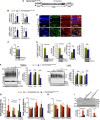
Selective proteostatic deficits in Tg-Ranbp2CLDm-HA::Ranbp2−/− and deregulation of selective ubiquitin-proteasome system activities in Tg-Ranbp2R2944A-HA::Ranbp2−/− and Tg-Ranbp2CLDm-HA::Ranbp2−/− mice. A, diagram of the transgenic BAC recombineering construct, Tg-Ranbp2CLDm, with a C-terminal hemagglutinin (HA) tag insertion at the end of the terminal exon encoding CY of Ranbp2 and the mutations, I2471K andV2472A, in CLD of Ranbp2. Note drawing not to scale. B, transcriptional expression of nontransgenic Ranbp2 and Tg-Ranbp2CLDm-HA in 4–6-week-old wild-type and Tg-Ranbp2CLDm-HA::Ranbp2−/− mice, respectively, by qRT-PCR. The liver and retina have increased 3′-transcriptional expression of Tg-Ranbp2CLDm-HAcompared with nontransgenic Ranbp2, but neither gene nor tissue have significant differences of 5′-transcriptional expression of Ranbp2. C, quantitation of protein expression levels of Ranbp2 and Tg-Ranbp2CLDm-HA was comparable between 4- and 6-week-old wild-type and Tg-Ranbp2CLDm-HA::Ranbp2−/−, respectively. Representative immunoblots and loading controls are shown below the graphs. Hsc70, the cytosolic heat shock protein 70, is used as loading control. D, immunolocalization of Tg-Ranbp2CLDm-HA protein in radial retinal sections (upper panel) and retinal flat mounts of cell bodies of ganglion neurons (lower panel) with an anti-HA antibody. Tg-Ranbp2CLDm-HA was distributed throughout cell bodies of retinal neurons with prominent expressions in the ciliary region of photoreceptors and cell bodies of ganglion neurons. There is an increased accumulation of Tg-Ranbp2CLDm-HA in the ciliary region of photoreceptors compared with the native Ranbp2 (see Fig. 3). Inset is a higher magnification of the boxed ciliary regions. Scale bar, 25 μm. E, quantitation of protein expression levels of sumoylated RanGAP (RanGAP*), HDAC4, and S1 subunit of the 19 S cap of the 26 S proteasome, and transcriptional levels (mRNA) of HDAC4. Compared with wild-type mice, the levels of sumoylated RanGAP and HDAC4, but not S1 subunit, are reduced in 10–12-week-old Tg-Ranbp2CLDm:Ranbp2−/− and without accompanying changes in the transcriptional levels of HDAC4. Representative immunoblots and loading controls are shown below the graphs. Hsc70 or GADPH are used as loading controls. F, there are no differences between 24-week-old wild-type and Tg-Ranbp2CLDm-HA::Ranbp2−/− mice of the levels of monoubiquitin (Ub), diubiquitin (Ub2), and conjugated polyubiquitin (P-Ubn) in the retina and liver. Quantitation analyses are shown next to the immunoblots. G, DUB activity is selectively increased in soluble proteasomal fractions of retinas of Tg-Ranbp2R2944A-HA::Ranbp2−/− mice. Compared with age-matched wild-type and Tg-Ranbp2CLDm-HA::Ranbp2−/− mice, the deubiquitylase activities toward Lys-63-linked (K63-Ub4, left) and Lys-48-linked (K48-Ub4, middle) tetraubiquitin are increased in the presence of the DUB inhibitors PR-619 and 1,10-phenanthroline, respectively, in 24-week-old Tg-Ranbp2R2944A-HA::Ranbp2−/−. H, compared with age-matched wild-type and Tg-Ranbp2R2944A-HA::Ranbp2−/− mice, the chymotrypsin-like activity of 20 S proteasome is selective reduced in 24-week-old Tg-Ranbp2CLDm-HA::Ranbp2−/− mice, although there are no changes in the trypsin- and caspase-like activities between any genotype (right). I, in comparison with age-matched wild-type and Tg-Ranbp2CLDm-HA::Ranbp2−/− mice, there is a robust accumulation of the S1 and S5b subunits of the 19 S cap of 26 S proteasome in the pellet of digitonin-permeabilized retinas of 24-week-old Tg-Ranbp2R2944A-HA::Ranbp2−/− mice, whereas no changes are observed in the soluble proteasomal fraction between any genotype. Quantitation analyses are shown below immunoblots. Acetylated α-tubulin (AcTub) is a loading control. Data shown in B, C, and E–I represent the mean ± S.D.; n = 4. ONL, outer nuclear layer (cell bodies of photoreceptors); INL, inner nuclear layer (cell bodies of second-order neurons); GC, ganglion cells; n.s., nonsignificant; HDAC4, histone deactylase-4; −/−, Ranbp2−/−; Tg-R2944A-HA, Tg-Ranbp2R2944A-HA. Scale bar, 10 μm.
FIGURE 10.
FIGURE 10.
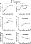
Electrophysiological responses of rod and cone photoreceptors and inner retinal neurons of Tg-Ranbp2R2944A-HA::Ranbp2−/− mice. Luminance-response functions for the dark-adapted (A) and light-adapted (B) ERGs obtained from 8-week-old mice. Luminance-response functions for VEP amplitudes (C and D) and implicit times (E and F) obtained from 24-week-old mice under dark-adapted (C and E) and light-adapted (D and F) stimulus conditions. No significant differences between genotypes were found in the scotopic (A) and photopic (B) a- and b-waves and VEP amplitudes (C and D). The VEP implicit times under dark-adapted (E), but not light-adapted (F) stimulus conditions, were reduced in Tg-Ranbp2R2944A-HA::Ranbp2−/− mice in comparison with Ranbp2R2944A-HA::Ranbp2+/− (p < 0.05) or Ranbp2+/− (p < 0.01) genotypes. Data points indicate the average ± S.E.; n = 9–10. −/−, Ranbp2−/−; +/−, Ranbp2+/−; Tg-R2944A-HA, Tg-Ranbp2R2944A-HA.
FIGURE 11.
FIGURE 11.
Model depicting idiosyncratic and physiological activities of CY of Ranbp2 toward the proteostasis of distinct substrates. The CY of Ranbp2 presents three distinct biological activities toward physiological substrates. First, the C-terminal domain of CY of Ranbp2 harbors chaperone activity selectively toward M-opsin. Impairment of the C-terminal chaperone activity of CY of Ranbp2 promotes M-opsin aggregation and accumulation in cone photoreceptors. Second, the PPIase activity of CY of Ranbp2 is required for the proteostasis of hnRNPA2/B1. Suppression of the PPIase activity of CY leads to the post-transcriptional down-regulation of hnRNPA2/B1. This is also accompanied by the down-regulation of diubiquitin, an effect that promotes the activation of selective deubiquitylases and a reduction of the levels of ubiquitylated substrates. Third, the CY of Ranbp2 presents another subdomain, which mediates the binding of latent and activated STAT3/STAT5. This STAT3/STAT5-binding domain in CY is distinct from its PPIase and C-terminal chaperone domains and has not yet been defined molecularly. Finally, this work supports that the PPIase activity of CY is modulated by phosphorylation of at least a residue (Ser-3036) near its active PPIase site. The phosphorylation of CY is likely modulated by extracellular stimuli (e.g. cytokines), and the physiological implications of the post-translational modification of CY have yet to be defined. CY of Ranbp2 is depicted with a ribbon representation. Residues Ser-3036 and Arg-2944 are shown as orange sticks and other residues of the catalytic PPIase site of CY are shown as green sticks.

References

    1. Brandts J. F., Halvorson H. R., Brennan M. (1975) Consideration of the possibility that the slow step in protein denaturation reactions is due to cis-trans isomerism of proline residues. Biochemistry 14, 4953–4963 - PubMed
    1. Cook K. H., Schmid F. X., Baldwin R. L. (1979) Role of proline isomerization in folding of ribonuclease A at low temperatures. Proc. Natl. Acad. Sci. U.S.A. 76, 6157–6161 - PMC - PubMed
    1. Weininger U., Jakob R. P., Eckert B., Schweimer K., Schmid F. X., Balbach J. (2009) A remote prolyl isomerization controls domain assembly via a hydrogen bonding network. Proc. Natl. Acad. Sci. U.S.A. 106, 12335–12340 - PMC - PubMed
    1. Fischer G., Bang H., Mech C. (1984) Determination of enzymatic catalysis for the cis-trans-isomerization of peptide binding in proline-containing peptides. Biomed. Biochim. Acta 43, 1101–1111 - PubMed
    1. Fischer G., Wittmann-Liebold B., Lang K., Kiefhaber T., Schmid F. X. (1989) Cyclophilin and peptidyl-prolyl cis-trans isomerase are probably identical proteins. Nature 337, 476–478 - PubMed

Publication types

MeSH terms

LinkOut - more resources