. 2014 Feb 21;289(8):5097-108.
doi: 10.1074/jbc.M113.533109. Epub 2014 Jan 8.

Monoubiquitination is critical for ovarian tumor domain-containing ubiquitin aldehyde binding protein 1 (Otub1) to suppress UbcH5 enzyme and stabilize p53 protein

Affiliations

Monoubiquitination is critical for ovarian tumor domain-containing ubiquitin aldehyde binding protein 1 (Otub1) to suppress UbcH5 enzyme and stabilize p53 protein

Yuhuang Li et al. J Biol Chem. .

Abstract

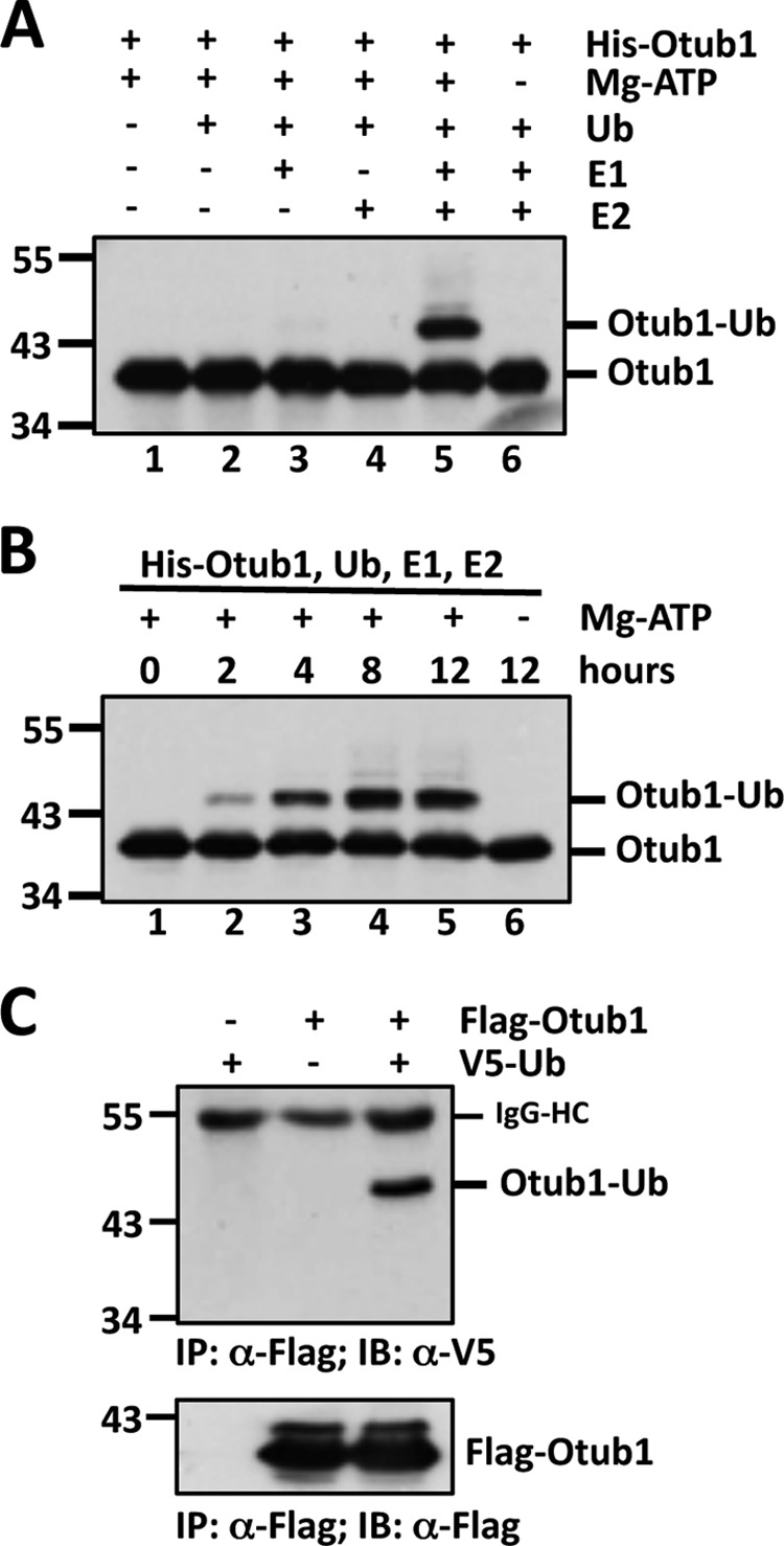
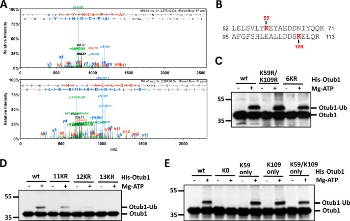
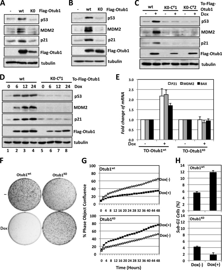
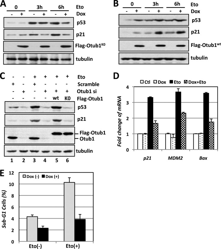
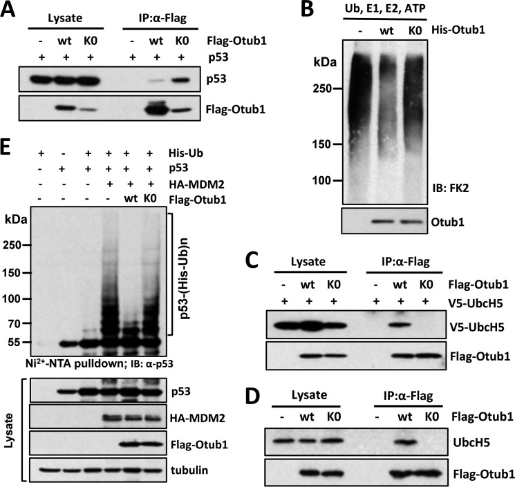
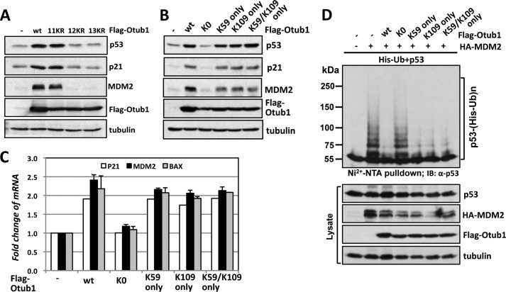
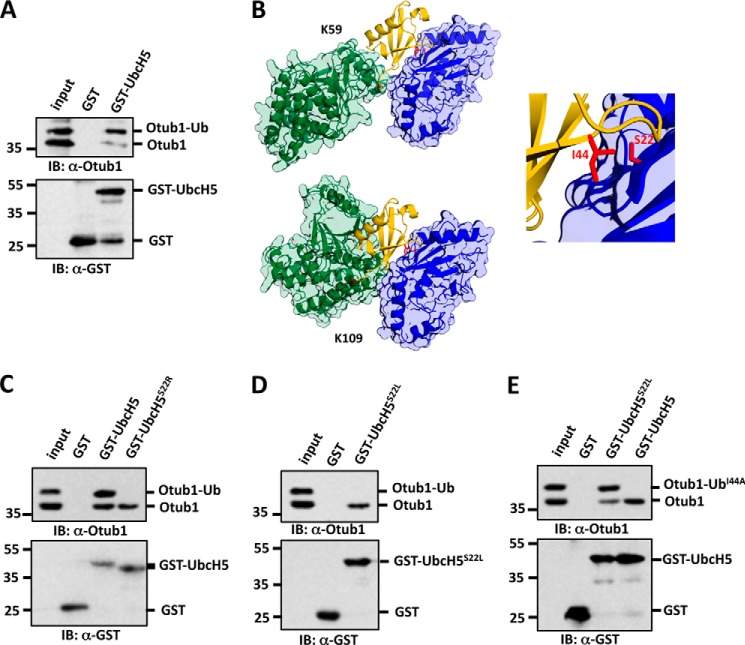
Ovarian tumor domain-containing ubiquitin (Ub) aldehyde binding protein 1 (Otub1) regulates p53 stability and activity via non-canonical inhibition of the MDM2 cognate Ub-conjugating enzyme (E2) UbcH5. However, it is not clear how this activity of Otub1 is regulated in cells. Here we report that Otub1 is monoubiquitinated by UbcH5 in cells and in vitro, primarily at the lysine 59 and 109 residues. This monoubiquitination, in turn, contributes to the activity of Otub1 to suppress UbcH5. The lysine-free Otub1 mutant (Otub1(K0)) fails to be monoubiquitinated and is unable to suppress the Ub-conjugating activity of UbcH5 in vitro and the MDM2-mediated p53 ubiquitination in cells. Consistently, this mutant is unable to stabilize p53, induce apoptosis, and suppress cell proliferation. Overexpression of Otub1(K0) inhibits DNA-damage induced apoptosis. Adding either Lys-59 or Lys-109 back to the Otub1(K0) mutant restores the monoubiquitination of Otub1 and its function to stabilize and activate p53. We further show that UbcH5 preferentially binds to the monoubiquitinated Otub1 via Ub interaction with its backside donor Ub-interacting surface, suggesting that this binding interferes with the self-assembly of Ub-charged UbcH5 (UbcH5∼Ub) conjugates, which is critical for Ub transfer. Thus, our data reveal novel insights into the Otub1 inhibition of E2 wherein monoubiquitination promotes the interaction of Otub1 with UbcH5 and the function to suppress it.

Keywords: Deubiquitinating Enzymes; Deubiquitination; Monoubiquitination; Otub1; UbcH5; Ubiquitin; Ubiquitin-conjugating Enzyme (Ubc); Ubiquitination; p53.


Figures

FIGURE 1.
FIGURE 1.
Otub1 is monoubiquitinated by UbcH5 in cells and in vitro. A and B, monoubiquitination of Otub1 by UbcH5 in vitro. His-Otub1 was incubated with different combinations of E1, UbcH5 (E2), Ub, and ATP, as indicated, for 8 h (A) or with E1, E2, and Ub in the presence or absence of ATP for different times, as indicated (B), followed by IB analysis with anti-Otub1. C, monoubiquitination of Otub1 in cells. H1299 cells transfected with FLAG-Otub1 with or without V5-Ub were subjected to co-IP with anti-FLAG antibody, followed by IB analysis with anti-V5 antibody (top panel) and anti-FLAG antibody (bottom panel). HC, heavy chain.
FIGURE 2.
FIGURE 2.
Otub1 is monoubiquitinated by UbcH5 primarily at Lys-59 and Lys-109. A, identification of the monoubiquitination sites by mass spectrometry. The MS/MS spectrum (+3 ion) of Otub152–71 indicating ubiquitination at residue Lys-59 (top panel) and the MS/MS spectrum (+3 ion) of Otub195–113 indicating ubiquitination at residue Lys-109 (bottom panel) are shown. Matched b-series fragment ions are indicated in red, matched y-ion series fragment ions are indicated in blue, and matched fragment ions (both y and b) undergoing loss of water are indicated in green. B, summary of the peptides containing Ub-modified lysines (Lys-59 and Lys-109) identified by LC-MS/MS analysis. C and D, Otub1wt, Otub1K59R/K109R, Otub16KR, and Otub111KR, but not Otub112KR and Otub113KR, were monoubiquitinated by UbcH5 in vitro. WT Otub1 or its mutants, as indicated, were incubated with E1, E2, and Ub in the presence or absence of ATP, followed by IB analysis using anti-Otub1. E, Otub1wt, Otub1K59 only, Otub1K109 only, and Otub1K59/K109 only, but not Otub1K0, were monoubiquitinated by UbcH5 in vitro. WT Otub1 or its mutants, as indicated, were incubated with E1, E2, and Ub in the presence or absence of ATP, followed by IB analysis using anti-Otub1.
FIGURE 3.
FIGURE 3.
Lysine-free Otub1 fails to induce and activate p53. A and B, Otub1K0 does not induce p53. U2OS cells were transfected with equal amounts (3 μg) of the FLAG-Otub1wt or FLAG-Otub1K0 plasmid (A) or 0.5 μg of the FLAG-Otub1wt or 3 μg of the FLAG-Otub1K0 plasmid (B), followed by IB analysis. C, induced expression of Otub1K0 fails to induce p53. Two representative U2OS-TO-FLAG-Otub1K0 clones and the U2OS-TO-FLAG-Otub1wt stable cell line were cultured in the absence or presence of 2 μg/ml of Dox for 24 h. The cell lysates were assayed by IB analysis. D, time course study of the p53 induction by Otub1wt but not Otub1K0. U2OS-TO-FLAG-Otub1wt or U2OS-TO-FLAG-Otub1K0 clone 1 (C#1) was cultured in the presence of 2 μg/ml of Dox and harvested at different time points, as indicated, followed by IB analysis. E, Otub1K0 does not induce p53 activity. U2OS-TO-FLAG-Otub1K0 or U2OS-TO-FLAG-Otub1wt cells were treated with or without Dox for 24 h, followed by RT-qPCR detection of p21, mdm2, and bax mRNA normalized to the expression of GAPDH. F, Otub1K0 does not inhibit cell proliferation. U2OS-TO-FLAG-Otub1wt or U2OS-TO-FLAG-Otub1K0 cells were cultured in the presence or absence of Dox for up to 3 weeks. The colonies were visualized by staining with crystal violet blue. G, T-Rex-U2OS-FLAG-Otub1wt or T-Rex-U2OS-FLAG-Otub1K0 cells were cultured in the presence or absence of 2 μg/ml doxycycline. The cell confluence was measured over time using the IncuCyte system. H, Otub1K0 does not induce apoptosis. U2OS-TO-FLAG-Otub1wt or U2OS-TO-FLAG-Otub1K0 cells were cultured with or without Dox for 24 h. The cells were stained with propidium iodide, followed by flow cytometry analysis. The average percentages of cells in sub-G1 are shown.
FIGURE 4.
FIGURE 4.
Lysine-free Otub1 attenuates p53 activation in response to DNA damage. A and B, Otub1K0, but not Otub1wt, attenuates p53 activation following Eto treatment. U2OS-TO-FLAG-Otub1K0 cells (A) and U2OS-TO-FLAG-Otub1wt cells (B) were cultured in the absence or presence of Dox for 24 h, followed by treatment with Eto (20 μm) or control dimethyl sulfoxide for the indicated times, followed by IB analysis. C, reintroduction of Otub1wt, but not the Otub1K0 mutant, rescues the p53 response following DNA damage in cells with Otub1 knockdown. U2OS cells transfected with control, the FLAG-Otub1wt or FLAG-Otub1K0 plasmid, and scrambled or Otub1 siRNA targeting the 3′ UTR of the Otub1 mRNA are shown, as indicated. The cells were treated with Eto (20 μm) for 6 h before harvesting 48 h after siRNA transfection, followed by IB analysis. D and E, Otub1K0 attenuates p53 activation in response to DNA damage. U2OS-TO-FLAG-Otub1K0 cells were cultured in the absence or presence of Dox for 24 h, followed by treatment with Eto (20 μm) or control (Ctl) dimethyl sulfoxide for 6 h, and assayed by RT-qPCR (D) and cell cycle profile analysis for sub-G1 phase cells (E).
FIGURE 5.
FIGURE 5.
Lysine-free Otub1 fails to interact with UbcH5 and suppress UbcH5 activity in vitro and MDM2-mediated p53 ubiquitination in cells. A, Otub1K0 binds to p53. H1299 cells transfected with p53 together with Otub1wt or Otub1K0 were subjected to co-IP using anti-FLAG antibodies, followed by IB analysis. B, Otub1K0 does not suppress UbcH5-dependent ubiquitin chain formation. The in vitro ubiquitination reactions were conducted in the presence of E1, E2, Ub, and ATP in the absence or presence of His-tagged Otub1wt or Otub1K0, as indicated. The reactions were assayed by IB analysis using anti-conjugated Ub antibody (clone FK2) (top panel). C and D, Otub1K0 does not interact with UbcH5 in cells. H1299 cells transfected with 0.25 μg of the FLAG-Otub1wt plasmid or 1.5 μg of the FLAG-Otub1K0 plasmid alone (D) or together with 1.5 μg of V5-UbcH5 (C) were subjected to co-IP with anti-FLAG antibodies, followed by IB analysis. E, Otub1K0 does not inhibit MDM2-mediated p53 ubiquitination in cells. H1299 cells transfected with indicated plasmids were subjected to pulldown using Ni-NTA beads under denaturing conditions, followed by IB analysis. The ubiquitinated species of p53 are indicated.
FIGURE 6.
FIGURE 6.
Monoubiquitination of Otub1 is required for its activity to induce and activate p53. A, monoubiquitination-competent but not monoubiquitination-defective mutants induce p53. U2OS cells were transfected with Otub1wt or its mutants (11KR, 12KR, and 13KR) followed by IB analysis. B and C, adding Lys-59 or Lys-109 back to Otub1K0 restores Otub1 function to regulate p53. U2OS cells were transfected with Otub1wt, Otub1K0, Otub1K59 only, Otub1K109 only, or Otub1K59/K109 only mutants, followed by IB analysis (B) and RT-qPCR (C) analysis. D, Lys-59-only or Lys-109-only Otub1 suppresses MDM2-mediated p53 ubiquitination in cells. H1299 cells transfected with His-Ub and p53 in the presence of the indicated plasmids were subjected to Ni-NTA pulldown under denaturing conditions, followed by IB analysis. The ubiquitinated species of p53 are indicated.
FIGURE 7.
FIGURE 7.
UbcH5 binds preferentially to monoubiquitinated Otub1 through backside UbcH5-Ub interaction. A, UbcH5 binds preferentially to the monoubiquitinated Otub1. His-Otub1 was subjected to an in vitro ubiquitination reaction as in Fig. 1. The reaction mixture containing both monoubiquitinated and unmodified Otub1 was then incubated with GST alone or GST-UbcH5c immobilized onto GSH beads. After washing, bead-bound proteins were assayed by IB analysis. B, modeling of the Ub-Otub1/UbcH5b interaction through docking of Ub linked to either Lys-59 (top left panel) or Lys-109 (bottom left panel) with the backside of UbcH5b. Otub1, UbcH5b, and Ub are colored green, blue, and yellow, respectively. Lys-59 and Lys-109 of Otub1, Ile-44 of Ub, and Ser-22 of UbcH5 are indicated in red. The enlarged view in the right panel shows the canonical Ub Ile-44 interacting with Ser-22 on UbcH5. C and D, UbcH5S22R and UbcH5S22L do not bind to monoubiquitinated Otub1. The in vitro ubiquitination reaction mixture containing both monoubiquitinated and unmodified Otub1 was incubated with GST alone, GST-UbcH5c, GST-UbcH5S22R, or GST-UbcH5S22L immobilized onto GSH beads. After washing, bead-bound proteins were assayed by IB analysis. E, mutating Ser-22 to Leu rescues the binding defect of UbcH5 with Otub1-UbI44A. His-Otub1 was subjected to an in vitro ubiquitination reaction using recombinant UbI44A. The reaction mixture containing both monoubiquitinated (Otub1-UbI44A) and unmodified Otub1 was incubated with GST alone, GST-UbcH5c, or GST-UbcH5S22L immobilized onto GSH beads. After washing, bead-bound proteins were assayed by IB analysis.

Similar articles

Cited by

References

    1. Devine T., Dai M. S. (2013) Targeting the ubiquitin-mediated proteasome degradation of p53 for cancer therapy. Curr. Pharm. Des. 19, 3248–3262 - PMC - PubMed
    1. Kruse J. P., Gu W. (2009) Modes of p53 regulation. Cell 137, 609–622 - PMC - PubMed
    1. Fang S., Jensen J. P., Ludwig R. L., Vousden K. H., Weissman A. M. (2000) Mdm2 is a RING finger-dependent ubiquitin protein ligase for itself and p53. J. Biol. Chem. 275, 8945–8951 - PubMed
    1. Momand J., Zambetti G. P., Olson D. C., George D., Levine A. J. (1992) The mdm-2 oncogene product forms a complex with the p53 protein and inhibits p53-mediated transactivation. Cell 69, 1237–1245 - PubMed
    1. Haupt Y., Maya R., Kazaz A., Oren M. (1997) Mdm2 promotes the rapid degradation of p53. Nature 387, 296–299 - PubMed

Publication types