. 2014 Feb 14;289(7):3991-4000.
doi: 10.1074/jbc.M113.514075. Epub 2014 Jan 8.

Evolution of mammalian Opn5 as a specialized UV-absorbing pigment by a single amino acid mutation

Affiliations

Evolution of mammalian Opn5 as a specialized UV-absorbing pigment by a single amino acid mutation

Takahiro Yamashita et al. J Biol Chem. .

Abstract

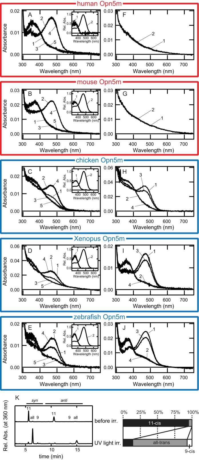
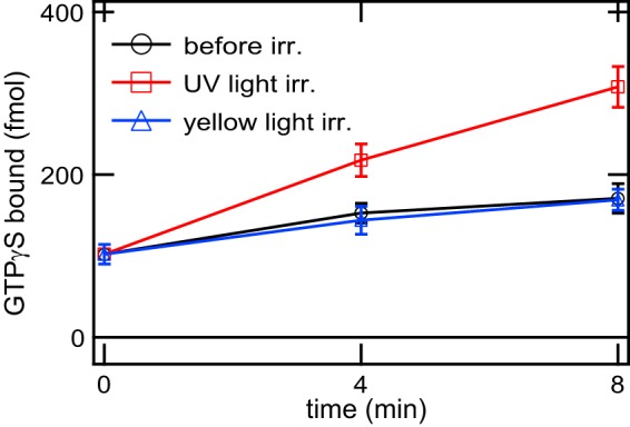
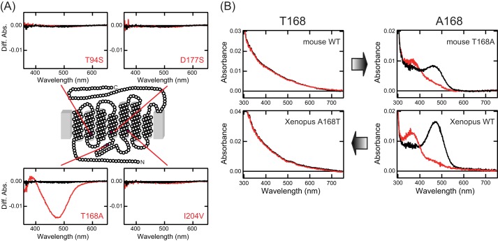
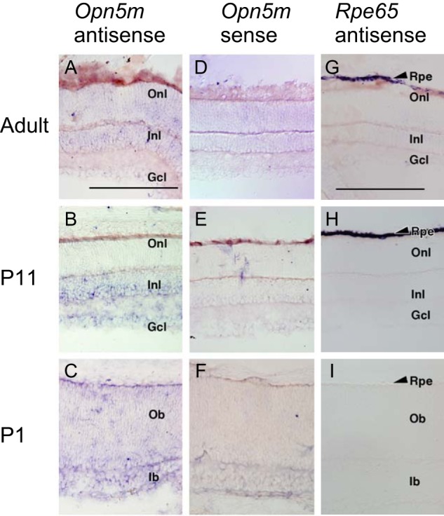
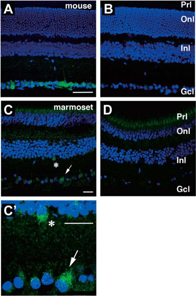
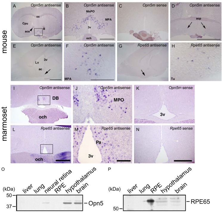
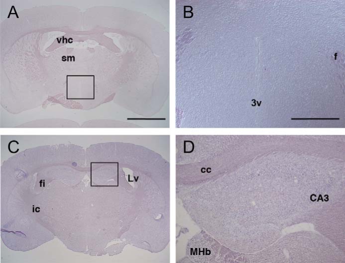
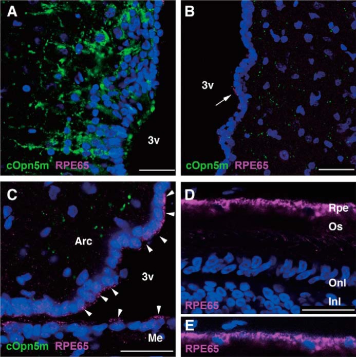
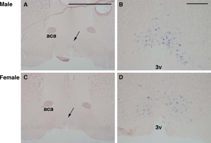
Opn5 is one of the recently identified opsin groups that is responsible for nonvisual photoreception in animals. We previously showed that a chicken homolog of mammalian Opn5 (Opn5m) is a Gi-coupled UV sensor having molecular properties typical of bistable pigments. Here we demonstrated that mammalian Opn5m evolved to be a more specialized photosensor by losing one of the characteristics of bistable pigments, direct binding of all-trans-retinal. We first confirmed that Opn5m proteins in zebrafish, Xenopus tropicalis, mouse, and human are also UV-sensitive pigments. Then we found that only mammalian Opn5m proteins lack the ability to directly bind all-trans-retinal. Mutational analysis showed that these characteristics were acquired by a single amino acid replacement at position 168. By comparing the expression patterns of Opn5m between mammals and chicken, we found that, like chicken Opn5m, mammalian Opn5m was localized in the ganglion cell layer and inner nuclear layer of the retina. However, the mouse and primate (common marmoset) opsins were distributed not in the posterior hypothalamus (including the region along the third ventricle) where chicken Opn5m is localized, but in the preoptic hypothalamus. Interestingly, RPE65, an essential enzyme for forming 11-cis-retinal in the visual cycle is expressed near the preoptic hypothalamus of the mouse and common marmoset brain but not near the region of the chicken brain where chicken Opn5m is expressed. Therefore, mammalian Opn5m may work exclusively as a short wavelength sensor in the brain as well as in the retina with the assistance of an 11-cis-retinal-supplying system.

Keywords: G Proteins; Molecular Evolution; Nonvisual Photoreception; Photoreceptors; Rhodopsin; Signal Transduction.


Figures

FIGURE 1.
FIGURE 1.
Characterization of the anti-mammalian Opn5m antibodies by Western blotting. Signals were detected in Opn5m-transfected cells but not in mock-transfected cells. Recombinant mammalian Opn5m proteins exhibited two bands, probably because of the heterogeneity of the post-translational modification within the cultured cells, such as the glycosylation in the N terminus of the protein, as shown for recombinant bovine rhodopsin (41).
FIGURE 2.
FIGURE 2.
Comparison of the ability to directly bind 11-cis- or all-trans-retinal in vertebrate Opn5m proteins. A–E, absorption spectra of human (A), mouse (B), chicken (C), X. tropicalis (D), and zebrafish (E) Opn5m proteins after incubation with 11-cis-retinal. Spectra were measured in the dark (curve 1) and after UV light irradiation (curve 2), subsequent yellow light (>500 nm) irradiation (curve 3), UV light reirradiation (curve 4), or yellow light reirradiation (curve 5). Insets, the calculated absorption spectra of each Opn5m protein in the dark (curve 1) and after UV light irradiation (curve 2). F–J, absorption spectra of human (F), mouse (G), chicken (H), Xenopus (I), and zebrafish (J) Opn5m proteins after incubation with all-trans-retinal. Spectra were measured in the dark (curve 1) and after yellow light (>500 nm) irradiation (curve 2), subsequent UV light irradiation (curve 3), or yellow light reirradiation (curve 4). K, retinal configuration changes by UV light irradiation of human Opn5m. Left-hand panel, the retinal configurations were analyzed with HPLC after extraction of the chromophore as retinal oximes (syn and anti forms of 9-cis-, 11-cis-, and all-trans-retinal oximes). Right-hand panel, isomeric compositions of retinal before and after light irradiation of human Opn5m.
FIGURE 3.
FIGURE 3.
Time courses of G protein activation ability of mouse Opn5m. Gi activation ability of mouse Opn5m reconstituted with 11-cis-retinal was measured in the dark (circles), after UV light irradiation (squares), and after subsequent yellow light (>500 nm) irradiation (triangles). The data are presented as the means ± S.E. of three independent experiments. irr., irradiation.
FIGURE 4.
FIGURE 4.
Alteration of the ability to directly bind all-trans-retinal in mouse and Xenopus Opn5m mutants. A, spectral changes of mouse Opn5m proteins caused by orange light (>550 nm) irradiation. Extracts containing wild type, T94S, T168A, D177S, and I204V of mouse Opn5m with 1% DM after reconstitution with all-trans-retinal were irradiated, and then the difference spectra before and after irradiation were calculated. Each panel contains the spectra of wild type (black curves) and mutant (red curves). B, modulation of the ability to directly bind all-trans-retinal by a single mutation at position 168. Absorption spectra of wild type and mutant of mouse and Xenopus Opn5m reconstituted with all-trans-retinal were measured in the dark (black curves) and after yellow light (>500 nm) irradiation (red curves).
FIGURE 5.
FIGURE 5.
Expression of Opn5m and Rpe65 mRNA in the mouse retina. A–F, detection of Opn5m mRNA in the retina of adult (ICR strain, 8 weeks, male), P11, and P1 mice. D–F, sense probes gave essentially no signals. G–I, detection of Rpe65 mRNA in the RPE of adult, P11, and P1 mouse. I, Rpe65 mRNA was undetectable in P1 retina. Rpe, retinal pigment epithelium; Onl, outer nuclear layer; Inl, inner nuclear layer; Gcl, ganglion cell layer; Ob, outer neuroblastic layer; Ib, inner neuroblastic layer. Scale bars, 0.2 mm in A–F and 0.2 mm in G–I.
FIGURE 6.
FIGURE 6.
Localization of Opn5m-expressing cells in the mammalian retina. Immunostaining of adult mouse (A and B) and common marmoset (C, C′, and D) retina is shown. B and D, when using the antibody preadsorbed with the antigenic peptide, there were no signals except weak signals in the ganglion cell layer in B and photoreceptor cell layer in D. Asterisk and arrow in C show Opn5m-expressing cells enlarged in C′. Onl, outer nuclear layer; Inl, inner nuclear layer; Gcl, Ganglion cell layer; Prl, photoreceptor layer. Scale bars, 50 μm in A and B and 25 μm in C, C′, and D.
FIGURE 7.
FIGURE 7.
Expression of Opn5m and Rpe65 in the mammalian brain. A–D, detection of Opn5m mRNA in the preoptic area. Coronal sections of mouse brain (ICR strain, female, 12 weeks) at the hypothalamus level. Approximate Bregma levels are 0.50 mm (A–C) and 0.14 mm (D). Large magnifications of A are shown in B as indicated. Opn5m mRNA was exclusively detected in the median preoptic nucleus and medial preoptic area at the anterior hypothalamus as shown in A, B, and D. C, sense probe did not give rise to any staining. D, a small number of Opn5m-expressing cells were detected in the medial preoptic area (arrows) at the level of the posterior part of the anterior commissure. Scale bars, 2 mm in A, 500 μm in B and C, and 2 mm in D. cc, corpus callosum; Cpu, caudate putamen (striatum); aca, anterior commissure, anterior part; MnPO, median preoptic nucleus; MPA, medial preoptic area; 3v, third ventricle; och, optic chiasm; acp, anterior commissure. E–H, detection of Rpe65 mRNA near the Opn5m expression domain in the mouse brain (approximate lateral level, 0.36 mm). The arrows show the localization of Opn5m mRNA in the medial optic area (E and F) and Rpe65 mRNA (G and H) in the olfactory tubercle. Scale bars, 2 mm in E and G and 0.2 mm in F and H. Lv, lateral ventricle; 3v, third ventricle; ac, anterior commissure; MPA, medial preoptic area; Tu, olfactory tubercle. I–N, detection of Opn5m and Rpe65 mRNA in the common marmoset anterior hypothalamus. Opn5m mRNA was expressed in the anterior part of the medial preoptic area (MPO; I and J) medially to the diagonal band (DB). The boxed area in I is magnified in J. Opn5m-positive cells were observed only in the anterior-most part of the MPO. Rpe65 mRNA was detected in the paraventricular nucleus (Pa) of the anterior hypothalamus, slightly caudal to the Opn5m-positive cells (L and M). The boxed area in L is magnified in M. Signals were not detected with sense probes (K and N). Scale bars, 1 mm in I and L, 200 μm in J and M, and 500 μm in K and N. O and P, expression of Opn5m and RPE65 proteins in the mouse brain analyzed by Western blotting. O, Opn5m protein was detected in the neural retina, hypothalamus, and other brain areas, not in the RPE, lung, or liver. P, REP65 protein was detected in the RPE, hypothalamus, and other brain areas, not in the lung or liver.
FIGURE 8.
FIGURE 8.
Expression of Opn5m mRNA in the mouse posterior hypothalamus and hippocampus. A–D, coronal sections of mouse brain (ICR strain, female, 12 weeks) at the posterior hypothalamus (A and B) and hippocampus (C and D) levels. Approximate Bregma levels are −0.58 mm (A and B) and −1.34 mm (C and D). Large magnifications of A and C are shown in B and D as indicated. A and B, at the posterior hypothalamus, Opn5m mRNA was not detected in the paraventricular nucleus along the third ventricle. C and D, Opn5m mRNA was not detectable in the hippocampus. vhc, ventral hippocampal commissure; sm, stria medullaris; 3v, third ventricle; f, fornix; fi, fimbria of the hippocampus; Lv, lateral ventricle; ic, internal capsule; cc, corpus callosum; CA3, field CA3 of the hippocampus; MHb, medial habenular nucleus. Scale bars, 2 mm in A and C and 500 μm in B and D.
FIGURE 9.
FIGURE 9.
Localization of RPE65-immunoreactive cells in the chick brain. A, Opn5m-expressing neurons in the PVO. There were no RPE65-immunoreactive cells in and near the PVO. B, in the anterior hypothalamus, a subset of ependymal cells were immunoreactive for RPE65. The arrow shows RPE65 protein localized to the apical side of an ependymal cell. C, more intense expression of RPE65 protein was observed in the ependymal cells lining the ventral region of the third ventricle (arrowheads). D, the anti-RPE65 antibody recognized retinal pigment epithelium (Rpe) in the retina at P10. E, to better visualize nuclei of RPE cells, the blue channel is enhanced. Nuclei are stained with DAPI. 3v, third ventricle; Arc, arcuate nucleus; Me, median eminence; Os, outer segments of photoreceptors; Onl, outer nuclear layer; Inl, inner nuclear layer. Scale bar, 25 μm.
FIGURE 10.
FIGURE 10.
Expression of Opn5m mRNA (arrows in A and C) in the mouse anterior hypothalamus (Bregma level, 0.50 mm). A and B, from male (C57BL6 strain, 8 weeks). C and D, from female (C58BL6, 8 weeks). Higher magnification of A and C (arrows) is shown in B and D, respectively. The expression pattern appeared similar between the two sexes. aca, anterior commissure, anterior part; 3v, third ventricle. Scale bars, 2 mm in A and C and 0.2 mm in B and D.

Similar articles

Cited by

References

    1. Koyanagi M., Terakita A. (2008) Gq-coupled rhodopsin subfamily composed of invertebrate visual pigment and melanopsin. Photochem. Photobiol. 84, 1024–1030 - PubMed
    1. Shichida Y., Matsuyama T. (2009) Evolution of opsins and phototransduction. Philos. Trans. R. Soc. Lond. B Biol. Sci. 364, 2881–2895 - PMC - PubMed
    1. Tarttelin E. E., Bellingham J., Hankins M. W., Foster R. G., Lucas R. J. (2003) Neuropsin (Opn5). A novel opsin identified in mammalian neural tissue. FEBS Lett. 554, 410–416 - PubMed
    1. Ohuchi H., Yamashita T., Tomonari S., Fujita-Yanagibayashi S., Sakai K., Noji S., Shichida Y. (2012) A non-mammalian type opsin 5 functions dually in the photoreceptive and non-photoreceptive organs of birds. PLoS One 7, e31534. - PMC - PubMed
    1. Tomonari S., Migita K., Takagi A., Noji S., Ohuchi H. (2008) Expression patterns of the opsin 5-related genes in the developing chicken retina. Dev. Dyn. 237, 1910–1922 - PubMed

Publication types

MeSH terms