. 2014 Feb 21;289(8):4798-812.
doi: 10.1074/jbc.M113.534149. Epub 2014 Jan 8.

Chironex fleckeri (box jellyfish) venom proteins: expansion of a cnidarian toxin family that elicits variable cytolytic and cardiovascular effects

Affiliations

Chironex fleckeri (box jellyfish) venom proteins: expansion of a cnidarian toxin family that elicits variable cytolytic and cardiovascular effects

Diane L Brinkman et al. J Biol Chem. .

Abstract

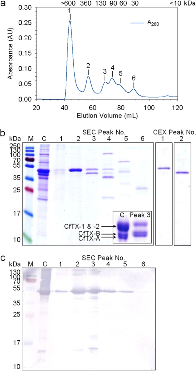
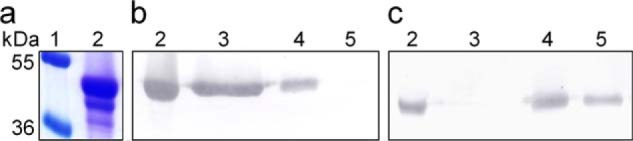
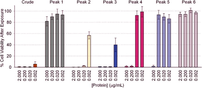
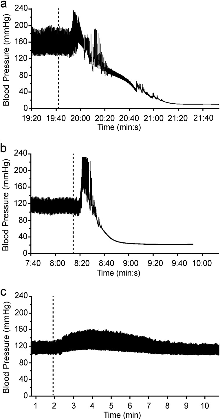
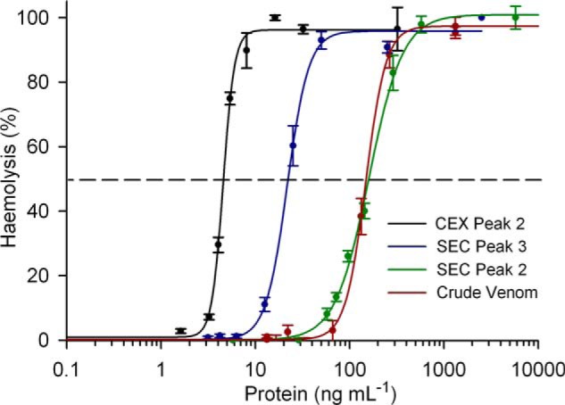
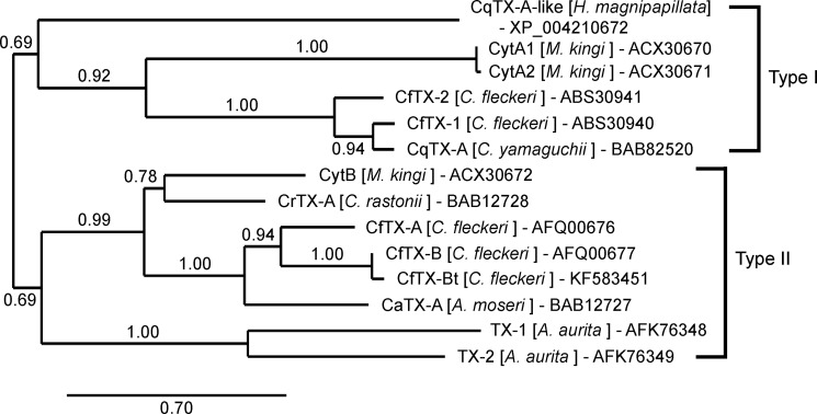
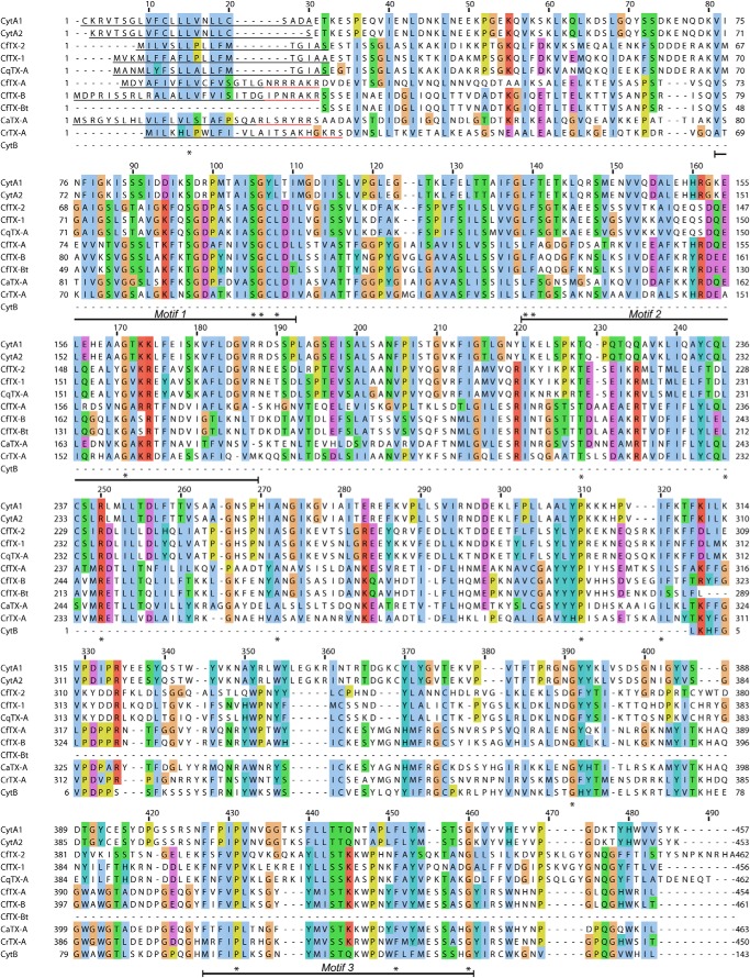
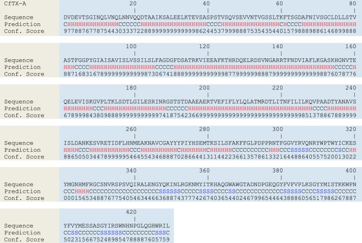
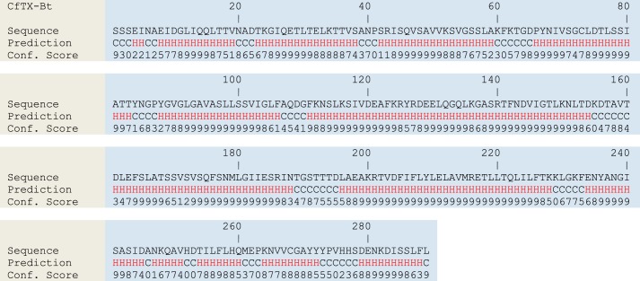
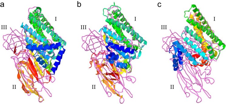
The box jellyfish Chironex fleckeri produces extremely potent and rapid-acting venom that is harmful to humans and lethal to prey. Here, we describe the characterization of two C. fleckeri venom proteins, CfTX-A (∼40 kDa) and CfTX-B (∼42 kDa), which were isolated from C. fleckeri venom using size exclusion chromatography and cation exchange chromatography. Full-length cDNA sequences encoding CfTX-A and -B and a third putative toxin, CfTX-Bt, were subsequently retrieved from a C. fleckeri tentacle cDNA library. Bioinformatic analyses revealed that the new toxins belong to a small family of potent cnidarian pore-forming toxins that includes two other C. fleckeri toxins, CfTX-1 and CfTX-2. Phylogenetic inferences from amino acid sequences of the toxin family grouped CfTX-A, -B, and -Bt in a separate clade from CfTX-1 and -2, suggesting that the C. fleckeri toxins have diversified structurally and functionally during evolution. Comparative bioactivity assays revealed that CfTX-1/2 (25 μg kg(-1)) caused profound effects on the cardiovascular system of anesthetized rats, whereas CfTX-A/B elicited only minor effects at the same dose. Conversely, the hemolytic activity of CfTX-A/B (HU50 = 5 ng ml(-1)) was at least 30 times greater than that of CfTX-1/2. Structural homology between the cubozoan toxins and insecticidal three-domain Cry toxins (δ-endotoxins) suggests that the toxins have a similar pore-forming mechanism of action involving α-helices of the N-terminal domain, whereas structural diversification among toxin members may modulate target specificity. Expansion of the cnidarian toxin family therefore provides new insights into the evolutionary diversification of box jellyfish toxins from a structural and functional perspective.

Keywords: Bioinformatics; Cardiovascular; Cubozoa; Cytolytic; Endotoxin; Jellyfish Toxin; Nematocyst; Protein Purification; Toxins; Venom.


Figures

FIGURE 1.
FIGURE 1.
Purification of C. fleckeri toxins using size exclusion and cation exchange chromatography. a, a typical chromatogram of crude venom fractionated on a Superdex 200pg column; protein elution was monitored at 280 nm. The estimated native molecular masses of SEC Peaks 1–6 are indicated above each peak in kDa. b, 15% denaturing SDS-PAGE protein profiles of crude venom (C) and chromatography fractions corresponding to SEC Peaks 1–6 and CEX Peaks 1 and 2. M, protein ladder; the molecular masses of the protein standards are shown alongside. Inset, 12.5% SDS-PAGE profile of crude venom and SEC Peak 3 showing increased resolution of CfTX-A and -B. c, Western blot of crude venom and SEC Peaks 1–6 using CfTX-1/2-specific antibodies.
FIGURE 2.
FIGURE 2.
Comparison of 12.5% SDS-PAGE and Western blot profiles during purification of CfTX-A/B. a, Coomassie-stained proteins; b, proteins bound to CfTX-1/2-specific antibodies; c, proteins bound to CfTX-A/B-specific antibodies. Lane 1, protein standards with molecular masses (kDa) indicated to the left; lane 2, crude C. fleckeri venom; lane 3, SEC Peak 2; lane 4, SEC Peak 3; lane 5, CEX Peak 2.
FIGURE 3.
FIGURE 3.
The in vitro cytotoxic effects of crude C. fleckeri venom and SEC-fractionated venom (Peaks 1–6) on cultured A7r5 cells, as determined by the MTS assay. The percentage of viable cells remaining in culture following exposure to the venom proteins compared with the negative control (100% cell viability) correlates inversely to cytotoxicity. Quadruplicate assays were performed at four different protein concentrations per sample. Error bars, S.E.
FIGURE 4.
FIGURE 4.
Representative arterial blood pressure traces from anesthetized rats injected with 25 μg kg−1 (intravenously) of crude C. fleckeri venom (a), SEC-purified CfTX-1/2 (Peak 2) (b), and CEX-purified CfTX-A/B (Peak 2) (c). A dotted line indicates the time of injection.
FIGURE 5.
FIGURE 5.
Hemolytic activity concentration-response curves for crude C. fleckeri venom and isolated CfTX toxins. The curve for crude venom is indicated in red. SEC Peak 2, containing purified CfTX-1/2, is indicated in green. SEC Peak 3, containing similar amounts of CfTX-1/2 and CfTX-A/B, is indicated in blue. CEX Peak 2, containing CfTX-A/B purified from SEC Peak 3, is indicated in black. Error bars, S.E. from four independent assays at each protein concentration.
FIGURE 6.
FIGURE 6.
Nucleotide and deduced amino acid sequences of CfTX-A (GenBankTM accession number JN695597). An 18-residue signal peptide is indicated with a dashed line; the start codon (ATG) is highlighted in boldface type. A 7-residue propeptide located between the signal peptide and the N terminus is shown in italic type. The N-terminal sequence (N) and two internal peptides (1 and 2) obtained by Edman sequencing are underlined. Peptide matches retrieved through LC-MS/MS analysis and Mascot searches are indicated in boldface type. An asterisk indicates the first stop codon in-frame with the start codon.
FIGURE 7.
FIGURE 7.
Nucleotide and deduced amino acid sequences of CfTX-B (GenBankTM accession number JN695598). A 24-residue signal peptide is indicated with a dashed line; the start codon (ATG) is highlighted in boldface type. A 7-residue propeptide located between the signal peptide, and the N terminus is shown in italic type. The N-terminal sequence (N) and an internal peptide (1) obtained by Edman sequencing are underlined. Peptide matches retrieved through LC-MS/MS analysis and Mascot searches are indicated in boldface type. An asterisk indicates the first stop codon in-frame with the start codon. Blue nucleotides correspond to a stretch of nucleotides that are absent in CfTX-Bt (Fig. 8); arrows indicate the deletion boundaries. Nucleotide variations between CfTX-B and CfTX-Bt are highlighted in red; for non-synonymous substitutions, the affected residue is also highlighted in red.
FIGURE 8.
FIGURE 8.
Nucleotide and deduced amino acid sequences of CfTX-Bt (GenBankTM accession number KF583451). Gene-specific primers for CfTX-B and CfTX-Bt (B-F3/B-R4) are underlined. An asterisk indicates the stop codon. Arrows correspond to nucleotide deletion boundaries in CfTX-B (Fig. 7). Nucleotide variations between CfTX-B and CfTX-Bt are indicated in red; for non-synonymous substitutions, the affected residue is also indicated in red.
FIGURE 9.
FIGURE 9.
Phylogenetic relationships of CfTX-like cnidarian toxins. The tree was constructed using the Phylogeny.fr pipeline (MUSCLE, PhyML, TreeDyn); branch support was evaluated using the approximate likelihood ratio test, Shimodaira-Hasegawa-like, statistical test. Sequence accession numbers are included after the species names.
FIGURE 10.
FIGURE 10.
Multiple sequence alignment of cubozoan toxin amino acid sequences. The sequences were aligned using MUSCLE, and the alignment was visualized using Jalview. Residue shading is based on the default Clustal protein color scheme. Identical residues are indicated with an asterisk, and dashes represent gaps introduced for better alignment. Signal peptides and proparts are underlined in black and red, respectively. Conserved sequence motifs are indicated below the alignment.
FIGURE 11.
FIGURE 11.
Secondary structure predictions for CfTX-A. H, S, and C, α-helical, β-strand, and coiled (loop) structures, respectively. Confidence scores range from 0 (low) to 9 (high).
FIGURE 12.
FIGURE 12.
Secondary structure predictions for CfTX-B. H, S, and C represent α-helical, β-strand, and coiled (loop) structures, respectively. Confidence scores range from 0 (low) to 9 (high).
FIGURE 13.
FIGURE 13.
Secondary structure predictions for CfTX-Bt. H and C represent α-helical and coiled (loop) structures, respectively. Confidence scores range from 0 (low) to 9 (high).
FIGURE 14.
FIGURE 14.
Predicted three-dimensional models of mature CfTX-A (a), CfTX-B (b), and CfTX-Bt (c), respectively, superimposed with the B. thuringiensis insecticidal 3d-Cry toxin, Cry8Ea1 (Protein Data Bank code 3EB7). Models were generated using the I-TASSER protocol and visualized using Jmol. The CfTXs are depicted in schematic form; the colors of secondary structures transition from blue (N terminus) to red (C terminus). Cry8Ea1 is depicted as a backbone trace (violet). The N-terminal, central, and C-terminal domains characteristic of 3d-Cry toxins are indicated as I–III, respectively.
FIGURE 15.
FIGURE 15.
Structural organization of Type I and Type II CfTX-like proteins. Signal peptides are indicated in green; the number of residues is indicated below. Putative N-terminal (N) and C-terminal domains (C) of the mature proteins are indicated in black and blue, respectively. The residue and theoretical molecular mass ranges of the mature toxins are indicated below each toxin type. A short propart (5–7 residues) present only in the Type II toxins is indicated in yellow. An arrow indicates the dibasic proteolytic cleavage site (KK/KR) at the C-terminal end of the propart.

References

    1. Currie B. J., Jacups S. P. (2005) Prospective study of Chironex fleckeri and other box jellyfish stings in the “Top End” of Australia's Northern Territory. Med. J. Aust. 183, 631–636 - PubMed
    1. Lumley J., Williamson J. A., Fenner P. J., Burnett J. W., Colquhoun D. M. (1988) Fatal envenomation by Chironex fleckeri, the north Australian box jellyfish. The continuing search for lethal mechanisms. Med. J. Aust. 148, 527–534 - PubMed
    1. Brinkman D. L., Burnell J. N. (2009) Biochemical and molecular characterisation of cubozoan protein toxins. Toxicon 54, 1162–1173 - PubMed
    1. Brinkman D., Burnell J. (2007) Identification, cloning and sequencing of two major venom proteins from the box jellyfish, Chironex fleckeri. Toxicon 50, 850–860 - PubMed
    1. Nagai H., Takuwa-Kuroda K., Nakao M., Oshiro N., Iwanaga S., Nakajima T. (2002) A novel protein toxin from the deadly box jellyfish (sea wasp, Habu-kurage) Chiropsalmus quadrigatus. Biosci. Biotechnol. Biochem. 66, 97–102 - PubMed

Publication types

MeSH terms

Associated data