. 2014 Jan 8;34(2):373-91.
doi: 10.1523/JNEUROSCI.0876-13.2014.

Rab5 and Rab4 regulate axon elongation in the Xenopus visual system

Affiliations

Rab5 and Rab4 regulate axon elongation in the Xenopus visual system

Julien Falk et al. J Neurosci. .

Abstract

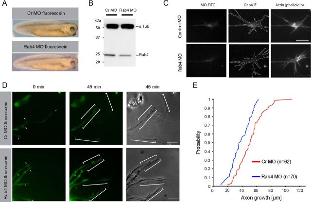
The elongation rate of axons is tightly regulated during development. Recycling of the plasma membrane is known to regulate axon extension; however, the specific molecules involved in recycling within the growth cone have not been fully characterized. Here, we investigated whether the small GTPases Rab4 and Rab5 involved in short-loop recycling regulate the extension of Xenopus retinal axons. We report that, in growth cones, Rab5 and Rab4 proteins localize to endosomes, which accumulate markers that are constitutively recycled. Fluorescence recovery after photo-bleaching experiments showed that Rab5 and Rab4 are recruited to endosomes in the growth cone, suggesting that they control recycling locally. Dynamic image analysis revealed that Rab4-positive carriers can bud off from Rab5 endosomes and move to the periphery of the growth cone, suggesting that both Rab5 and Rab4 contribute to recycling within the growth cone. Inhibition of Rab4 function with dominant-negative Rab4 or Rab4 morpholino and constitutive activation of Rab5 decreases the elongation of retinal axons in vitro and in vivo, but, unexpectedly, does not disrupt axon pathfinding. Thus, Rab5- and Rab4-mediated control of endosome trafficking appears to be crucial for axon growth. Collectively, our results suggest that recycling from Rab5-positive endosomes via Rab4 occurs within the growth cone and thereby supports axon elongation.


Figures

Figure 1.
Figure 1.
Constitutive recycling exhibited by Xenopus RGC growth cones is necessary for axon elongation in vitro. A–C, Biotinylated cell surface proteins are internalized and recycled in RGC growth cones. A, Biotinylated proteins were allowed to internalize for 20 min, after which the biotin still present at the surface was removed by MESNA and intracellular accumulations of biotinylated protein were detected in permeabilized growth cones. B, As expected, biotin labeling was barely detected just after MESNA treatment in nonpermeabilized growth cones. C, Twenty minutes later, however, some biotin labeling is recovered at the surface of nonpermeabilized growth cones. D–F, Primaquine reduces local recycling. D, FM dye is internalized in vesicles of different sizes that accumulate in the central domain of the growth cone; the white line delineates the outline of the growth cone. E, The endosomes in growth cones were loaded with FM4–64 and time-lapse imaging was performed. The total loss of FM4–64 fluorescence over time represents the speed of local recycling as individual FM dye-loaded vesicles release their content to the environment. Note that primaquine (0.068 mm) reduces local recycling, as more FM dye is retained in primaquine-treated growth cones. The outlines of growth cones are shown in the white broken line. F, The histogram shows the quantification of the total FM4–64 fluorescence intensity in the growth cones over time. Note that the difference in recycling between control and primaquine-treated growth cones becomes significant after 2 min. G, H, Primaquine blocks axon extension in vitro. G, The histogram shows the quantification of the extension rate of axons before, during, and after primaquine treatment. Numbers above bars indicate the number of axons analyzed. H, Snapshots from an in vitro time-lapse sequence illustrate the axon elongation rate of the same GFP-expressing axons in the absence of (left), in the presence of (middle), and after the dilution (right) of primaquine. For each axon, the dash and the arrowhead indicate the growth cone positions at the beginning and the end of the 45 min recording period, respectively. The dotted lines represent the distances covered by the growth cones during that time and exemplify the sharp inhibition of elongation observed in presence of primaquine. Error bars represent the SEM. *p < 0.05; ***p < 0.001. Scale bars: A–C, 5 μm; D, 4 μm; E, 5 μm; H, 30 μm.
Figure 2.
Figure 2.
RGC growth cones contain Rab5-, Rab4-, and Rab11-positive endosomes. A, Rab5 endogenous protein is detected in axon shaft and growth cone of retinal ganglion cells by immunocytochemistry. Rab5 labeling appears as focal puncta suggestive of vesicles in both the central (filled green arrowheads) and peripheral (open red arrowheads) domains of the growth cone. B–E, Xenopus Rab5c fused to GFP labels vesicles in both domains of RGC growth cones in vitro (B–D) and in vivo (E). C, D, Small vesicles are frequently observed in filopodia or lamellipodia. F, RFP-Rab5c labels vesicles that recruit FYVE-GFP (another marker of early endosomes). G, Rab4 is detected in the axon and growth cone of RGCs. Immunoreactivity exhibits vesicular-appearing signals in both domains of the growth cone. H–J, Xenopus GFP-Rab4 accumulates on vesicle-like structures present in both domains of the growth cone. I, J, Rab4 vesicles are found in filopodia and lamellipodia. K, GFP-Rab11b associates with small vesicles present in the axon shaft and both domains of the growth cone. Filled green and open red arrowheads point to vesicles located in the central and peripheral domains respectively. L, Confocal images of growth cone coexpressing RFP-Rab4 and GFP-Rab5c show that small Rab4-positive endosomes in the periphery are distinct from Rab5 ones (arrowheads). Rab4 and Rab5c colocalize, however, on the large vesicles frequently found in the central domain (asterisk). M, Confocal images show that Rab4 endosomes are distinct from the acidic LysoTracker-stained late endosomes and lysosomes. B, F, J, L, and M were acquired with a confocal spinning disk. Scale bars: A–L, 2 μm; M, 3 μm.
Figure 3.
Figure 3.
FRAP analysis indicates that Rab5- and Rab4-positive endosomes could form locally. A, The schematic illustrates the use of FRAP to define the origin of the Rab-positive endosomes present in the growth cone. The GFP-positive endosomes that reappear after photobleaching can be (1) unbleached endosomes that moved from the axon or (2) endosomes that have recruited unbleached cytosolic Rab5c-GFP that diffused into the growth cone. B–F, Representative images of the GFP signal before bleaching, immediately after, and during the recovery in growth cone expressing GFP-Rab5c (B–D), GFP-Rab4 (E), or GFP-Rab11 (F). B, Rab5c-positive endosomes have reappeared in the bleached growth cone, although many of the axonal Rab5c-positive endosomes remained in the axon (white arrowheads). C, Tracking of the endosomes present in the growth cone suggests that the majority of the Rab5c-positive endosomes does not come from the axons as only one of five endosomes labeled could be traced back to the axon. D, Rab5 signal is recovered on both long-lasting large endosomes and small dynamic endosomes. The small endosomes in the lamellipodia (open arrowhead at −18 s) move or disappear within 10 s. In contrast, large endosomes (filled arrowhead) remain stable and show little displacement. During the recovery, the large endosomes progressively recover Rab5 signal (filled arrowhead). In addition, small endosomes appear suddenly in the peripheral domain (open arrowheads at 180 and 190 s). E, Still images illustrating the GFP-Rab4 signal during the FRAP experiment. As with GFP-Rab5, GFP-Rab4 signal is recovered in the growth cone. Similarly, many Rab4-positive endosomes that appear in the growth cone after photobleaching could not be related to axonal endosomes (E). In contrast, most of the Rab11-positive endosomes come from the axon (F). Open arrowheads identify endosomes that appear to have formed within the growth cone. Filled arrowheads point to the endosomes that have moved from the axon. Dotted lines outline the bleached area. ″ denotes seconds Scale bars: A, C–F, 2 μm; B, 4 μm.
Figure 4.
Figure 4.
Dynamic analysis of Rab4-positive endosomes suggests that they support local recycling in the growth cone. A, Time-lapse confocal images show a Rab4-positive tubule (arrowhead) that buds and splits from a large Rab5c- and Rab4-positive endosome. The 2 Hz time-lapse imaging was performed on growth cones coexpressing wild-type Xenopus GFP-Rab5c and RFP-Rab4. B, After photobleaching, the RFP-Rab4 signal recovers on large GFP-Rab5c-positive endosomes in RGC growth cone. Left, Prebleaching signal (−4 s). Middle, The RFP signal, but not the GFP signal, is lost after photobleaching (0 s). As illustrated in the left panel (150 s), during recovery, the RFP signal is progressively recovered on the large GFP-positive endosome (red arrowhead). Rab4 is not recruited to Rab5 endosomes that were Rab4-negative (white arrowhead). C, Bleaching of the peripheral domain reveals the shipment of small Rab4-positive endosomes from the central to the peripheral domain (green arrowheads). D, Time-lapse images illustrate the entry of a small Rab4-positive (Rab5-negative) endosome into a filopodium and its movement toward the tip. Arrowheads mark the position of the endosome at different time points. E, F, Confocal images show that internalized biotinylated proteins (red) partially colocalize with vesicles labeled by GFP-Rab4 (green). The small Rab4-positive endosomes present in filopodia also contain biotinylated proteins (arrowhead, F). ″ denotes seconds. Scale bars, 2 μm.
Figure 5.
Figure 5.
Constitutively active Rab5 slows the axon extension rate in vitro. A, The electroporation of CA-Rab5c in RGCs induces the formation of enlarged endosomes. In the RGC soma, CA-Rab5c that lacks GTPase activity (QL mutant) labels endosomes that are considerably larger than those labeled by wild-type GFP-Rab5c. B, C, Still images from time-lapse sequences illustrate axon extension of GFP-expressing (B) and constitutively active GFP-Rab5c-expressing (C) axons. Time-lapse imaging was performed on whole eyes from stage 33/34 embryos that had been cultured overnight. Arrowheads indicate the positions of the growth cone at 0 and 45 min. Dotted lines exemplify the distance extended by the growth cones during the 45 min recording period. D, The histogram shows the average extension rate of axons expressing GFP, the Xenopus CA-Rab5c we generated (xRab5QL), and the previously characterized zebrafish CA-Rab5c (zRab5c) constructs. E, Cumulative distributions of the apparent diameter of the endosomes labeled by GFP-CA-Rab5c (black line; N = 64) and wild-type GFP-Rab5c (gray dotted line; N = 404) suggest that the CA-Rab5c leads to an enlargement of the Rab5-positive endosomes in the growth cone. Numbers above the bars indicate the number of axons analyzed. Error bars represent the SEM. ***p < 0.001 vs the control (GFP). ′ denotes minutes. Scale bars: A, 2 μm; B, C, 30 μm.
Figure 6.
Figure 6.
Dominant-negative Rab4 slows the axon extension rate in vitro. A, Primaquine treatment alters Rab4-positive endosomes. Images of Rab4-expressing axons before (left) and 45 min after (right) primaquine application (0.05 mm) illustrate the appearance of large abnormal Rab4-positive tubules (arrowheads). B, An example of RGC growth cone expressing the dominant-negative Xenopus GFP-tagged Rab4-NI mutant. Note that, in contrast to wild type (A, left), the mutant does not display a punctate distribution. C–E, DN-Rab4 construct impairs axon extension. C, D, Images of GFP-positive axons at the start and the end of the time-lapse recording illustrate the extension of GFP-expressing (C) and DN-xRab4-expressing (D) axons. Arrowheads indicate the positions of the growth cones at 0 and 45 min. Dotted lines highlight the distance extended by the growth cones during this period. E, The histograms show the average extension rate measured from time-lapse recording of axons that expressed GFP, DN-xRab4 (NI mutant), or CA-xRab4 (QL mutant). F, Rab7 mutants do not slow axon extension in RGCs. The extension rate of that axon that expressed DN-Rab7 (TN mutation) or CA-Rab7 (QL mutation) was measured from time-lapse recording and compared with the extension rate of GFP-expressing axons from the same electroporation batch. Neither Xenopus (xRab7) nor human (hRab7) DN-Rab7 decreases the axon extension rate. Numbers above the bars indicate the number of axons (E, F). Error bars represent the SEM. ***p < 0.001 vs the control. Scale bars: A, B, 5 μm; C, D, 30 μm.
Figure 7.
Figure 7.
Rab4 knockdown impairs axon elongation in vitro. A, The assessment of the effect of Rab4 morpholino on developing Xenopus embryos. Shown are typical examples of embryos at stage 33–34, injected with 12 ng/blastomere fluorescein-tagged control morpholino (top) or Rab4 morpholino (bottom). Note that, at this dose, the Rab4 morpholino does not cause any gross developmental abnormalities. B, Rab4 morpholino reduces the Rab4 protein levels approximately by half. Shown is a representative example of a Western blot of brain-and-eye protein extracts from stage 37 embryos. C, Rab4 morpholino reduces the levels of Rab4 protein in RGC growth cones of cultured eye explants by 50.1 ± 6.2% relative to control morpholino. All quantified growth cones contain green fluorescence from the morpholino tag. Actin (phalloidin) staining was used to show the outline of the growth cone. D, Rab4 morpholino impairs axonal outgrowth in vitro. The extension of axons containing either control morpholino (green, top row) or Rab4 morpholino (green, bottom row) was monitored over 45 min. Corresponding phase-contrast images (grayscale) were used for actual measurements. Scale bars: C, 10 μm; D, 25 μm. E, Quantification of D. Axon outgrowth is significantly reduced in cells containing Rab4 morpholino. Cumulative distribution plots show that both datasets: control morpholino (red line) and Rab4 morpholino (blue line) are significantly different from each other (p = 5.15 × 10⁻⁶, Student's t test).
Figure 8.
Figure 8.
Constitutively active Rab5 delays axon arrival at the tectum in vivo. A, Schematic representation of the experiment; retinas of Xenopus embryos were electroporated when RGCs are generated, and embryos were killed before or after RGC axons have reached the tectum (i.e., stage 37/38 or 39). B, Right, 3D representation of the retino-tectal projection in Xenopus embryo. RGC axons from the contralateral eye (black) enter the brain at the optic chiasm and grow dorsally in the diencephalon before turning caudally to enter their target, the tectum. Left, Phase image of a stage 39 hemi-brain depicts how brain and tectum outlines (dashed lines) are drawn. The normal course and length of the RGC axons is presented (black lines). C–E, RGC axons that express CA-Rab5c are shorter than the control. C, D, Lateral views of whole-mount hemi-brain from stage 37/38 embryos show that GFP-expressing axons approach the tectum (C), while CA-GFP-Rab5-expressing ones are still far away (D). The length of the longest axons corresponding to the dotted lines (C, D) was measured, and the quantifications are presented in E. The experiments were performed with the Xenopus CA-Rab5c (xRab5QL) and the zebrafish CA-Rab5c (zRab5c) constructs, and were analyzed at stage 37/38 and stage 39 in both cases. Numbers above the bars indicate the number of embryos analyzed. Error bars represent the SEM. ***p < 0.001 vs the control. Scale bars: B, D, 100 μm. OC, Optic chiasm; Di., diencephalon; Tel., telencephalon; Pin., pineal gland.
Figure 9.
Figure 9.
DN-Rab4 delays axon arrival at the tectum and slows axon extension in vivo. A–C, Lateral views of stage 37/38 contralateral brains illustrate the length of GFP-positive (A), DN-GFP-Rab4-positive (B), and CA-GFP-Rab4-positive (C) axons. The tectum (dashed line) and the brain (plain line) outlines were drawn from the phase image. Dotted lines between arrowheads exemplify the length of the longest axon. Axons that express GFP (A) or GFP-CA-Rab4 (QL mutant; C) extend equally well along the optic tract. In contrast, DN-Rab4 (NI mutant)-expressing axons are much shorter (B). Lengths of GFP-positive, CA-Rab4-positive, DN-Rab4-positive, and DN-Rab7-positive (Xenopus, human) axons were measured in stage 37/38 and 39 brains. D–F, Summary of the quantifications shows that DN-Rab4 is the only construct tested here that significantly reduces axon length. G, H, DN-Rab4 decreases axon extension rate in vivo. G, Pictures from in vivo time-lapse sequences show DN-GFP-Rab4-expressing (left) and GFP-expressing (right) axons that extend along the optic tract. Images are z-projections at the 0 and 60 min time points. Open and filled arrowheads indicate the growth cone positions at the start and the end of the 1 h recording, respectively. Dotted lines exemplify the distance traveled by the growth cones during the 1 h monitoring. Quantifications of the in vivo time-lapse recordings are presented in H. Numbers above bars indicate the number of embryos (D–F) or axons (H) analyzed. Error bars represent the SEM. ***p < 0.001 vs the control. Scale bars: C, 100 μm; G, 15 μm.

Similar articles

Cited by

References

    1. Archer KJ, Reese SE. Detection call algorithms for high-throughput gene expression microarray data. Brief Bioinform. 2010;11:244–252. doi: 10.1093/bib/bbp055. - DOI - PMC - PubMed
    1. Ascaño M, Richmond A, Borden P, Kuruvilla R. Axonal targeting of Trk receptors via transcytosis regulates sensitivity to neurotrophin responses. J Neurosci. 2009;29:11674–11685. doi: 10.1523/JNEUROSCI.1542-09.2009. - DOI - PMC - PubMed
    1. Bananis E, Murray JW, Stockert RJ, Satir P, Wolkoff AW. Regulation of early endocytic vesicle motility and fission in a reconstituted system. J Cell Sci. 2003;116:2749–2761. doi: 10.1242/jcs.00478. - DOI - PubMed
    1. Barbieri MA, Roberts RL, Gumusboga A, Highfield H, Alvarez-Dominguez C, Wells A, Stahl PD. Epidermal growth factor and membrane trafficking. EGF receptor activation of endocytosis requires Rab5a. J Cell Biol. 2000;151:539–550. doi: 10.1083/jcb.151.3.539. - DOI - PMC - PubMed
    1. Bergtold A, Desai DD, Gavhane A, Clynes R. Cell surface recycling of internalized antigen permits dendritic cell priming of B cells. Immunity. 2005;23:503–514. doi: 10.1016/j.immuni.2005.09.013. - DOI - PubMed

Publication types

LinkOut - more resources