. 2014 Jan 8;34(2):586-95.
doi: 10.1523/JNEUROSCI.4257-13.2014.

Amygdala inputs to the ventral hippocampus bidirectionally modulate social behavior

Affiliations

Amygdala inputs to the ventral hippocampus bidirectionally modulate social behavior

Ada C Felix-Ortiz et al. J Neurosci. .

Abstract

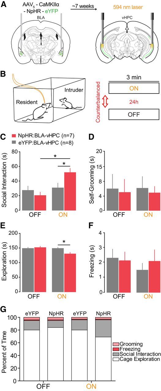
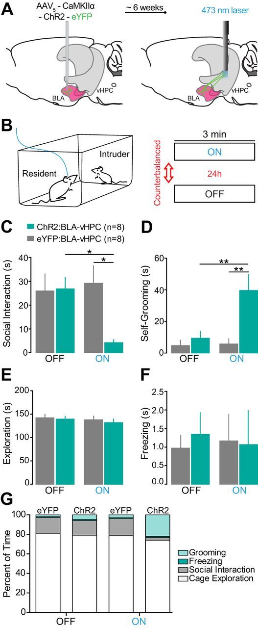
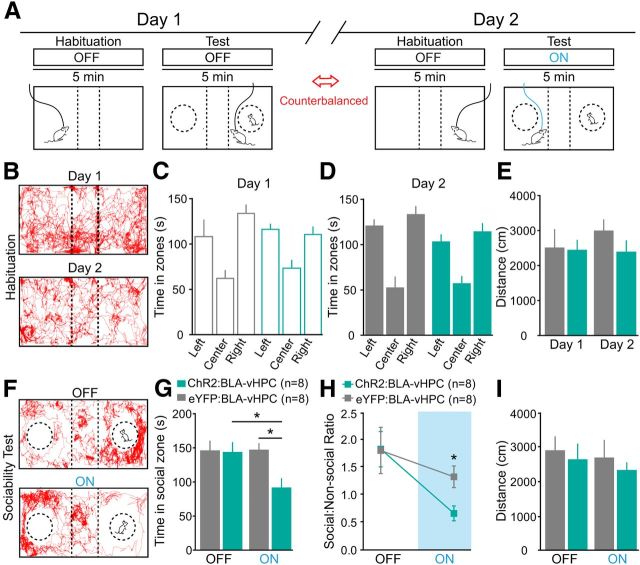
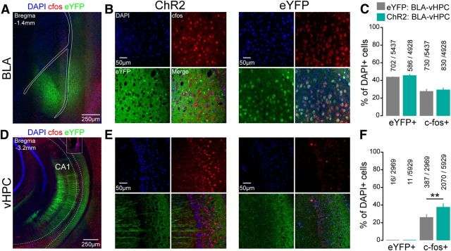
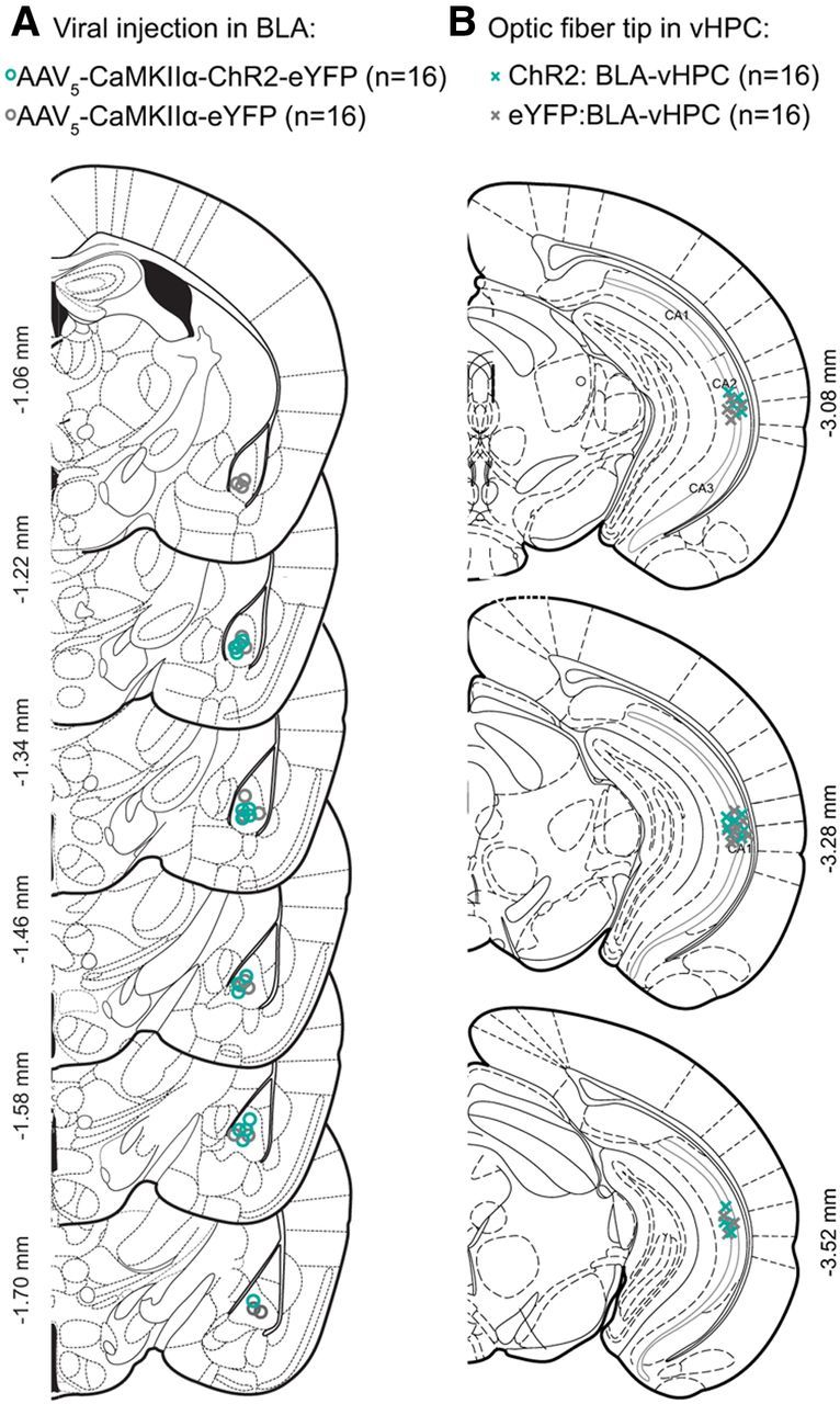
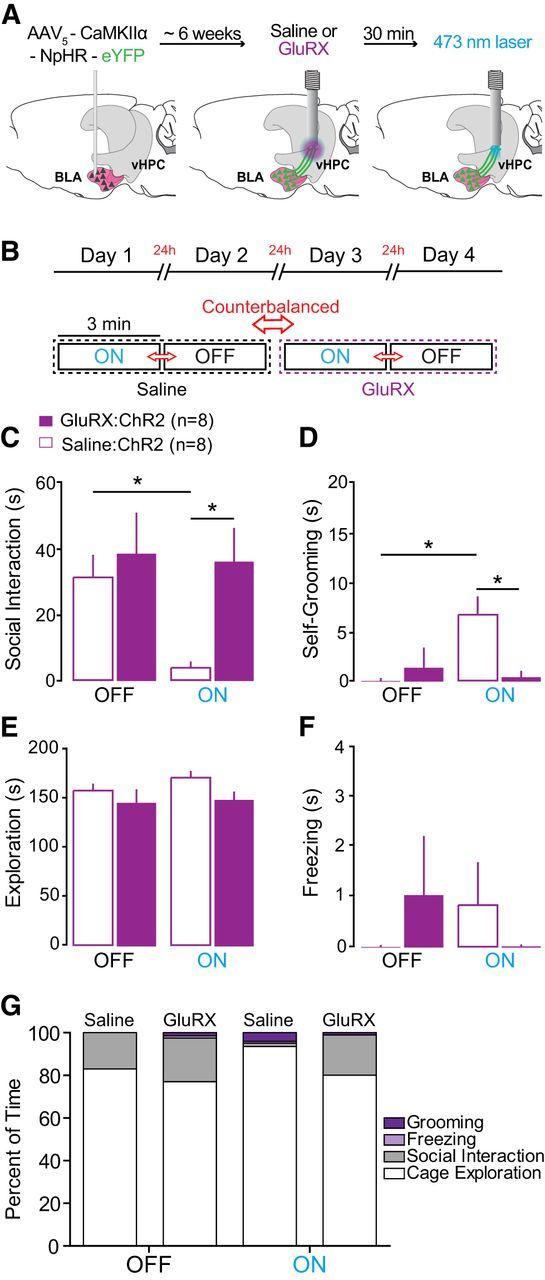
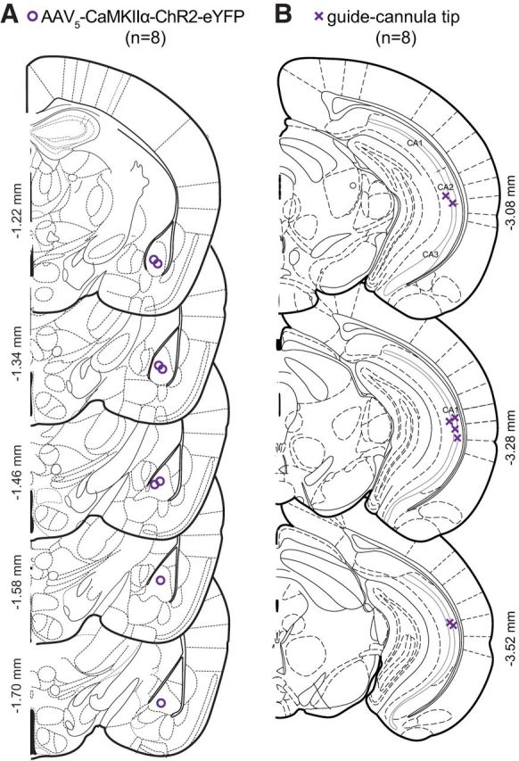
Impairments in social interaction represent a core symptom of a number of psychiatric disease states, including autism, schizophrenia, depression, and anxiety. Although the amygdala has long been linked to social interaction, little is known about the functional role of connections between the amygdala and downstream regions in noncompetitive social behavior. In the present study, we used optogenetic and pharmacological tools in mice to study the role of projections from the basolateral complex of the amygdala (BLA) to the ventral hippocampus (vHPC) in two social interaction tests: the resident-juvenile-intruder home-cage test and the three chamber sociability test. BLA pyramidal neurons were transduced using adeno-associated viral vectors (AAV5) carrying either channelrhodopsin-2 (ChR2) or halorhodopsin (NpHR), under the control of the CaMKIIα promoter to allow for optical excitation or inhibition of amygdala axon terminals. Optical fibers were chronically implanted to selectively manipulate BLA terminals in the vHPC. NpHR-mediated inhibition of BLA-vHPC projections significantly increased social interaction in the resident-juvenile intruder home-cage test as shown by increased intruder exploration. In contrast, ChR2-mediated activation of BLA-vHPC projections significantly reduced social behaviors as shown in the resident-juvenile intruder procedure as seen by decreased time exploring the intruder and in the three chamber sociability test by decreased time spent in the social zone. These results indicate that BLA inputs to the vHPC are capable of modulating social behaviors in a bidirectional manner.

Keywords: ChR2; NpHR; amygdala; hippocampus; optogenetics; social.


Figures

Figure 1.
Figure 1.
Inhibition of BLA terminals projecting to the vHPC with NpHR increases social interaction in a resident-juvenile intruder procedure. BLA glutamatergic neurons were transduced with either NpHR-eYFP (n = 7) or eYFP alone as a control (n = 8). Yellow light was delivered (3 min, constant) via bilateral optical fibers implanted in the vHPC, after ∼7–8 weeks of viral incubation. A, Left, Coronal brain schematic indicating the site of viral injection into the BLA. Right, Coronal schematic indicating optic fiber location into the vHPC. Top, Experimental timeline. B, Left, Schematic indicating the home-cage resident-juvenile (3–4 weeks) intruder behavioral procedure. Right, Schematic indicating the 3 min epochs with 24 h, counterbalanced between the social tasks. Different intruders were used for each epoch. C, NpHR mice spent significantly more time performing social interaction than eYFP mice during the yellow light illumination epoch. *p = 0.046. D, No significant effect of light stimulation or group was detected on time spent self-grooming compared with eYFP mice. E, NpHR mice spent less time (seconds) exploring their home cage during light stimulation compared with eYFP control mice. *p = 0.035. F, No significant effect of light stimulation or group was detected on freezing behavior in the presence of a juvenile intruder. G, Percentage of total time (3 min), showing social interaction, self-grooming, cage exploration, and freezing. Data are mean values. Error bars indicate SEM.
Figure 2.
Figure 2.
Histologically verified placements of viral injections and optical fiber tips in NpHR:BLA-vHPC and eYFP:BLA-vHPC groups. A, Coronal sections from bregma containing the BLA. Center of the viral injections in the BLA for all the mice injected with NpHR (n = 7; orange circles) and eYFP (n = 8; gray circles). B, BLA confocal image from a representative NpHR mouse. Right, Confocal images of individual cells in the BLA of a mouse in the NpHR group. C, Coronal sections from bregma containing the vHPC. Location of the optical fiber tips above the pyramidal layer of vHPC for NpHR:BLA-vHPC (orange crosses) and eYFP (gray crosses). D, vHPC confocal image indicating the optical fiber placement in a representative mouse from the NpHR group. Right, Confocal images of individual cells in the vHPC of a mouse expressing NpHR in BLA axon terminals.
Figure 3.
Figure 3.
Activation of BLA axon terminals in the vHPC with ChR2 decreases social interaction in a resident-juvenile intruder procedure. Glutamatergic neurons from the BLA were transduced with either ChR2-eYFP (n = 8) or eYFP control (n = 8). After ∼6–7 weeks of incubation after viral transduction of BLA cell somata, blue light was delivered (3 min, 20 Hz, 5 ms pulses) via a unilateral optical fiber implanted above the vHPC. A, Left, Sagittal view brain schematic indicating viral injection into the BLA. Right, Schematic indicating unilateral optical fiber location into the vHPC. Top, Experimental timeline. B, Schematic indicating the home-cage resident-juvenile (3–4 weeks) intruder behavioral procedure. Three minute epochs were counterbalanced for order with a 24 h interval between ON and OFF light conditions. Novel juvenile intruders were used for each epoch. C, ChR2 mice spent significantly less time (seconds) performing social interaction than eYFP mice during the blue light illumination epoch. *p = 0.034. D, ChR2 mice also spent significantly more time (seconds) performing self-grooming than eYFP mice in the presence of a juvenile intruder. **p = 0.008. E, No significant effect of light stimulation or group was detected for the time spent exploring their home cage. F, No significant effect of light stimulation or group was detected in freezing behavior in the presence of a juvenile intruder. G, Percentage of total time (3 min), showing social interaction, self-grooming, cage exploration, and freezing. Data are mean values; error bars indicate ± SEM.
Figure 4.
Figure 4.
Activation of BLA projections to the vHPC with ChR2 decreases sociability in a three chamber sociability test. In a different group of animals from Figure 3, glutamatergic neurons from the BLA were transduced with either ChR2-eYFP (n = 8) or eYFP control (n = 8). After ∼6–7 weeks of viral transduction of BLA cell somata, blue light was delivered (5 min, 20 Hz, 5 ms pulses) via unilateral optical fiber implanted in the vHPC. A, Top, Timeline across 2 d of testing. Bottom, Schematic indicating the three chamber sociability test and 5 min experimental epochs. Each testing day was composed of habituation to the arena followed by 5 min of laser ON or laser OFF stimulation (counterbalanced for order). Different juvenile mice were used for each testing epoch, counterbalanced for side of the testing arena. B, Representative animal tracks during the habituation phase for day 1 (top) and day 2 (bottom). C, Time spent in the different zones of the arena during day 1. No significant differences were found between the times spent on the right chamber versus the left chamber. D, Time spent in the different zones of the arena during day 2. No significant differences were found between the times spent on the right chamber versus the left chamber. E, No significant effect on locomotion during the habituation phase during the 2 d of testing. F, Representative animal tracks during the testing sessions for OFF (top) and ON (bottom). G, ChR2 mice spent significantly less time (seconds) in the social zone relative to eYFP mice during the blue light illumination epoch. *p = 0.033. H, ChR2 also showed a significantly lower social/nonsocial ratio during laser stimulation compared with eYFP mice. *p = 0.023. I, No significant effect of locomotion during the habituation phase during the laser stimulation. Data are mean values; error bars indicate ± SEM.
Figure 5.
Figure 5.
Activation of BLA axon terminals in the vHPC using ChR2 increases c-fos expression in the pyramidal layer of vHPC. Blue represents DAPI; green, eYFP; red, c-fos. A, Confocal image of the BLA of representative ChR2 animal. B, Confocal images of the BLA from two representative mice. Representative ChR2:BLA-vHPC animal (left) and representative eYFP:BLA-vHPC mice (right). C, Percentage of DAPI-positive (+) cells expressing eYFP or c-fos in the BLA (n = 8 ChR2 mice and n = 8 eYFP mice). No differences between groups were found in c-fos+ or eYFP+ cells in the BLA. D, Confocal image of the vHPC from representative ChR2 mice. E, vHPC confocal images of two representative mice. Representative ChR2:BLA-vHPC animal (left) and representative eYFP:BLA-vHPC mice (right). F, Percentage of DAPI-positive (+) cells expressing eYFP or c-fos in the vHPC (n = 8 ChR2 mice and n = 8 eYFP mice). Compared with eYFP:BLA-vHPC controls, light stimulation of BLA terminals in the vHPC increased the percentage of c-fos(+) cells in the vHPC of ChR2:BLA-vHPC group. Data are mean ± SEM. **p = 0.0028.
Figure 6.
Figure 6.
Histologically verified placements of viral injections and optical fiber tips in ChR2:BLA-vHPC and eYFP:BLA-vHPC groups. A, Coronal sections from bregma of the BLA. Center of the viral injections in the BLA for all the mice injected with ChR2 (n = 16; green circles) and eYFP (n = 16; gray circles). B, Coronal sections from bregma of the vHPC. Location of the optic fibers tip above the pyramidal layer of vHPC for ChR2:BLA-vHPC (green crosses) and eYFP (gray crosses).
Figure 7.
Figure 7.
Activation of BLA inputs to the vHPC is sufficient to mediate changes in social interaction without affecting the ability to explore the environment. A, Glutamate receptor antagonists (GluRX:AP5+NBQX, purple) or saline (black) were unilaterally infused into the vHPC using the same guide cannula subsequently used for light delivery via an acutely inserted optical fiber. Top, Experimental timeline. Left, Sagittal brain schematic indicating viral injections into the BLA. Middle, Sagittal schematic indicating unilateral cannula location into the vHPC. Right, Sagittal schematic of removable optical fiber used for light delivery 30 min after GluRX delivery. B, Top, Testing took place over 4 consecutive days. Schematic of 3 min epochs where mice received either drug or saline treatment. All experiments where counterbalanced for treatment and stimulation (ON or OFF epoch) order. Novel juvenile intruders were used for each session. C, GluRX attenuated the light stimulation effect. GluRX:ChR2 mice (n = 8) spent significantly more time (seconds) performing social interaction than Saline:ChR2 mice (n = 8) during the blue light illumination epoch. *p = 0.048. D, GluRX:ChR2 mice also spent significantly less time performing self-grooming compared with Saline:ChR2 mice. *p = 0.036. E, No significant effect of light stimulation or group was detected on the time spent exploring their home cage. F, No significant effect of light stimulation or group was detected for freezing behavior in the presence of a juvenile intruder. G, Percentage of total time (3 min), showing social interaction, self-grooming, cage exploration, and freezing. Data are mean ± SEM.
Figure 8.
Figure 8.
Histologically verified placements of viral injections and optical fiber tips in ChR2 animals used for pharmacology experiments. A, Coronal sections of the BLA with coordinates reflecting anteroposterior distance from bregma. Center of viral injections in the BLA for all mice expressing ChR2 (n = 8; purple circles). B, Coronal sections of the vHPC with coordinates from bregma. Location of cannulae above the pyramidal layer of vHPC for ChR2 animals (purple crosses).

References

    1. Adhikari A, Topiwala MA, Gordon JA. Synchronized activity between the ventral hippocampus and the medial prefrontal cortex during anxiety. Neuron. 2010;65:257–269. doi: 10.1016/j.neuron.2009.12.002. - DOI - PMC - PubMed
    1. Amaral DG, Corbett BA. The amygdala, autism and anxiety. In: Bock Organizer G, Goode J, editors. Autism: neural basis and treatment possibilities. New York: Wiley; 2008. pp. 177–197.
    1. Amaral DG. The primate amygdala and the neurobiology of social behavior: implications for understanding social anxiety. Biol Psychiatry. 2002;51:11–17. doi: 10.1016/S0006-3223(01)01307-5. - DOI - PubMed
    1. American Psychiatric Association. Diagnostic and statistical manual of mental disorders. Ed 5. Arlington, VA: American Psychiatric Association; 2013.
    1. Ansseau M, Legros JJ, Mormont C, Cerfontaine JL, Papart P, Geenen V, Adam F, Franck G. Intranasal oxytocin in obsessive-compulsive disorder. Psychoneuroendocrinology. 1987;12:231–236. doi: 10.1016/0306-4530(87)90009-6. - DOI - PubMed

Publication types