. 2014 Jan 8;34(2):629-45.
doi: 10.1523/JNEUROSCI.3859-13.2014.

Axon regeneration genes identified by RNAi screening in C. elegans

Affiliations

Axon regeneration genes identified by RNAi screening in C. elegans

Paola Nix et al. J Neurosci. .

Abstract

Axons of the mammalian CNS lose the ability to regenerate soon after development due to both an inhibitory CNS environment and the loss of cell-intrinsic factors necessary for regeneration. The complex molecular events required for robust regeneration of mature neurons are not fully understood, particularly in vivo. To identify genes affecting axon regeneration in Caenorhabditis elegans, we performed both an RNAi-based screen for defective motor axon regeneration in unc-70/β-spectrin mutants and a candidate gene screen. From these screens, we identified at least 50 conserved genes with growth-promoting or growth-inhibiting functions. Through our analysis of mutants, we shed new light on certain aspects of regeneration, including the role of β-spectrin and membrane dynamics, the antagonistic activity of MAP kinase signaling pathways, and the role of stress in promoting axon regeneration. Many gene candidates had not previously been associated with axon regeneration and implicate new pathways of interest for therapeutic intervention.

Keywords: C. elegans; DLK; MAP kinases; axon regeneration; laser axotomy; neural regeneration.


Figures
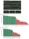
Figure 1.
Figure 1.
Axon regeneration genes identified by genetic screening. AF, GABA neurons visualized with Punc-47:GFP. Animals are arranged with posterior at left, ventral side down. For axotomy, five to six posterior commissures were severed at the midline. Scale bar, 20 μm. A, Wild-type. B, unc-70 mutant fed empty vector control RNAi bacteria. C, unc-70 mutant fed an RNAi clone targeted against a candidate regeneration gene. D, Wild-type axon regeneration after axotomy. E, Axotomy in a mutant with improved regeneration. F, Axotomy in a mutant with blocked regeneration. GH, Percentage of growth cone formation in candidate genes characterized by axotomy. Mutants showing significantly improved regeneration are in green and mutants showing significantly decreased regeneration are in red. G, Candidate genes identified in the unc-70 RNAi screen. pxn-2 was characterized by Gotenstein et al. (2010) and is not included on this graph. H, Additional candidate genes identified by axotomy. Error bars indicate 95% confidence interval.
Figure 2.
Figure 2.
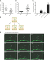
Axon regeneration defects in unc-70/β-spectrin mutants. A, Timing of first growth cone (GC) appearance in unc-70 mutants compared with wild-type. Each point represents an individual time-lapse experiment. B, Axon retraction distance after axotomy. Each point represents an individual cut axon. C, Quantification of spontaneous growth cone formation in unc-70 mutants per hour per commissure. ***p < 0.001; *p < 0.05, t test. Error bars indicate SEM. D, Schematic drawing showing spontaneous growth cone formation in unc-70 and possible consequences to outgrowth: (a) the formerly intact axon breaks, (b) the new growth cone extends to a nearby, intact axon causing it to break, and (c) the migrating growth cone itself breaks and generates a new growth cone. E, Time-lapse recording shows the unc-70 migrating growth cone (arrowhead) extending across the adjacent commissural axon (arrow) and causing it to break. The intact commissure (asterisk) dynamically extends and retracts branches over the course of the time lapse. At 48 minutes, the growth cone has broken the adjacent axon and the proximal end rapidly retracts (lower arrow), leaving behind fragments that are slowly cleared (top arrow, 48–159 min). At 132–159 minutes, the axon behind the migrating growth cone breaks (arrowhead, 132 min) and the proximal axon fragment retracts back to the branch point (left arrowhead, 159 min), leaving the distal process to degenerate (right arrowhead, 159–225 min). At 291–528 minutes, both stumps regenerate new growth cones. Scale bar, 10 μm.
Figure 3.
Figure 3.
Microvesicle release is an early step in axon regeneration. A, Time-lapse images of an axotomized wild-type axon. The retraction bulb is formed within 12 minutes (arrowhead). At 201 minutes, there is an expulsion of numerous microvesicles, which are then cleared and mostly removed by 321 min. The growth cone forms shortly after at 363 min. Scale bar, 5 μm. B, Microvesicle release from an uninjured wild-type axon. At 0 minutes, a bulge predicts the site of vesicle release. At 3 minutes, the vesicle is connected by a faint neck and, by 6 minutes, the vesicle is separated from the axon. Scale bar, 1 μm. C, Microvesicle shedding per hour per commissure from intact axons is increased in unc-70 mutants. p-values were determined by t test. Error bars indicate SEM. D, nex-1 is required for normal GABA neuron regeneration. ***p < 0.001, Fisher's exact test. Error bars indicate 95% confidence interval. E, Time-lapse images of axotomized nex-1 axons suggest defects in damaged membrane repair. At 228 minutes, membrane blebs are visible and appear to interfere with normal growth cone formation. Scale bar, 5 μm.
Figure 4.
Figure 4.
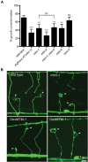
Axon regeneration defects in myotubularin mutants by loss-of-function and overexpression. A, mtm-1 and other myotubularin family members are required for normal GABA neuron regeneration. **p < 0.01; ***p < 0.001, NS, Fisher's exact test. Error bars indicate 95% confidence interval. B, mtm-1 loss-of-function and overexpression in GABA neurons causes axonal membrane defects. Arrowheads indicate the highly branched wild-type growth cones seen at L4. The mtm-1 growth cones are often large and unbranched (left growth cone), but many fail (right growth cone). Overexpression of mtm-1 may cause failure to initiate growth cones (lower left). Bottom left, right arrowhead, Successful growth cones often leave behind growth cone remnants. Bottom right, Failed growth cone collapsed around a large vacuole (left arrowhead) and a large growth cone remnant along the axon of a successful commissure (right arrowhead). Asterisk shows a large vacuole in a neuron cell body. Scale bar, 5 μm.
Figure 5.
Figure 5.
Multiple MAPK pathways antagonistically influence axon regeneration. A, dlk-1 and kgb-1 mutants represent two MAPK modules essential for axon regeneration. MAPK signaling via JNK-1 inhibits axon regeneration and requires functional kinase activity. Improved regeneration by loss of jnk-1 weakly suppresses loss of kgb-1, but is not sufficiently to suppress the loss of dlk-1. B, The MAP2Ks jkk-1 and sek-6 may function upstream of JNK-1 to inhibit axon regeneration. Loss of jkk-1 and sek-6 partially suppresses the effects of JNK-1 overexpression. C, The MAP3K kin-18 may inhibit axon regeneration via JNK-1. D, FOS-1 transcription factor is necessary for regeneration and is a target of KGB-1 MAPK signaling. E, Summary of MAPK signaling components affecting axon regeneration. *p < 0.05; **p < 0.01; ***p < 0.001, NS, Fisher's exact test. Error bars indicate 95% confidence interval.
Figure 6.
Figure 6.
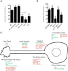
Stress response pathways are necessary for efficient axon regeneration. A, Stress-inducing conditions improve regeneration outcomes and require hsf-1. Regeneration in hif-1 is also reduced, but was not tested under hypoxic conditions. WT, Wild-type; HS, heat shock. B, Increased IRE-1 activity in xbp-1 mutants and by ire-1 overexpression inhibits regeneration. *p < 0.05; **p < 0.01; ***p < 0.001, NS, Fisher's exact test. Error bars indicate 95% confidence interval. C, Example of candidates affecting axon regeneration. Proteins “positively” required for axon regeneration are shown in green; those that “inhibit” axon regeneration are shown in red. Mammalian orthologs are in parentheses where known and different from the C. elegans name.

References

    1. Abe N, Cavalli V. Nerve injury signaling. Curr Opin Neurobiol. 2008;18:276–283. doi: 10.1016/j.conb.2008.06.005. - DOI - PMC - PubMed
    1. Anckar J, Sistonen L. Regulation of HSF1 function in the heat stress response: implications in aging and disease. Annu Rev Biochem. 2011;80:1089–1115. doi: 10.1146/annurev-biochem-060809-095203. - DOI - PubMed
    1. Ben-Yaakov K, Dagan SY, Segal-Ruder Y, Shalem O, Vuppalanchi D, Willis DE, Yudin D, Rishal I, Rother F, Bader M, Blesch A, Pilpel Y, Twiss JL, Fainzilber M. Axonal transcription factors signal retrogradely in lesioned peripheral nerve. EMBO J. 2012;31:1350–1363. doi: 10.1038/emboj.2011.494. - DOI - PMC - PubMed
    1. Ben-Zvi A, Miller EA, Morimoto RI. Collapse of proteostasis represents an early molecular event in Caenorhabditis elegans aging. Proc Natl Acad Sci U S A. 2009;106:14914–14919. doi: 10.1073/pnas.0902882106. - DOI - PMC - PubMed
    1. Bloom OE, Morgan JR. Membrane trafficking events underlying axon repair, growth, and regeneration. Mol Cell Neurosci. 2011;48:339–348. doi: 10.1016/j.mcn.2011.04.003. - DOI - PubMed

Publication types

Substances