. 2014 Jan 8;281(1777):20133011.
doi: 10.1098/rspb.2013.3011. Print 2014 Feb 22.

Visual navigation in starfish: first evidence for the use of vision and eyes in starfish

Affiliations

Visual navigation in starfish: first evidence for the use of vision and eyes in starfish

Anders Garm et al. Proc Biol Sci. .

Abstract

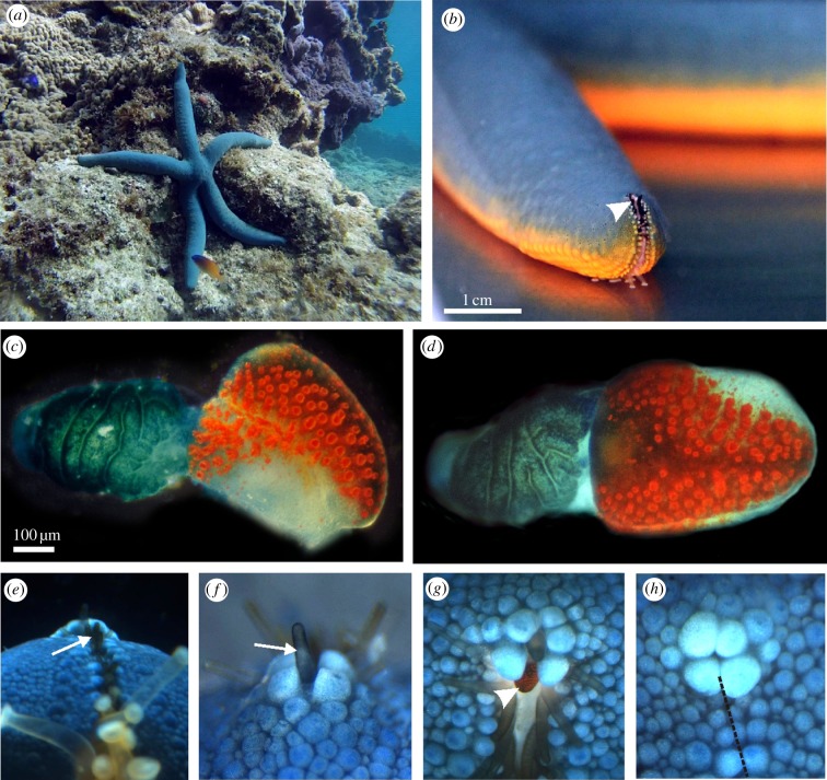
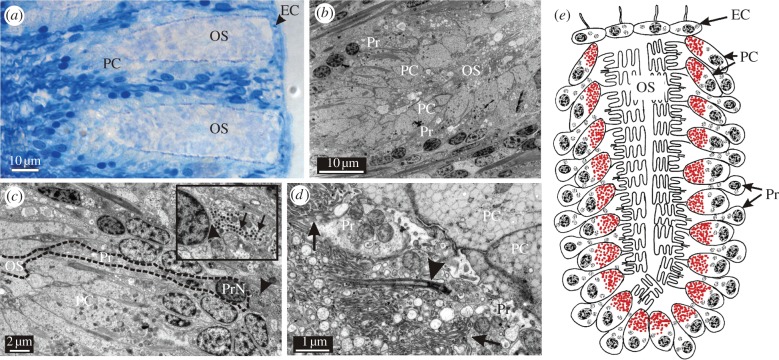
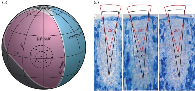
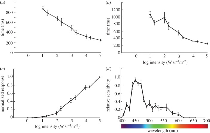
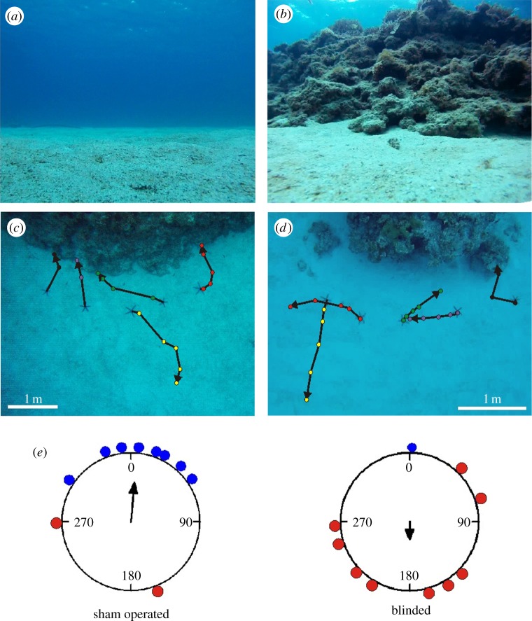
Most known starfish species possess a compound eye at the tip of each arm, which, except for the lack of true optics, resembles an arthropod compound eye. Although these compound eyes have been known for about two centuries, no visually guided behaviour has ever been directly associated with their presence. There are indications that they are involved in negative phototaxis but this may also be governed by extraocular photoreceptors. Here, we show that the eyes of the coral-reef-associated starfish Linckia laevigata are slow and colour blind. The eyes are capable of true image formation although with low spatial resolution. Further, our behavioural experiments reveal that only specimens with intact eyes can navigate back to their reef habitat when displaced, demonstrating that this is a visually guided behaviour. This is, to our knowledge, the first report of a function of starfish compound eyes. We also show that the spectral sensitivity optimizes the contrast between the reef and the open ocean. Our results provide an example of an eye supporting only low-resolution vision, which is believed to be an essential stage in eye evolution, preceding the high-resolution vision required for detecting prey, predators and conspecifics.

Keywords: Linckia; compound eye; coral reef; echinoderm; navigation.


Figures

Figure 1.
Figure 1.
Visual system of the starfish L. laevigata. (a) Linckia laevigata in its natural coral reef habitat at Akajima, Japan, where it feeds on detritus and algae. (b) As in other starfish species, the compound eye of L. laevigata is situated on the tip of each arm (arrowhead). It sits in the ambulaceral groove which continues to the top of the arm tip. (c) Lateral view of the compound eye, also called the optical cushion, which is sitting on the base of a modified tube foot. The eye has approximately 150 separate ommatidia with bright red screening pigment. (d) Frontal view of the compound eye showing its bilateral symmetry. (e) The tip of the arm seen from below. The view of the compound eye is obscured by a double row of modified black tube feet (arrow). (f) The arm tip seen straight from above. Note that the eye is again obscured from view by a modified black tube foot (arrow). (g) The compound eye (arrowhead) seen from 45° above horizontal in a freely behaving animal. When the animal is active, the modified black tube feet spread out to allow vision. (h) If the animal is disturbed, it closes the ambulaceral groove (broken line) at the arm tip and withdraws the modified tube feet. The compound eye is then completely covered, leaving the animal blind.
Figure 2.
Figure 2.
Morphology of the starfish eye. (a) LM of two ommatidia sectioned longitudinally. Each of the fully developed ommatidia is composed of 100–150 photoreceptors and about the same number of pigment cells (PC). Note the thin layer of epithelial cells (EC, arrowhead) covering the outer segments (OS). The receptors are arranged in seven to eight layers perpendicular to the surface (b). (c) Longitudinal section of a photoreceptor (Pr, dashed black line). Interestingly, it receives feedback from the nervous system as indicated by afferent synapses (insert, arrowhead indicates synapse, arrows indicate synaptic vesicles). PrN, photoreceptor nucleus. (d) The outer segments of the starfish photoreceptors are morphologically a mixture of the rhabdomeric- and ciliary-type receptors. They are formed by microvilli coming both directly from the cell membrane (arrows) and from a modified cilium (arrowhead). (e) Schematic drawing of an ommatidium showing the layered arrangement of the photoreceptors and pigment cells.
Figure 3.
Figure 3.
The visual field of a starfish compound eye. (a) Visual field measured with a goniometer. The left (pink) and right (blue) halves of the eye are symmetric around the midline. Altogether, the eye covers about 170° vertically and 120°–210° horizontally. To illustrate the sampling array, optical axes from a central and six neighbouring ommatidia are plotted. The average separation between the central ommatidia and the neighbours is 16°. The dashed circles illustrate the estimated acceptance cone of individual receptors in the middle and bottom of an ommatidium, respectively. (b) Estimates of acceptance angles from the middle and bottom part of the ommatidia.
Figure 4.
Figure 4.
Temporal resolution and spectral sensitivity. (a) The temporal resolution of the photoreceptors was examined with ERGs. The half-width of the impulse response had a minimum of approximately 250 ms, which is comparatively very slow. (b) The slow response is supported by the time to peak measurements of the impulse response. Again the minimal time to peak was about 250 ms. (c) It is seen from the V/logI-curve that the dynamic range of the eyes was at least 4 log units, but it is also seen from the curve that the receptors were not saturated at the maximum test intensity (1.1 × 10⁵ W sr−1 m2). (d) The spectral sensitivity of the receptors showed a single and relatively narrow peak in the deep blue (450 nm) part of the spectrum. The half-width is about 50 nm, which is somewhat narrower than the typical opsin, indicating the presence of spectral filters.
Figure 5.
Figure 5.
Navigation experiments. In the behavioural experiments, the test animals were removed from their natural reef habitat and placed 1 m from the reef edge. (a) The visual scene away from the coral reef habitat was the open ocean. (b) The visual scene towards the natural habitat (coral reef) at a distance of 1 m. (c) Trajectories from the sham-operated animals tested at locality 1 (reef front to the east). Most of the animals quickly found their way back to the habitat. (d) Trajectories from the eye-ablated animals tested at locality 2 (reef front to the south). They moved at the same pace as the intact and sham-operated animals but in random directions. (e) Circular statistics of the behavioural experiments show that the direction of movement of the sham-operated animals differed significantly from random and correlated with the direction to the reef front. The direction of movement of the eye-ablated animals did not differ significantly from random. Blue dots represent animals reaching the reef within the 25 min, red dots represent animals that did not. See experimental procedure for details of the statistical analysis.

Comment in

Similar articles

Cited by

References

    1. Arendt D, Hausen H, Purschke G. 2009. The ‘division of labour’ model of eye evolution. Phil. Trans. R. Soc. B 364, 2809–2817 (doi:10.1098/rstb.2009.0104) - DOI - PMC - PubMed
    1. Kozmic Z, Swamynathan SK, Ruzickova J, Jonasova K, Paces V, Vlcek C, Piatigorsky J. 2008. Cubozoan crystallins: evidence for convergent evolution of pax regulatory sequences. Evol. Dev. 10, 52–61 (doi:10.1111/j.1525-142X.2007.00213.x) - DOI - PubMed
    1. Nilsson DE. 2004. Eye evolution: a question of genetic promiscurity. Curr. Opin. Neurobiol. 14, 407–414 (doi:10.1016/j.conb.2004.07.004) - DOI - PubMed
    1. O'Connor M, Garm A, Nilsson DE. 2009. Structure and optics of the eyes of the box jellyfish Chiropsella bronzie. J. Comp. Physiol. A 195, 557–569 (doi:10.1007/s00359-009-0431-x) - DOI - PubMed
    1. Nilsson DE, Gislén L, Coates MM, Skogh C, Garm A. 2005. Advanced optics in a jellyfish eye. Nature 435, 201–205 (doi:10.1038/nature03484) - DOI - PubMed

Publication types

LinkOut - more resources