Intrauterine growth restriction caused by underlying congenital cytomegalovirus infection
- PMID: 24403553
- PMCID: PMC3997585
- DOI: 10.1093/infdis/jiu019
Intrauterine growth restriction caused by underlying congenital cytomegalovirus infection
Abstract
Background: Human cytomegalovirus (HCMV) is the major viral etiology of congenital infection and birth defects. Fetal transmission is high (30%-40%) in primary maternal infection, and symptomatic babies have permanent neurological, hearing, and vision defects. Recurrent infection is infrequently transmitted (2%) and largely asymptomatic. Congenital infection is also associated with intrauterine growth restriction (IUGR).
Methods: To investigate possible underlying HCMV infection in cases of idiopathic IUGR, we studied maternal and cord sera and placentas from 19 pregnancies. Anti-HCMV antibodies, hypoxia-related factors, and cmvIL-10 were measured in sera. Placental biopsy specimens were examined for viral DNA, expression of infected cell proteins, and pathology.
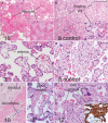
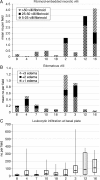
Results: Among 7 IUGR cases, we identified 2 primary and 3 recurrent HCMV infections. Virus replicated in glandular epithelium and lymphatic endothelium in the decidua, cytotrophoblasts, and smooth muscle cells in blood vessels of floating villi and the chorion. Large fibrinoids with avascular villi, edema, and inflammation were significantly increased. Detection of viral proteins in the amniotic epithelium indicated transmission in 2 cases of IUGR with primary infection and 3 asymptomatic recurrent infections.
Conclusions: Congenital HCMV infection impairs placental development and functions and should be considered as an underlying cause of IUGR, regardless of virus transmission to the fetus.
Keywords: HCMV; IUGR; amnion; blood vessels; chorion; congenital; fetus; placenta; pregnancy; villi.
Figures


Comment in
-
IUGR and congenital cytomegalovirus infection.J Infect Dis. 2014 May 15;209(10):1497-9. doi: 10.1093/infdis/jiu092. Epub 2014 Feb 12. J Infect Dis. 2014. PMID: 24526739 No abstract available.
Similar articles
-
Human cytomegalovirus infection interferes with the maintenance and differentiation of trophoblast progenitor cells of the human placenta.J Virol. 2015 May;89(9):5134-47. doi: 10.1128/JVI.03674-14. Epub 2015 Mar 4. J Virol. 2015. PMID: 25741001 Free PMC article.
-
Persistent Cytomegalovirus Infection in Amniotic Membranes of the Human Placenta.Am J Pathol. 2016 Nov;186(11):2970-2986. doi: 10.1016/j.ajpath.2016.07.016. Epub 2016 Sep 13. Am J Pathol. 2016. PMID: 27638253 Free PMC article.
-
APOBEC3A Is Upregulated by Human Cytomegalovirus (HCMV) in the Maternal-Fetal Interface, Acting as an Innate Anti-HCMV Effector.J Virol. 2017 Nov 14;91(23):e01296-17. doi: 10.1128/JVI.01296-17. Print 2017 Dec 1. J Virol. 2017. PMID: 28956761 Free PMC article.
-
Diagnosis and management of human cytomegalovirus infection in the mother, fetus, and newborn infant.Clin Microbiol Rev. 2002 Oct;15(4):680-715. doi: 10.1128/CMR.15.4.680-715.2002. Clin Microbiol Rev. 2002. PMID: 12364375 Free PMC article. Review.
-
Primary Human Cytomegalovirus (HCMV) Infection in Pregnancy.Dtsch Arztebl Int. 2017 Jan 27;114(4):45-52. doi: 10.3238/arztebl.2017.0045. Dtsch Arztebl Int. 2017. PMID: 28211317 Free PMC article. Review.
Cited by
-
Congenital cytomegalovirus infection: Clinical presentation, epidemiology, diagnosis and prevention.Obstet Med. 2014 Dec;7(4):140-6. doi: 10.1177/1753495X14552719. Epub 2014 Sep 25. Obstet Med. 2014. PMID: 27512442 Free PMC article. Review.
-
Neutralizing Antibodies to Human Cytomegalovirus Recombinant Proteins Reduce Infection in an Ex Vivo Model of Developing Human Placentas.Vaccines (Basel). 2022 Jul 4;10(7):1074. doi: 10.3390/vaccines10071074. Vaccines (Basel). 2022. PMID: 35891239 Free PMC article.
-
Human cytomegalovirus infection interferes with the maintenance and differentiation of trophoblast progenitor cells of the human placenta.J Virol. 2015 May;89(9):5134-47. doi: 10.1128/JVI.03674-14. Epub 2015 Mar 4. J Virol. 2015. PMID: 25741001 Free PMC article.
-
Findings of Reduced Head Circumference with COVID-19 Infection in the Third Trimester: A Retrospective Cohort Study.Biomedicines. 2025 Mar 31;13(4):832. doi: 10.3390/biomedicines13040832. Biomedicines. 2025. PMID: 40299415 Free PMC article.
-
Persistent Cytomegalovirus Infection in Amniotic Membranes of the Human Placenta.Am J Pathol. 2016 Nov;186(11):2970-2986. doi: 10.1016/j.ajpath.2016.07.016. Epub 2016 Sep 13. Am J Pathol. 2016. PMID: 27638253 Free PMC article.
References
-
- Stagno S, Pass RF, Cloud G, et al. Primary cytomegalovirus infection in pregnancy. Incidence, transmission to fetus, and clinical outcome. JAMA. 1986;256:1904–8. - PubMed
-
- Pass RF, Fowler KB, Boppana SB, Britt WJ, Stagno S. Congenital cytomegalovirus infection following first trimester maternal infection: symptoms at birth and outcome. J Clin Virol. 2006;35:216–20. - PubMed
-
- Enders G, Daiminger A, Bader U, Exler S, Enders M. Intrauterine transmission and clinical outcome of 248 pregnancies with primary cytomegalovirus infection in relation to gestational age. J Clin Virol. 2011;52:244–6. - PubMed
-
- Garcia AG, Fonseca EF, Marques RL, Lobato YY. Placental morphology in cytomegalovirus infection. Placenta. 1989;10:1–18. - PubMed
Publication types
MeSH terms
Substances
Grants and funding
LinkOut - more resources
Full Text Sources
Other Literature Sources
Medical

