. 2014 Mar;88(6):3516-26.
doi: 10.1128/JVI.03331-13. Epub 2014 Jan 8.

HIV DNA subspecies persist in both activated and resting memory CD4+ T cells during antiretroviral therapy

Affiliations

HIV DNA subspecies persist in both activated and resting memory CD4+ T cells during antiretroviral therapy

John M Murray et al. J Virol. 2014 Mar.

Abstract

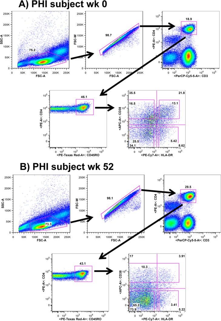
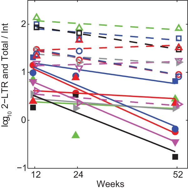
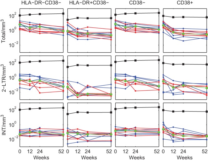
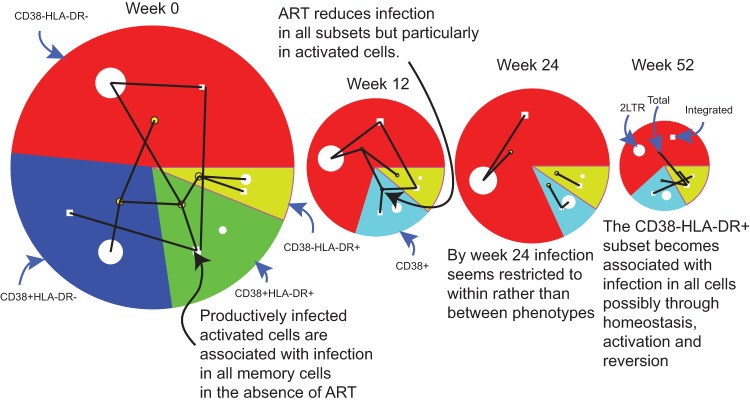
The latent HIV reservoir is a major impediment to curing HIV infection. The contribution of CD4(+) T cell activation status to the establishment and maintenance of the latent reservoir was investigated by enumerating viral DNA components in a cohort of 12 individuals commencing antiretroviral therapy (ART) containing raltegravir, an integrase inhibitor. Prior to ART, the levels of total HIV DNA were similar across HLA-DR(+) and HLA-DR(-) (HLA-DR(±)) CD38(±) memory CD4(+) T cell phenotypes; episomal two-long terminal repeat (2-LTR) HIV DNA levels were higher in resting (HLA-DR(-) CD38(-)) cells, and this phenotype exhibited a significantly higher ratio of 2-LTR to integrated HIV DNA (P = 0.002). After 1 year of ART, there were no significant differences across each of the memory phenotypes of any HIV DNA component. The decay dynamics of integrated HIV DNA were slow within each subset, and integrated HIV DNA in the resting HLA-DR(-) CD38(-) subset per mm(3) of peripheral blood exhibited no significant decay (half-life of 25 years). Episomal 2-LTR HIV DNA decayed relative to integrated HIV DNA in resting cells with a half-life of 134 days. Surprisingly, from week 12 on, the decay rates of both total and episomal HIV DNA were lower in activated CD38(+) cells. By weeks 24 and 52, HIV RNA levels in plasma were most significantly correlated with the numbers of resting cells containing integrated HIV DNA. On the other hand, total HIV DNA levels in all subsets were significantly correlated with the numbers of HLA-DR(+) CD38(-) cells containing integrated HIV DNA. These results provide insights into the interrelatedness of cell activation and reservoir maintenance, with implications for the design of therapeutic strategies targeting HIV persistence.

Importance: It is generally believed that HIV is not cleared by extensive antiretroviral therapy (ART) due to the difficulty in eradicating the latent reservoir in resting CD4(+) T cells. New therapies that attempt to activate this reservoir so that immune or viral cytopathic mechanisms can remove those infected cells are currently being investigated. However, results obtained in this research indicate that activation, at least on some level, already occurs within this reservoir. Furthermore, we are the first to describe the dynamics of different HIV DNA species in resting and activated memory CD4+ T cell subsets that point to the role different levels of activation play in maintaining the HIV reservoir.


Figures

FIG 1
FIG 1
Sorting strategy for resting and activated memory CD4+ T cells for a typical PHI patient at two different times. (A) Sorting strategy for resting and activated memory CD4+ T cells for a typical PHI patient at week 0 into the 4 memory CD4+ T cell subsets, HLA-DR CD38, HLA-DR+ CD38−, HLA-DR CD38+, and HLA-DR+ CD38+ cells. (B) Sorting strategy for resting and activated memory CD4+ T cells for a typical PHI patient at week 52 into the 3 memory subsets, HLA-DR CD38, HLA-DR+ CD38, and CD38+ cells. Abbreviations: 50K, 50,000; FSC, forward scatter; SSC, side scatter; PerCP, peridinin chlorophyll protein; PE, phycoerythrin.
FIG 2
FIG 2
Week 0 and week 52 levels of total, 2-LTR, and integrated HIV DNA copies per 106 cells for each cell subset, separated into PHI and CHI. Each symbol represents the value for an individual. The short horizontal lines show the median values for groups. The only statistically significant difference for all patients (combined PHI and CHI) across all cell subsets and at either time point was for 2-LTR HIV DNA at week 0 (P = 0.02), where levels were higher in the resting HLA-DR CD38 subset than in the activated HLA-DR+ CD38+ subset (median value of 8,906 versus 1,963 copies per 106 cells). Within each subset, levels differed significantly for PHI versus CHI only for 2-LTR HIV DNA levels in the activated HLA-DR+ CD38+ subset at week 0 (125 copies per 106 cells for PHI versus 3,875 copies per 106 cells for CHI; P = 0.03) and for integrated HIV DNA levels in the CD38+ subset at week 52 (263 copies per 106 cells for PHI versus 700 copies per 106 cells for CHI; P = 0.048).
FIG 3
FIG 3
HIV DNA dynamics in resting/activated memory CD4+ T cell subsets. HIV DNA components (total, 2-LTR, and integrated [INT] HIV DNA) are shown as the number of copies per 106 cells of memory CD4+ T cell subsets: HLA-DR CD38, HLA-DR+ CD38, CD38, and CD38+ (PHI are shown in blue and CHI in red). The limits of quantification (LOQ) were 125 copies/106 cells. Values below the LOQ were set to the LOQ. The median values are shown in green. The last two rows display medians and interquartile ranges of the number of cells/mm3 and proliferating (Ki-67+) percentages of each subset.
FIG 4
FIG 4
Decay of 2-LTR and total HIV DNA relative to integrated HIV DNA in resting memory CD4+ T cells. Decay of log10 of ratios of 2-LTR to integrated HIV DNA copies (solid lines and symbols), and total to integrated HIV DNA copies (dashed lines and open symbols) in the resting HLA-DR CD38 memory CD4+ T cell phenotype from week 12. Individuals with integrated HIV DNA below the assay LOQ were omitted.
FIG 5
FIG 5
HIV DNA per mm3 dynamics in resting/activated memory CD4+ T cell subsets. HIV DNA components are shown as the number of copies per mm3 of memory CD4+ T cell subsets in peripheral blood: HLA-DR CD38, HLA-DR+ CD38, CD38, and CD38+ (PHI shown in blue and CHI in red). The median values are shown in green. The median values of the numbers of each cell subset per mm3 are shown as black markers and lines.
FIG 6
FIG 6
Size of each HIV DNA component in resting and activated memory CD4+ T cells during ART and significant correlations between them at each time point. The size of each sector represents median values of total HIV DNA copies/mm3 in each of the CD38± HLA-DR± subsets. The median 2-LTR HIV DNA copies/mm3 is represented by the area of the white circle within each sector. The median integrated HIV DNA copies/mm3 is represented by the area of the white square within each sector. Significant (reduced as described in Materials and Methods) rank correlations between HIV DNA components and subsets at each time are shown as black lines, where a correlation with total HIV DNA is represented by a connection to that sector's more centrally located yellow node. For example, at week 0, the number of total HIV DNA copies/mm3 in the activated CD38+ HLA-DR+ cells (represented by the green sector with correlations to total HIV DNA in this phenotype represented by lines connecting the inner node of this sector) shows the most correlations, significantly correlated with integrated HIV DNA within the same subset but also with total HIV DNA in CD38+ HLA-DR and CD38 HLA-DR+ cells, plus with 2-LTR levels in resting CD38 HLA-DR cells.
FIG 7
FIG 7
Decline in cellular activation with ART. Biphasic decrease in activated HLA-DR+ CD38+ CD8+ T cells/mm3 with ART (PHI shown in blue and CHI in red).

References

    1. Finzi D, Blankson J, Siliciano JD, Margolick JB, Chadwick K, Pierson T, Smith K, Lisziewicz J, Lori F, Flexner C, Quinn TC, Chaisson RE, Rosenberg E, Walker B, Gange S, Gallant J, Siliciano RF. 1999. Latent infection of CD4+ T cells provides a mechanism for lifelong persistence of HIV-1, even in patients on effective combination therapy. Nat. Med. 5:512–517. 10.1038/8394 - DOI - PubMed
    1. Swiggard WJ, O'Doherty U, McGain D, Jeyakumar D, Malim MH. 2004. Long HIV type 1 reverse transcripts can accumulate stably within resting CD4+ T cells while short ones are degraded. AIDS Res. Hum. Retroviruses 20:285–295. 10.1089/088922204322996527 - DOI - PubMed
    1. Zack JA, Arrigo SJ, Weitsman SR, Go AS, Haislip A, Chen IS. 1990. HIV-1 entry into quiescent primary lymphocytes: molecular analysis reveals a labile, latent viral structure. Cell 61:213–222. 10.1016/0092-8674(90)90802-L - DOI - PubMed
    1. Swiggard WJ, Baytop C, Yu JJ, Dai J, Li C, Schretzenmair R, Theodosopoulos T, O'Doherty U. 2005. Human immunodeficiency virus type 1 can establish latent infection in resting CD4+ T cells in the absence of activating stimuli. J. Virol. 79:14179–14188. 10.1128/JVI.79.22.14179-14188.2005 - DOI - PMC - PubMed
    1. Siliciano RF, Greene WC. 2011. HIV latency. Cold Spring Harb. Perspect. Med. 1:a007096. 10.1101/cshperspect.a007096 - DOI - PMC - PubMed

Publication types