. 2014 Apr;88(8):3986-96.
doi: 10.1128/JVI.03203-13. Epub 2014 Jan 8.

Insights into the structure and assembly of the bacteriophage 29 double-stranded DNA packaging motor

Affiliations

Insights into the structure and assembly of the bacteriophage 29 double-stranded DNA packaging motor

Sheng Cao et al. J Virol. 2014 Apr.

Abstract

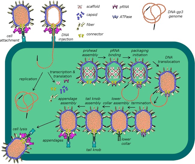
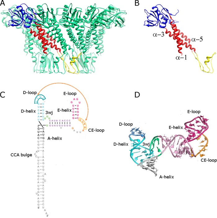
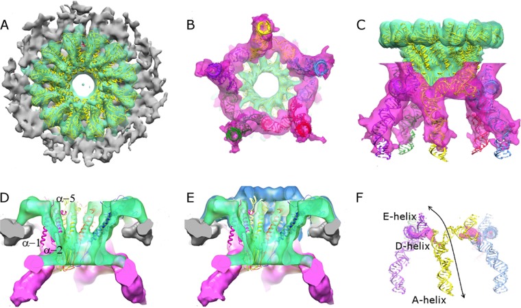
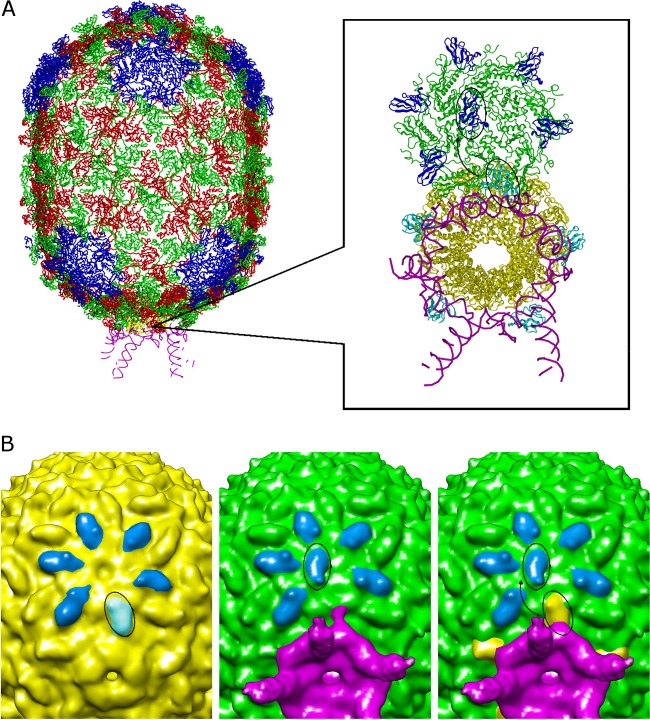
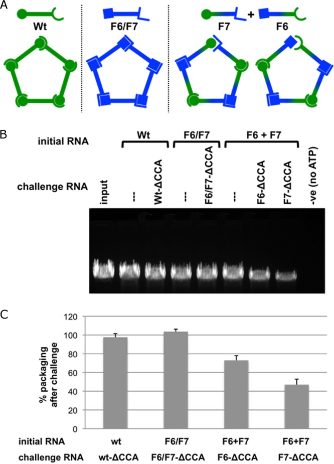
The tailed double-stranded DNA (dsDNA) bacteriophage 29 packages its 19.3-kbp genome into a preassembled procapsid structure by using a transiently assembled phage-encoded molecular motor. This process is remarkable considering that compaction of DNA to near-crystalline densities within the confined space of the capsid requires that the packaging motor work against significant entropic, enthalpic, and DNA-bending energies. The motor consists of three phage-encoded components: the dodecameric connector protein gp10, an oligomeric RNA molecule known as the prohead RNA (pRNA), and the homomeric ring ATPase gp16. Although atomic resolution structures of the connector and different pRNA subdomains have been determined, the mechanism of self-assembly and the resulting stoichiometry of the various motor components on the phage capsid have been the subject of considerable controversy. Here a subnanometer asymmetric cryoelectron microscopy (cryo-EM) reconstruction of a connector-pRNA complex at a unique vertex of the procapsid conclusively demonstrates the pentameric symmetry of the pRNA and illuminates the relative arrangement of the connector and the pRNA. Additionally, a combination of biochemical and cryo-EM analyses of motor assembly intermediates suggests a sequence of molecular events that constitute the pathway by which the motor assembles on the head, thereby reconciling conflicting data regarding pRNA assembly and stoichiometry. Taken together, these data provide new insight into the assembly, structure, and mechanism of a complex molecular machine.

Importance: Viruses consist of a protein shell, or capsid, that protects and surrounds their genetic material. Thus, genome encapsidation is a fundamental and essential step in the life cycle of any virus. In dsDNA viruses, powerful molecular motors essentially pump the viral DNA into a preformed protein shell. This article describes how a viral dsDNA packaging motor self-assembles on the viral capsid and provides insight into its mechanism of action.


Figures

FIG 1
FIG 1
Assembly pathway of bacteriophage ϕ29.
FIG 2
FIG 2
Structures of the ϕ29 connector and pRNA. (A) Side view of the connector. Eleven of the 12 monomers are shown in green, and one monomer is colored by domain, with the wide, central, and narrow domains shown in blue, red and yellow, respectively. (B) Single monomer of the connector structure, shown in the same color scheme and orientation as in panel A, with central-domain α-helices α-1, α-3, and α-5 labeled at their N-terminal ends. (C) Secondary structural diagram of the pRNA. The A-, D-, and E-helices are labeled, along with the CE-, D-, and E-loops and the U-rich 3-way junction (3wj; bases shown in green). (D) Atomic structure of the prohead-binding domain of the pRNA (PDB 3R4F) (18), labeled and colored as in panel C. (Reprinted from the Proceedings of the National Academy of Sciences of the United States of America [18] with permission of the publisher.)
FIG 3
FIG 3
Asymmetric reconstruction of the connector-pRNA vertex. (A through C) Top (A), bottom (B), and side (C) views of the asymmetric reconstruction of connector-pRNA complex are shown. Densities corresponding to the capsid, connector, and pRNA are rendered in gray, green, and magenta, respectively. The fitted X-ray structure of the connector is shown in yellow. A model of the pRNA based on fitting the X-ray structure of the prohead-binding domain of the pRNA (18), the NMR structure of the CCA bulge (29), and an ideal RNA helix as the A-helix was also fitted; the five subunits are colored red, green, blue, yellow, and purple. (D) Reconstruction of the connector-pRNA vertex rendered at a higher contour level (∼4 standard deviations above the mean), where it is possible to recognize α-helices in the central region of the connector. Central domain α-helices α-1, α-3, and α-5 labeled in one magenta colored connector subunit. (E) A rendering of the density corresponding to the connector at a lower contour level superimposed on the rendering of the complex presented in panel D shows extra density corresponding to the crown region of the connector (blue). (F) Density corresponding to the pRNA rendered at a very high contour level (magenta), indicating that pRNA bases involved in the intermolecular Watson-Crick base pairing are highly ordered in the reconstruction. The arrow indicates an RNA superhelix that spans two pRNA subunits, and the contributing E-, D-, and A-helical elements are labeled.
FIG 4
FIG 4
Cryo-EM reconstructions of proheads produced in E. coli before and after the addition of in vitro-transcribed pRNA. (A) Pseudoatomic structure of the prohead. Pentameric subunits are colored blue, and quasiequivalent alternating subunits in hexamers are shown in red and green. (Reprinted from Molecular Cell [19] with permission of the publisher.) (Inset) Zoomed-in view of the connector-pRNA vertex looking from the bottom of the head at a slightly skewed angle. The connector is shown in yellow and the pRNA in magenta. One of the five hexamers circumscribing the vertex is also shown, with HK97 domains colored green, observed positions of Ig-like domains shown in blue, and the “expected” positions of Ig-like domains shown in cyan, demonstrating that if the domain had not moved, it would have clashed with pRNA. The approximately 180° degree rotation is indicated by the arrow connecting two circled Ig-like domains. (B) (Left) Cryo-EM reconstruction of a pRNA-naïve E. coli-produced prohead, with Ig-like domains colored blue. The Ig-like domain that rotates upon the addition of pRNA is circled. (Center) Reconstruction of E. coli-produced proheads after incubation with in vitro-transcribed pRNA. Ig-like domains are colored blue. The rotated Ig-like domain is circled. (Right) Superposition of structures of proheads before (yellow) and after (green) the addition of in vitro-transcribed pRNA. The approximately 180° rotation is indicated by the arrow connecting two circled Ig-like domains. The pRNA is shown in magenta.
FIG 5
FIG 5
Effect of a mismatched interface in the pRNA ring on pRNA assembly and DNA packaging. (A) Schematic illustrating pentameric pRNA ring formation. The wt and the double mutant F6/F7 generate closed pRNA rings, while the dimer pair F6+F7 generates a single mismatched interface. The open symbol end represents the CE-loop of pRNA, and the closed symbol end represents the D-loop. The loop sequences (CE-45–48 and D-82–85 loops) for the pRNAs are as follows: for the wt, AACC and GGUU, respectively; for F6/F7, GCGA and UCGC, respectively; for F6, GCGA and GGUU, respectively; and for F7, AACC and UCGC, respectively) (17). (B) Effect on DNA packaging using assembled pRNA-prohead complexes challenged with pRNA containing the lethal ΔCCA mutation. The initial RNA is the pRNA form used to reconstitute RNA-free proheads. The challenge RNA (ΔCCA mutation-containing pRNA in corresponding pseudoknot sequence backgrounds) is then added to the prohead-pRNA complexes, and after incubation, the mixture is assessed in the in vitro DNA-packaging assay (see Materials and Methods). After the assay is completed, unpackaged DNA is digested by the addition of DNase I. The packaged, protected DNA is then extracted and analyzed by agarose gel electrophoresis (34) and the effect on DNA packaging assessed (see Materials and Methods). Vertical lines above lanes indicate nonchallenged samples. (C) Histogram showing packaging efficiency in the presence of the challenge pRNA containing the ΔCCA mutation compared to that for the nonchallenged sample (taken as 100%). For wt pRNA challenged with wt-ΔCCA, the packaging efficiency was 97.6% ± 3.9% (compare the second and third lanes); for the F6/F7 double mutant challenged with F6/F7-ΔCCA, it was 103.6% ± 2.7% (compare the fourth and fifth lanes); and for the dimer pair F6 +F7, it was 73.0% ± 5.0% for F6-ΔCCA challenge (compare the sixth and seventh lanes) and 47.0% ± 5.9% for F7-ΔCCA challenge (compare the sixth and eight lanes). n = 3 experiments; standard deviations are reported, and a single representative gel is shown.

References

    1. Earnshaw WC, Casjens SR. 1980. DNA packaging by the double-stranded DNA bacteriophages. Cell 21:319–331. 10.1016/0092-8674(80)90468-7 - DOI - PubMed
    1. Johnson JE, Chiu W. 2007. DNA packaging and delivery machines in tailed bacteriophages. Curr. Opin. Struct. Biol. 17:237–243. 10.1016/j.sbi.2007.03.011 - DOI - PubMed
    1. Rao VB, Feiss M. 2008. The bacteriophage DNA packaging motor. Annu. Rev. Genet. 42:647–681. 10.1146/annurev.genet.42.110807.091545 - DOI - PubMed
    1. Grimes S, Jardine PJ, Anderson D. 2002. Bacteriophage ϕ29 DNA packaging. Adv. Virus Res. 58:255–294. 10.1016/S0065-3527(02)58007-6 - DOI - PubMed
    1. Morais MC. 2012. The dsDNA packaging motor in bacteriophage ϕ29. Adv. Exp. Med. Biol. 726:511–547. 10.1007/978-1-4614-0980-9_23 - DOI - PubMed

Publication types

LinkOut - more resources