. 2014 Apr;88(7):3653-63.
doi: 10.1128/JVI.03263-13. Epub 2014 Jan 8.

The human papillomavirus E7 proteins associate with p190RhoGAP and alter its function

Affiliations

The human papillomavirus E7 proteins associate with p190RhoGAP and alter its function

Biljana Todorovic et al. J Virol. 2014 Apr.

Abstract

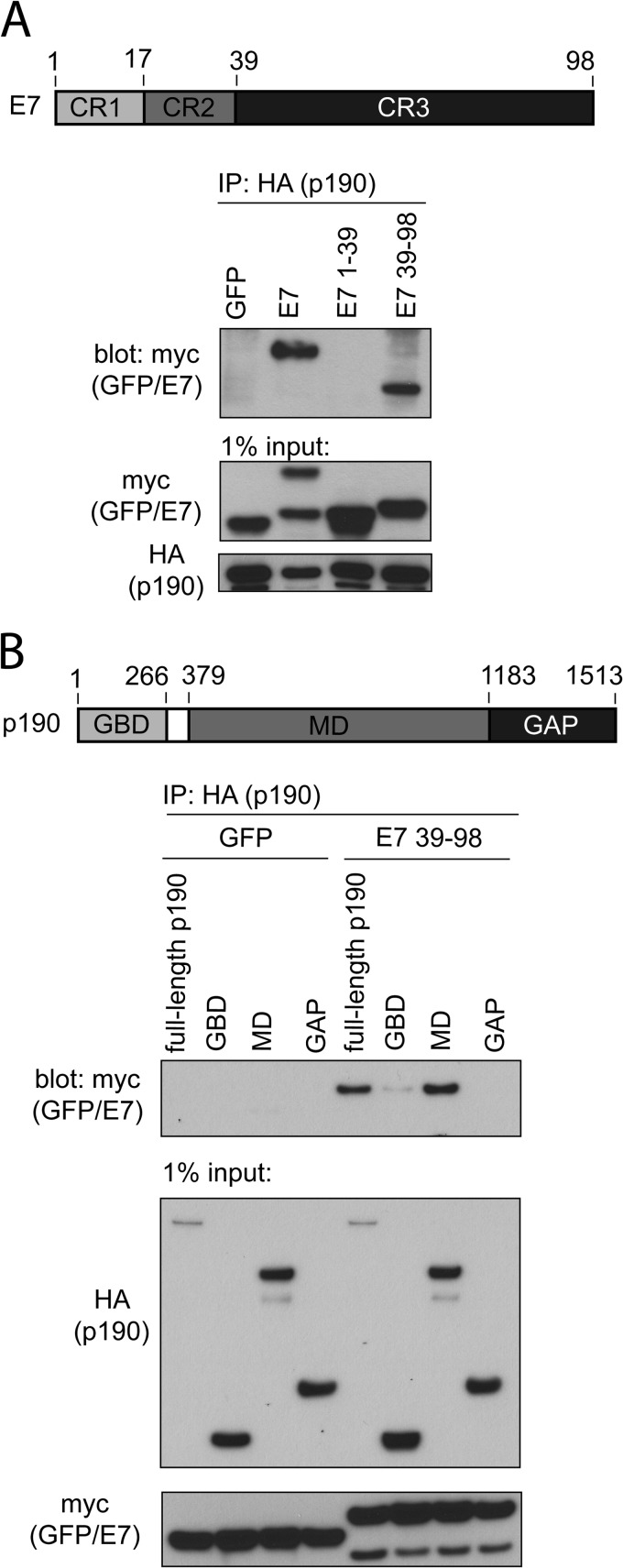
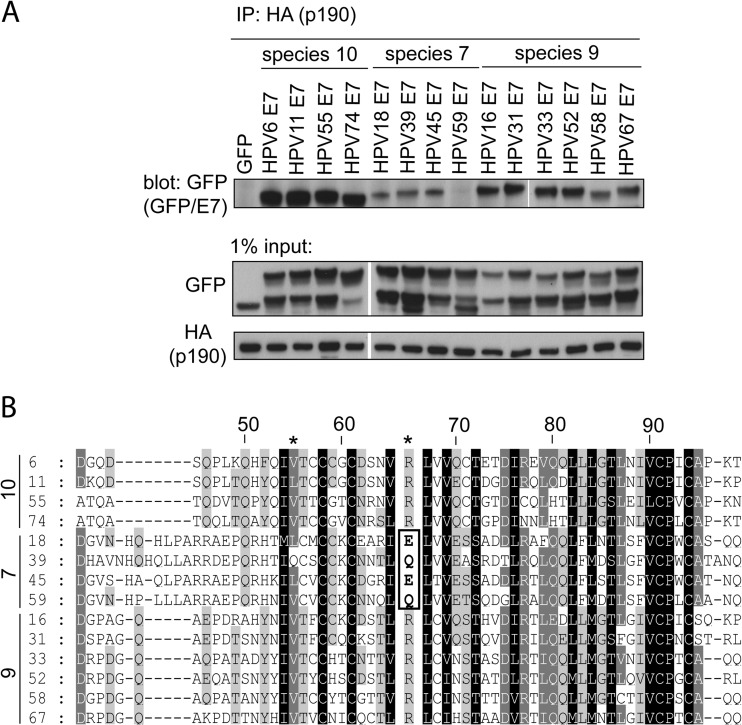
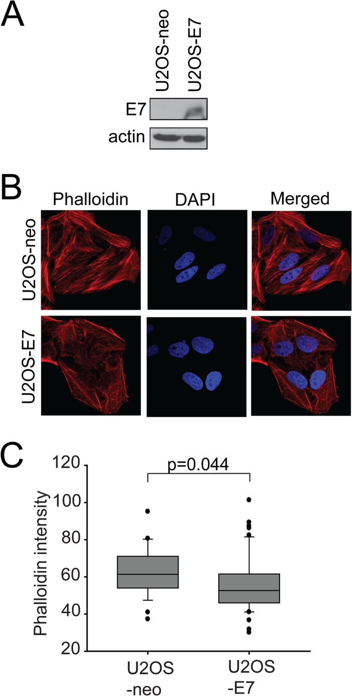
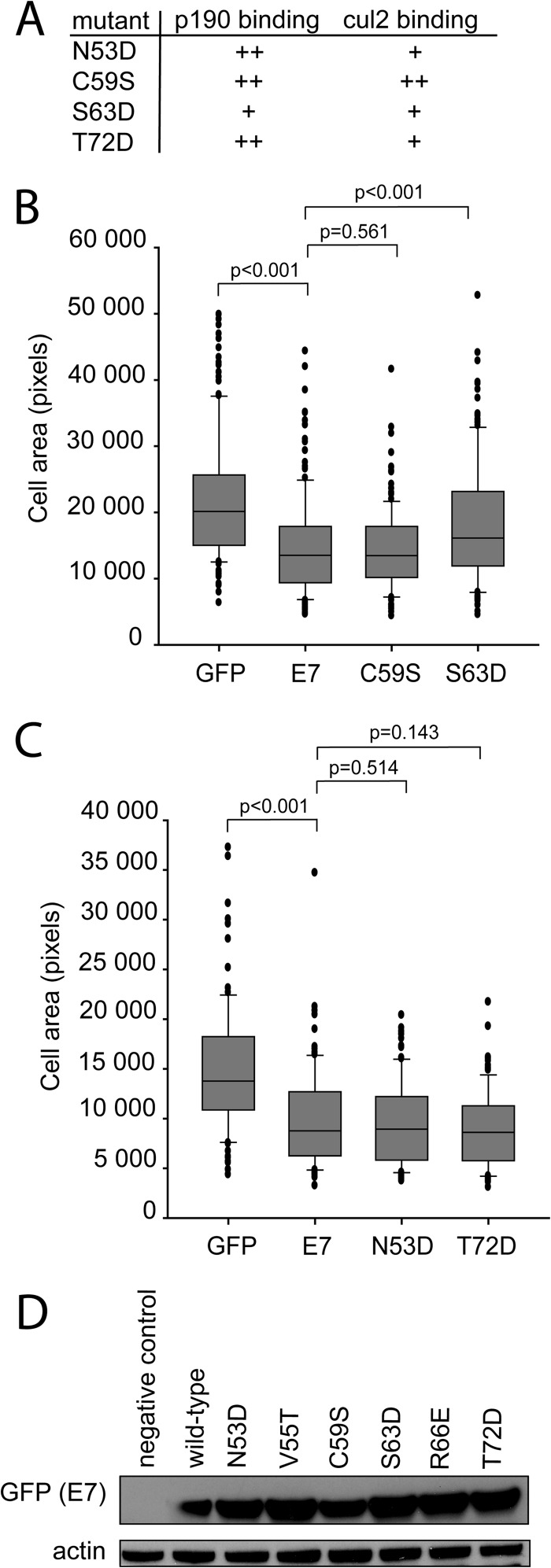
Using mass spectrometry, we identified p190RhoGAP (p190) as a binding partner of human papillomavirus 16 (HPV16) E7. p190 belongs to the GTPase activating protein (GAP) family and is one of the primary GAPs for RhoA. GAPs stimulate the intrinsic GTPase activity of the Rho proteins, leading to Rho inactivation and influencing numerous biological processes. RhoA is one of the best-characterized Rho proteins and is specifically involved in formation of focal adhesions and stress fibers, thereby regulating cell migration and cell spreading. Since this is the first report that E7 associates with p190, we carried out detailed interaction studies. We show that E7 proteins from other HPV types also bind p190. Furthermore, we found that conserved region 3 (CR3) of E7 and the middle domain of p190 are important for this interaction. More specifically, we identified two residues in CR3 of E7 that are necessary for p190 binding and used mutants of E7 with mutations of these residues to determine the biological consequences of the E7-p190 interaction. Our data suggest that the interaction of E7 with p190 dysregulates this GAP and alters the actin cytoskeleton. We also found that this interaction negatively regulates cell spreading on a fibronectin substrate and therefore likely contributes to important aspects of the HPV life cycle or HPV-induced tumorigenesis.

Importance: This study identifies p190RhoGAP as a novel cellular binding partner for the human papillomavirus (HPV) E7 protein. Our study shows that a large number of different HPV E7 proteins bind p190RhoGAP, and it identifies regions in both E7 and p190RhoGAP which are important for the interaction to occur. This study also highlights the likelihood that the E7-p190RhoGAP interaction may have important biological consequences related to actin organization in the infected cell. These changes could be an important contributor to the viral life cycle and during progression to cancer in HPV-infected cells. Importantly, this work also emphasizes the need for further study in a field which has largely been unexplored as it relates to the HPV life cycle and HPV-induced transformation.


Figures

FIG 1
FIG 1
HPV16 E7 associates with p190RhoGAP (p190). Lysates from HPV16-positive (CaSki) and HPV-negative (C33A) cells were prepared and immunoprecipitated (IP) for E7 as described in Materials and Methods. Samples were immunoblotted as indicated.
FIG 2
FIG 2
Conserved region 3 (CR3) of HPV16 E7 and the middle domain (MD) of p190 are sufficient for interaction. (A) CR3 of HPV16 E7 (residues 39 to 98) is necessary and sufficient to associate with p190. HT1080 cells were transfected with full-length HA-tagged p190 and the indicated GFP or GFP-E7 fusion construct (E7, E7 1–39, and E7 39–98 are all GFP fusions). At 24 h posttransfection, cell lysates were prepared and subjected to coimmunoprecipitation as indicated. (B) The middle domain of p190 is sufficient to associate with E7 (residues 39 to 98). HT1080 cells were transfected with the indicated HA-tagged p190 constructs and GFP or GFP-E7 39–98. Samples were subjected to coimmunoprecipitation as indicated.
FIG 3
FIG 3
Residues V55 and R66 in HPV16 E7 are required for association with p190. (A) HT1080 cells were transfected with full-length HA-tagged p190 and GFP or GFP fused to residues 39 to 98 of HPV16 E7 containing the indicated mutations. Coimmunoprecipitation experiments were carried out at 24 h posttransfection, as indicated. (B) HT1080 cells were transfected with full-length HA-tagged p190 and full-length wild-type GFP-E7 or the V55T or R66E mutant, and samples were processed for coimmunoprecipitation as indicated for panel A.
FIG 4
FIG 4
The E7 proteins of multiple HPV species interact with p190. (A) E7 proteins from other HPV species and types associate with p190. HT1080 cells were transfected with HA-tagged p190 and GFP or GFP-E7 fusions, as indicated. At 24 h posttransfection, cell lysates were prepared and subjected to coimmunoprecipitation using anti-HA antibody. The samples were immunoblotted as indicated. (B) Multiple-sequence alignment of HPV E7 CR3 sequences. The residue numbering indicated on the top line corresponds to HPV16 E7 numbering. Indicated on the left of the alignment are the species and HPV type. Darker shading corresponds to more highly conserved residues. The positions of residues V55 and R66 are indicated at the top of the alignment (*). The E and Q residues of species 7 types at the position of R66 (HPV16) are highlighted with a box.
FIG 5
FIG 5
Interaction with p190 is required for HPV16 E7 to disrupt actin stress fiber formation. (A) Dominant negative p190 disrupts actin stress fibers. U2OS cells were transfected with GFP or GFP-p190RA (dominant negative p190) and then stained with phalloidin-Alexa 568 to visualize F-actin. The nuclei were stained with DAPI, and images were acquired on a confocal microscope. (B) Quantification of phalloidin intensities in GFP- and p190RA-expressing U2OS or HaCat cells. Data are representative results from three independent experiments. (C) HPV16 E7 mutants unable to bind p190 do not efficiently disrupt actin stress fibers. U2OS cells were transfected with the indicated plasmids and stained for F-actin by use of phalloidin-Alexa 568. Nuclei were stained with DAPI. (D) Quantification of phalloidin staining in transfected U2OS or HaCat cells. Phalloidin intensities were quantified from images captured with a fluorescence microscope as described in Materials and Methods. Data are representative of three independent experiments.
FIG 6
FIG 6
E7 affects F-actin levels in stable cell lines. (A) Expression of E7 in U2OS-neo and U2OS-E7 cells. Equal amounts of cell extracts from U2OS-neo and U2OS-E7 cells were analyzed by immunoblotting as indicated. (B) U2OS-neo and U2OS-E7 cells were stained with phalloidin-Alexa 568 to visualize F-actin. The nuclei were stained with DAPI, and images were acquired on a confocal microscope. (C) Quantification of phalloidin intensities in U2OS-neo and U2OS-E7 cells. Data are representative results from three independent experiments.
FIG 7
FIG 7
E7 affects cell spreading. (A) U2OS cells were transiently transfected with GFP or GFP fusions of wild-type, V55T, or R66E E7 or dominant negative p190 (p190RA), plated on fibronectin for 20 min, and then stained for F-actin by use of phalloidin. Images were captured with a fluorescence microscope using a ×40 objective; representative images are shown. (B) The relative areas of individual U2OS, HaCat, and CaSki cells (transfected as indicated and processed as described for panel A) in digital images were measured with ImageJ. Data are representative of at least 75 cells and three independent experiments.
FIG 8
FIG 8
The ability of E7 to associate with p190, but not cullin 2, appears to be necessary to reduce cell spreading. (A) p190 and cullin 2 (cul2) binding phenotypes of the mutants used in this study. The N53D, C59S, and T72D mutants bound p190 as well as wild-type E7 (++); the S63D mutant had reduced p190 binding (+). The C59S mutant bound cul2 comparably to the wild type (++), whereas the N53D, S63D, and T72D mutants all had reduced cul2 binding (+). (B) U2OS cells were transiently transfected with GFP or GFP fusions of wild-type, C59S, or S63D E7, plated on fibronectin for 20 min, and then stained for F-actin by use of phalloidin. Images were captured with a fluorescence microscope using a ×40 objective. The relative areas of individual cells in digital images were measured with ImageJ. Data are representative of at least 75 cells and two independent experiments. (C) U2OS cells were transiently transfected as indicated and processed as described for panel B. (D) U2OS cells were transfected with wild-type E7 or the indicated E7 mutant. At 24 h posttransfection, cell lysates were examined for E7 expression levels via Western blotting.
FIG 9
FIG 9
Effects on RhoA-GTP levels and p190 tyrosine phosphorylation. (A) Expression of wild-type p190, but not wild-type E7 or dominant negative p190, affects RhoA-GTP levels. U2OS cells were transfected with GFP, wild-type E7, p190, or dominant negative p190. At 24 h posttransfection, the cells were trypsinized, kept in suspension for 1 h, and then plated on fibronectin-coated tissue culture dishes for 20 min. The cells were collected and analyzed for RhoA-GTP levels as described in Materials and Methods. There was no statistical significance between wild-type E7 or p190RA and GFP. (B) E7 does not affect p190 tyrosine phosphorylation. U2OS cells were transfected with GFP or E7, and p190 tyrosine phosphorylation was assessed as described in Materials and Methods.

References

    1. zur Hausen H. 2002. Papillomaviruses and cancer: from basic studies to clinical application. Nat. Rev. Cancer 2:342–350. 10.1038/nrc798 - DOI - PubMed
    1. Snijders PJ, Steenbergen RD, Heideman DA, Meijer CJ. 2006. HPV-mediated cervical carcinogenesis: concepts and clinical implications. J. Pathol. 208:152–164. 10.1002/path.1866 - DOI - PubMed
    1. Gillison ML, Koch WM, Capone RB, Spafford M, Westra WH, Wu L, Zahurak ML, Daniel RW, Viglione M, Symer DE, Shah KV, Sidransky D. 2000. Evidence for a causal association between human papillomavirus and a subset of head and neck cancers. J. Natl. Cancer Inst. 92:709–720. 10.1093/jnci/92.9.709 - DOI - PubMed
    1. Grussendorf-Conen EI. 1997. Anogenital premalignant and malignant tumors (including Buschke-Lowenstein tumors). Clin. Dermatol. 15:377–388. 10.1016/S0738-081X(96)00159-9 - DOI - PubMed
    1. Masih AS, Stoler MH, Farrow GM, Wooldridge TN, Johansson SL. 1992. Penile verrucous carcinoma: a clinicopathologic, human papillomavirus typing and flow cytometric analysis. Mod. Pathol. 5:48–55 - PubMed

Publication types

MeSH terms

Substances

LinkOut - more resources