. 2014 Mar;25(5):643-57.
doi: 10.1091/mbc.E13-07-0429. Epub 2014 Jan 8.

The fate of the primary cilium during myofibroblast transition

Affiliations

The fate of the primary cilium during myofibroblast transition

Matthew Rozycki et al. Mol Biol Cell. 2014 Mar.

Abstract

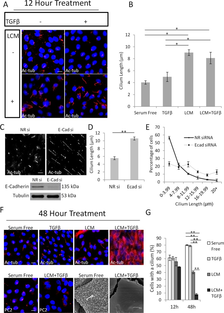
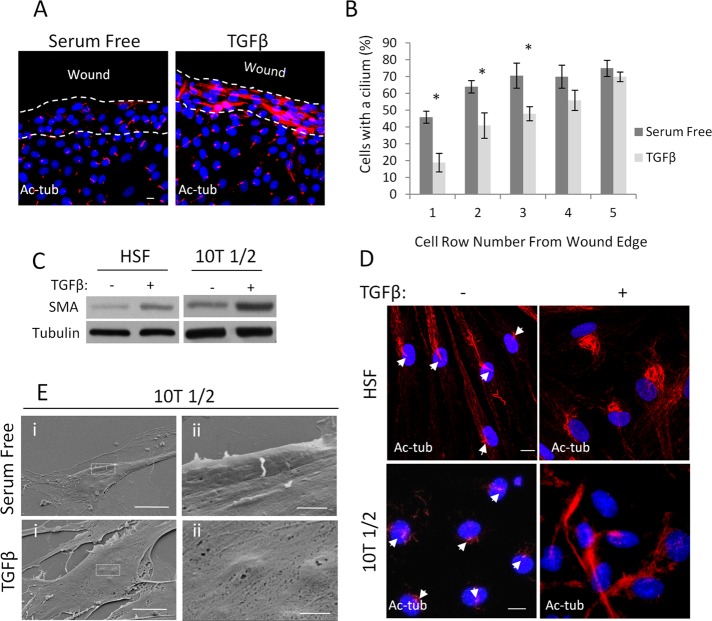
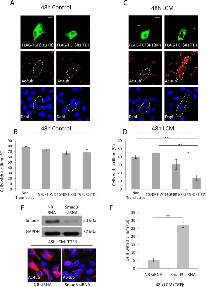
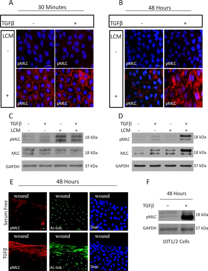
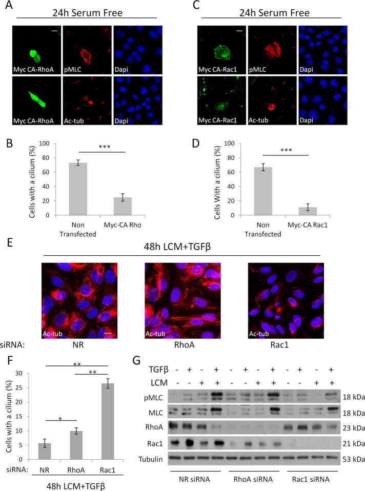
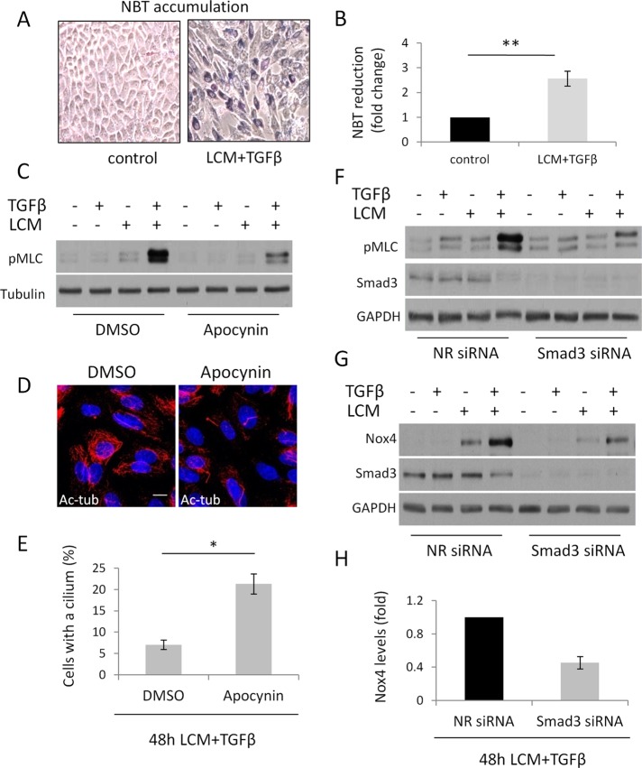
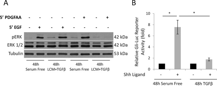
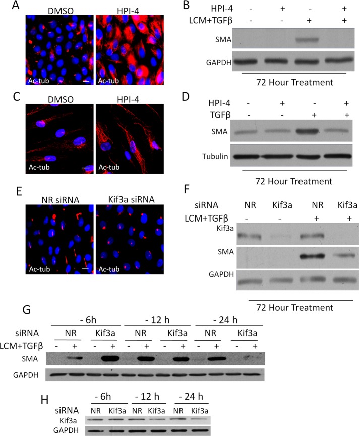
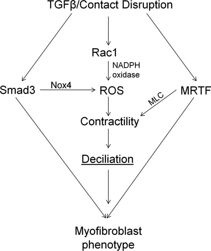
Myofibroblasts, the culprit of organ fibrosis, can originate from mesenchymal and epithelial precursors through fibroblast-myofibroblast and epithelial-myofibroblast transition (EMyT). Because certain ciliopathies are associated with fibrogenesis, we sought to explore the fate and potential role of the primary cilium during myofibroblast formation. Here we show that myofibroblast transition from either precursor results in the loss of the primary cilium. During EMyT, initial cilium growth is followed by complete deciliation. Both EMyT and cilium loss require two-hit conditions: disassembly/absence of intercellular contacts and transforming growth factor-β1 (TGFβ) exposure. Loss of E-cadherin-dependent junctions induces cilium elongation, whereas both stimuli are needed for deciliation. Accordingly, in a scratch-wounded epithelium, TGFβ provokes cilium loss exclusively along the wound edge. Increased contractility, a key myofibroblast feature, is necessary and sufficient for deciliation, since constitutively active RhoA, Rac1, or myosin triggers, and down-regulation of myosin or myocardin-related transcription factor prevents, this process. Sustained myosin phosphorylation and consequent deciliation are mediated by a Smad3-, Rac1-, and reactive oxygen species-dependent process. Transitioned myofibroblasts exhibit impaired responsiveness to platelet-derived growth factor-AA and sonic hedgehog, two cilium-associated stimuli. Although the cilium is lost during EMyT, its initial presence contributes to the transition. Thus myofibroblasts represent a unique cilium-less entity with profoundly reprogrammed cilium-related signaling.


Figures

FIGURE 1:
FIGURE 1:
The primary cilium undergoes a biphasic change during EMyT. (A) Confluent LLC-PK1 cells were treated as indicated for 12 h (TGFβ, 4 ng/ml) and stained for acetylated tubulin (Ac-tub). (B) Quantification of cilium length for the indicated conditions (mean ± SEM, n = 3, >350 cells/experiment). (C) LLC-PK1 cells were transfected with nonrelated (NR) or E-cadherin (E-cad) siRNA at 40% confluence, serum starved for 24 h after reaching confluence, and then stained for Ac-tub. E-cad knockdown was confirmed by Western blotting. (D) Quantification of cilium length as in B. (E) Distribution of cilium length in control and E-cadherin–depleted cells (n = 3, >350 cells/experiment). (F) LLC-PK1 cells were treated as indicated for 48 h and stained for Ac-tub or polycystin-2 (PC2) or processed for scanning electron microscopy. (G) Quantification of ciliated cells by Ac-tub staining (n = 3, ∼400 cells/experiment).
FIGURE 2:
FIGURE 2:
Epithelial cells along a wound edge and nonepithelial progenitors lose their primary cilium as they transition into myofibroblasts. (A) Confluent LLC-PK1 layers were wounded, treated as indicated for 48 h, and stained for Ac-tub. (B) Percentage of cells with a primary cilium was determined 24 h after the indicated treatment as a function of the cell row (1–5) from the wound edge (n = 4, 25 cells/experiment for each row). (C) Human skin fibroblasts (HSFs; left) and 10T1/2 cells (right) were serum starved or treated with TGFβ (5 ng/ml) for 72 h, followed by Western blotting for the indicated proteins. (D) The same two cell types were treated as in C and then stained for Ac-tub. Nuclei were visualized with 4′,6-diamidino-2-phenylindole. (E) 10T1/2 cells were treated as in C and then processed for scanning electron microscopy and visualized using magnification 2500× (left) and 15,000× (right). Right, enlarged area indicated by boxes on the left. Bars, 10 and 1 μm for upper and lower panels, respectively.
FIGURE 3:
FIGURE 3:
Smad3 signaling is necessary for deciliation. (A–D) LLC-PK1 cells transfected with kinase-dead (KR; A) or constitutively active (TD; C) TGFβR1 grown to confluence, serum starved for 48 h in normal or LCM medium, and stained for the FLAG epitope and Ac-tub. For B and D, ciliation was quantified also for nontransfected and WT TGFβR1-expressing cells (n = 4, 20 cells/condition). (E, F) Cells transfected with nonrelated or Smad3 siRNA, grown to confluence, and then treated with LCM plus TGFβ for 48 h, stained for Ac-tub (A), and quantified for ciliation (F; n = 3, ∼150 cells/condition).
FIGURE 4:
FIGURE 4:
Sustained increase in contractility correlates with loss of the primary cilium. (A, B) LLC-PK1 cells were treated as indicated for 30 min (A) or 48 h (B) and stained for pMLC. (C, D) Cells treated as in A and B, respectively, were processed for Western blotting for the indicated proteins. (E) Confluent monolayers were wounded, treated as indicated for 48 h, and stained for pMLC and Ac-tub. (F) 10T1/2 cells were serum starved or treated with TGFβ for 48 h, followed by Western blotting for the indicated proteins.
FIGURE 5:
FIGURE 5:
Prolonged increase in myosin activity is sufficient and necessary for cilium loss. (A) Confluent LLC-PK1 cells were transfected with the constitutively active Myc-tagged (DD) MLC II in serum-free medium and stained 24 h later for Myc and Ac-tub. (B) The presence of the cilium was quantified in DD-MLC–expressing cells and their nontransfected neighbors on the same coverslip (mean ± SEM, n = 3, 25 cells/condition). (C) LLC-PK1 cells were transfected with nonrelated (NR) or myosin heavy chain II (MHC II)–specific siRNA, grown to confluence, treated as indicated for 48 h, and then stained for Ac-tub. MHC II knockdown was verified by Western blotting. (D) Ciliation percentage was quantified (mean ± SEM, n = 3, ∼150 cells/experiment). (E) LLC-PK1 cells were transfected with siRNAs against MRTF A and MRTF B or NR siRNA. After reaching confluence, cells were treated as shown for 48 h and then subjected to Western blotting for the indicated proteins. (F) LLC-PK1 cells treated as in E were stained for Ac-Tub. (G) Ciliation percentage for E was quantified (mean ± SEM, n = 3, ≈150 cells/experiment).
FIGURE 6:
FIGURE 6:
Rac1 is required for increased myosin activity and deciliation during EMyT. (A–D) Confluent LLC-PK1 cells were transfected with constitutively active, Myc-tagged Q63L RhoA (Myc CA-RhoA; A, B) or active, Myc-tagged Q61L Rac1 (Myc CA-Rac1; B, D) and 24 h later doubly stained for the Myc epitope and either pMLC or Ac-tub. Quantification of percentages of ciliated cells for A and C are shown in B and D, respectively. Myc-positive cells were compared with their Myc-negative neighbors on the same coverslips (n = 3, 20 cells/experiment). (E, F) LLC-PK1 cells were transfected at 60% confluence with siRNA against RhoA or Rac1 or nonrelated (NR) siRNA and after reaching confluence treated with LCM plus TGFβ for 48 h, stained for Ac-tub (E), and quantified for ciliation (F; n = 3, ∼150 cells/experiment). (G) LLC-PK1 cells were transfected with Rac1, RhoA, or NR siRNA at 60% confluence and, when confluent, treated as indicated for 48 h and subjected to Western blotting for the indicated proteins.
FIGURE 7:
FIGURE 7:
Reactive oxygen species are necessary for enhanced contractility and deciliation. (A, B) LLC-PK1 cells were grown to confluence and treated with serum-free medium (control) or LCM plus TGFβ for 48 h. Cells were then exposed to NBT for 45 min and processed as described in Materials and Methods. Reduced NBT (formazan) particles were first visualized by light microscopy (A) and then extracted, solubilized, and quantified (B; n = 5). (C) LLC-PK1 cells were treated as in A in the presence of dimethyl sulfoxide (DMSO) or 600 μM apocynin and processed for Western blotting for the indicated proteins. (D) Confluent LLC-PK1 cells were treated with LCM plus TGFβ for 48 h in the presence of DMSO or 600 μM apocynin and stained for Ac-tub. (E) Ciliation for B was quantified (n = 3, ∼150 cells/experiment). (F, G) LLC-PK1 cells were transfected at 60% confluence with nonrelated or Smad3 siRNA and, upon reaching confluence, treated as indicated for 48 h and processed for Western blotting for the indicated proteins. (H) Densitometric quantification of Nox4 expression in control and Smad3-depleted cells upon LCM plus TGFβ treatment, as shown in E (n = 6).
FIGURE 8:
FIGURE 8:
Cilium-related signaling is altered in myofibroblasts. (A) Confluent LLC-PK1 cells were serum starved or treated with LCM plus TGFβ for 48 h. Subsequently, cells were incubated for 5 min with EGF or PDGF-AA and subjected to Western blotting for the indicated proteins. (B) 10T1/2 cells were transfected with the Gli-luciferase reporter construct at 60% confluence. After reaching full confluence, cells were serum starved for 24 h and then left in serum-free medium or treated with TGFβ for 72 h. During the last 24 h of the treatment, Shh ligand was added as indicated, and luciferase activity was measured. Bars, fold increase in firefly luciferase activity normalized to Renilla activity (n = 3).
FIGURE 9:
FIGURE 9:
Efficient induction of EMyT requires the primary cilium. (A) LLC-PK1 cells were incubated for 24 h with DMSO or 30 μM HPI-4 and then stained for Ac-tub. (B) Confluent LLC-PK1 were incubated with DMSO or 30 μM HPI-4 for 24 h in serum-free medium and then left untreated or exposed to LCM plus TGFβ supplemented with DMSO or HPI-4. Western blotting was then performed for the indicated proteins. (C) Human skin fibroblasts were grown to confluence, incubated as in A, and then fixed and stained for Ac-tub. (D) Confluent human skin fibroblasts were incubated as in B and then either left in serum-free medium or treated with TGFβ for 72 h. Subsequently Western blotting was performed for the indicated proteins. (E) LLC-PK1 cells were transfected with nonrelated (NR) or Kif3a siRNA at 40% confluence, grown to confluence, serum starved for 24 h, and stained for Ac-tub. (F) LLC-PK1 cells were transfected as in E, treated as indicated for 72 h, and subjected to Western blotting for the indicated proteins. (G, H) The process of deciliation may facilitate SMA expression. (G) LLC-PK1 cells were transfected with NR or Kif3a siRNA at varying times (6, 12, and 24 h) before treatment (labeled as -6, -12, and -24 h, respectively) with serum-free medium or LCM plus TGFβ for an additional 72 h. Subsequently, Western blotting was performed for SMA and GAPDH as loading control. Note the reversal (early stimulation and late inhibition) of the SMA response. (H) To check for Kif3a expression at the time of stimulation, cells were transfected as shown and after the indicated time processed for Western blotting for Kif3A and GAPDH.
FIGURE 10:
FIGURE 10:
The major processes and their mediators underlying EMyT-associated loss of the cilium. For further explanation see the Discussion.

Similar articles

Cited by

References

    1. Barnes EA, Heidtman KJ, Donoghue DJ. Constitutive activation of the shh-ptc1 pathway by a patched1 mutation identified in BCC. Oncogene. 2005;24:902–915. - PubMed
    1. Bell PD, et al. Loss of primary cilia upregulates renal hypertrophic signaling and promotes cystogenesis. J Am Soc Nephrol. 2011;22:839–848. - PMC - PubMed
    1. Bershteyn M, Atwood SX, Woo WM, Li M, Oro AE. Mim and cortactin antagonism regulates ciliogenesis and hedgehog signaling. Dev Cell. 2010;19:270–283. - PMC - PubMed
    1. Blitzer AL, Panagis L, Gusella GL, Danias J, Mlodzik M, Iomini C. Primary cilia dynamics instruct tissue patterning and repair of corneal endothelium. Proc Natl Acad Sci USA. 2011;108:2819–2824. - PMC - PubMed
    1. Bondi CD, Manickam N, Lee DY, Block K, Gorin Y, Abboud HE, Barnes JL. NAD(P)H oxidase mediates TGF-beta1-induced activation of kidney myofibroblasts. J Am Soc Nephrol. 2010;21:93–102. - PMC - PubMed

Publication types

MeSH terms