Evaluation of marginal leakage of different temporary restorative materials in Endodontics
- PMID: 24403791
- PMCID: PMC3883326
- DOI: 10.4103/0976-237X.123045
Evaluation of marginal leakage of different temporary restorative materials in Endodontics
Abstract
Aim: The aim of this study is to assess the coronal marginal leakage of three temporary restorative materials used for root canal sealing after endodontic treatment.
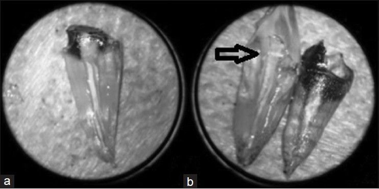
Materials and methods: A total of 88 single-rooted teeth were submitted to biomechanical preparation and filled by lateral condensation technique. After obturation process, the teeth were randomly separated into four groups, being two teeth of each group used as positive and negative control. Temporary sealing was performed as follows: GI - Clip F (VOCO); GII - Bioplic (Biodinβmica); GIII - Vitremer (3M ESPE) and GIV - Ketak N100 (3M ESPE). Next, the specimens were immersed into Indian ink for 30 and 60- days, being 10 specimens for each time interval and then submitted to diaphanization to verify the amount of coronal leakage using a measuring microscope.
Results: Leakage mean values within the 30-day period were as follows: Vitremer (0.3 mm), Ketak N100 and Clip F (0.6 mm) and Bioplic (1.7 mm). Within the 60-day period, leakage means were 1.1 mm, 1.5 mm, 2.2 mm and 2.6 mm, respectively.
Conclusions: None of the materials was capable of preventing marginal leakage within the 30- and 60-day period. In both time intervals, Bioplic presented the highest mean of leakage and Vitremer the lowest.
Keywords: Endodontics; marginal microleakage; temporary restoration.
Conflict of interest statement
Figures
Similar articles
-
Coronal microleakage with five different temporary restorative materials following walking bleach technique: An ex-vivo study.Contemp Clin Dent. 2012 Oct;3(4):421-6. doi: 10.4103/0976-237X.107431. Contemp Clin Dent. 2012. PMID: 23633802 Free PMC article.
-
Sealing ability, water sorption, solubility and toothbrushing abrasion resistance of temporary filling materials.Int Endod J. 2009 Oct;42(10):893-9. doi: 10.1111/j.1365-2591.2009.01590.x. Epub 2009 Jun 22. Int Endod J. 2009. PMID: 19549149 Clinical Trial.
-
Microleakage of different temporary filling materials in primary teeth.J Clin Pediatr Dent. 2009 Winter;34(2):157-60. doi: 10.17796/jcpd.34.2.922360t276015347. J Clin Pediatr Dent. 2009. PMID: 20297708
-
Coronal microleakage of four endodontic temporary restorative materials: an in vitro study.Oral Surg Oral Med Oral Pathol Oral Radiol Endod. 2009 Oct;108(4):e67-70. doi: 10.1016/j.tripleo.2009.05.015. Epub 2009 Aug 28. Oral Surg Oral Med Oral Pathol Oral Radiol Endod. 2009. PMID: 19716727 Clinical Trial.
-
An In-Vitro Evaluation and Comparison of Apical Sealing Ability of Three Different Obturation Technique - Lateral Condensation, Obtura II, and Thermafil.J Int Oral Health. 2013 Apr;5(2):35-43. J Int Oral Health. 2013. PMID: 24155589 Free PMC article. Review.
Cited by
-
Assessing correlation between different temporary restorative materials for microleakage following endodontic treatment: an in-vitro study.BMC Oral Health. 2024 Dec 19;24(1):1505. doi: 10.1186/s12903-024-05302-6. BMC Oral Health. 2024. PMID: 39702142 Free PMC article.
-
Microleakage of Restorative Materials Used for Temporization of Endodontic Access Cavities.J Clin Med. 2023 Jul 18;12(14):4762. doi: 10.3390/jcm12144762. J Clin Med. 2023. PMID: 37510877 Free PMC article.
-
Evaluation of physical-mechanical properties, antibacterial effect, and cytotoxicity of temporary restorative materials.J Appl Oral Sci. 2018;26:e20170562. doi: 10.1590/1678-7757-2017-0562. Epub 2018 Aug 20. J Appl Oral Sci. 2018. PMID: 30133673 Free PMC article.
References
-
- Siqueira JF, Jr, Araújo MC, Garcia PF, Fraga RC, Dantas CJ. Histological evaluation of the effectiveness of five instrumentation techniques for cleaning the apical third of root canals. J Endod. 1997;23:499–502. - PubMed
-
- Sasaki EW, Versiani MA, Perez DE, Sousa-Neto MD, Silva-Sousa YT, Silva RG. Ex vivo analysis of the debris remaining in flattened root canals of vital and nonvital teeth after biomechanical preparation with Ni-Ti rotary instruments. Braz Dent J. 2006;17:233–6. - PubMed
-
- Gonçalves LC, Sponchiado EC, Jr, Marques AA, Frota MF, Garcia Lda F. Morphometrical analysis of cleaning capacity of a hybrid instrumentation in mesial flattened root canals. Aust Endod J. 2010;36:1–6. - PubMed
-
- Sponchiado EC, Jr, Azevedo LK, Marchesan MA, Brugnera Júnior A, Alfredo E, Sousa Neto MD. Cervical microleakage in root canals treated with Er: YAG and Nd: YAG laser. Proc SPIE Lasers Dent. 2005;6:140–3.
LinkOut - more resources
Full Text Sources
Other Literature Sources