

Skin, fascias, and scars: symptoms and systemic connections
- PMID: 24403836
- PMCID: PMC3883554
- DOI: 10.2147/JMDH.S52870
Skin, fascias, and scars: symptoms and systemic connections
Abstract
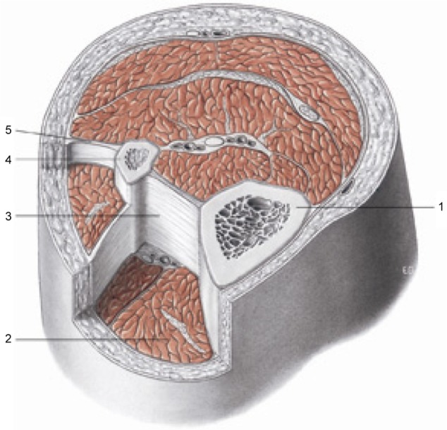
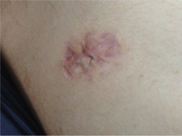
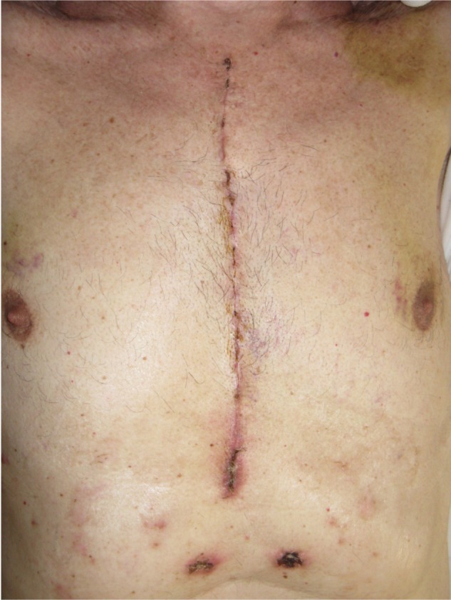
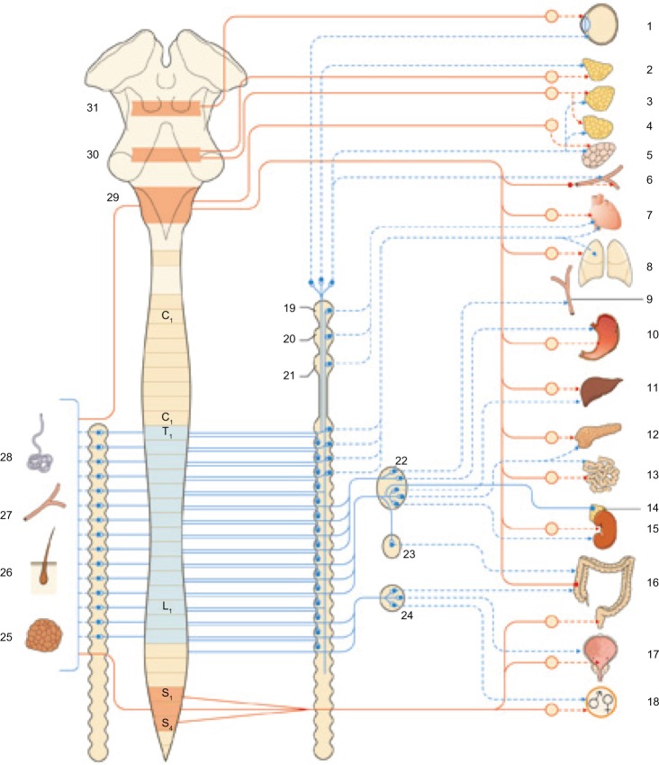
Every element or cell in the human body produces substances that communicate and respond in an autocrine or paracrine mode, consequently affecting organs and structures that are seemingly far from each other. The same also applies to the skin. In fact, when the integrity of the skin has been altered, or when its healing process is disturbed, it becomes a source of symptoms that are not merely cutaneous. The skin is an organ, and similar to any other structure, it has different functions in addition to connections with the central and peripheral nervous system. This article examines pathological responses produced by scars, analyzing definitions and differences. At the same time, it considers the subcutaneous fascias, as this connective structure is altered when there is a discontinuous cutaneous surface. The consequence is an ample symptomatology, which is not limited to the body area where the scar is located, such as a postural or trigeminal disorder.
Keywords: fascia; osteopathic; scar; scarring; skin.
Figures
Similar articles
-
Osteopathic Approach for Keloids and Hypertrophic Scars.Cureus. 2023 Sep 7;15(9):e44815. doi: 10.7759/cureus.44815. eCollection 2023 Sep. Cureus. 2023. PMID: 37692181 Free PMC article. Review.
-
Scar-free healing: from embryonic mechanisms to adult therapeutic intervention.Philos Trans R Soc Lond B Biol Sci. 2004 May 29;359(1445):839-50. doi: 10.1098/rstb.2004.1475. Philos Trans R Soc Lond B Biol Sci. 2004. PMID: 15293811 Free PMC article. Review.
-
Controlling Inflammation Pre-Emptively or at the Time of Cutaneous Injury Optimises Outcome of Skin Scarring.Front Immunol. 2022 May 27;13:883239. doi: 10.3389/fimmu.2022.883239. eCollection 2022. Front Immunol. 2022. PMID: 35711461 Free PMC article. Review.
-
Endothelial dysfunction may play a key role in keloid and hypertrophic scar pathogenesis - Keloids and hypertrophic scars may be vascular disorders.Med Hypotheses. 2016 Nov;96:51-60. doi: 10.1016/j.mehy.2016.09.024. Epub 2016 Sep 28. Med Hypotheses. 2016. PMID: 27959277
-
[Precise application of Traditional Chinese Medicine in minimally-invasive techniques].Zhongguo Gu Shang. 2018 Jun 25;31(6):493-496. doi: 10.3969/j.issn.1003-0034.2018.06.001. Zhongguo Gu Shang. 2018. PMID: 29945400 Chinese.
Cited by
-
Post-sternotomy pain syndrome following cardiac surgery: case report.J Pain Res. 2017 May 15;10:1163-1169. doi: 10.2147/JPR.S129394. eCollection 2017. J Pain Res. 2017. PMID: 28553137 Free PMC article.
-
Adapted Physical Activity Protocol for Lower Limb Functional and Strength Recovery in a Young Athlete with Cutaneous Melanoma: Feasibility and Efficacy during COVID-19 Pandemic.Int J Environ Res Public Health. 2022 Aug 4;19(15):9590. doi: 10.3390/ijerph19159590. Int J Environ Res Public Health. 2022. PMID: 35954946 Free PMC article.
-
Neck-to-shoulder pain as an unusual presentation of pulmonary embolism in a patient with cervical spinal cord injury: A case report.Medicine (Baltimore). 2017 Oct;96(42):e8288. doi: 10.1097/MD.0000000000008288. Medicine (Baltimore). 2017. PMID: 29049229 Free PMC article.
-
Impaired Lymphatic Drainage and Interstitial Inflammatory Stasis in Chronic Musculoskeletal and Idiopathic Pain Syndromes: Exploring a Novel Mechanism.Front Pain Res (Lausanne). 2021 Aug 23;2:691740. doi: 10.3389/fpain.2021.691740. eCollection 2021. Front Pain Res (Lausanne). 2021. PMID: 35295453 Free PMC article.
-
Osteopathic Approach for Keloids and Hypertrophic Scars.Cureus. 2023 Sep 7;15(9):e44815. doi: 10.7759/cureus.44815. eCollection 2023 Sep. Cureus. 2023. PMID: 37692181 Free PMC article. Review.
References
-
- Miletich I, Sharpe PT. Neural crest contribution to mammalian tooth formation. Birth Defects Res C Embryo Today. 2004;72(2):200–212. - PubMed
LinkOut - more resources
Full Text Sources
Other Literature Sources
