. 2013 Dec;15(12):1330-46.
doi: 10.1593/neo.131688.

Inflammatory factors of the tumor microenvironment induce plasticity in nontransformed breast epithelial cells: EMT, invasion, and collapse of normally organized breast textures

Affiliations

Inflammatory factors of the tumor microenvironment induce plasticity in nontransformed breast epithelial cells: EMT, invasion, and collapse of normally organized breast textures

Tal Leibovich-Rivkin et al. Neoplasia. 2013 Dec.

Abstract

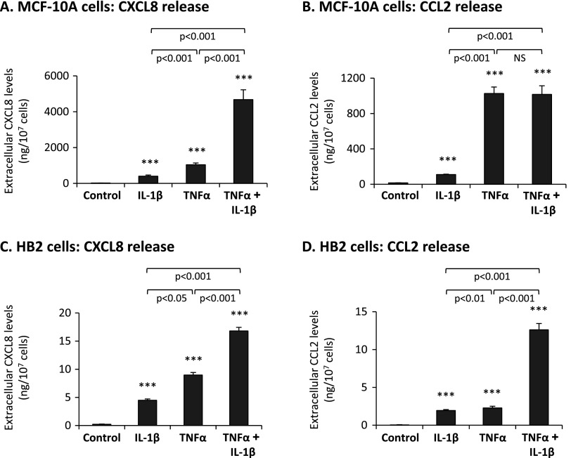
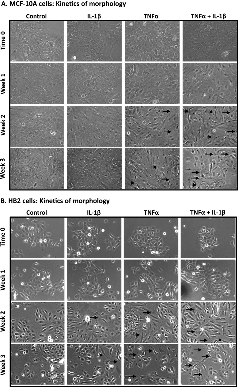
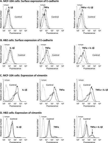
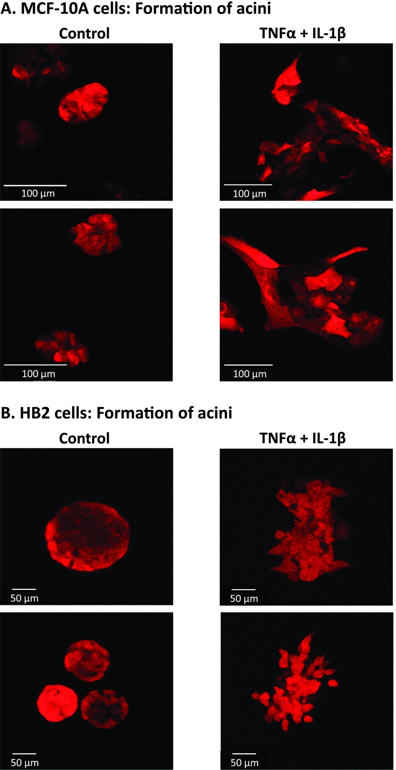
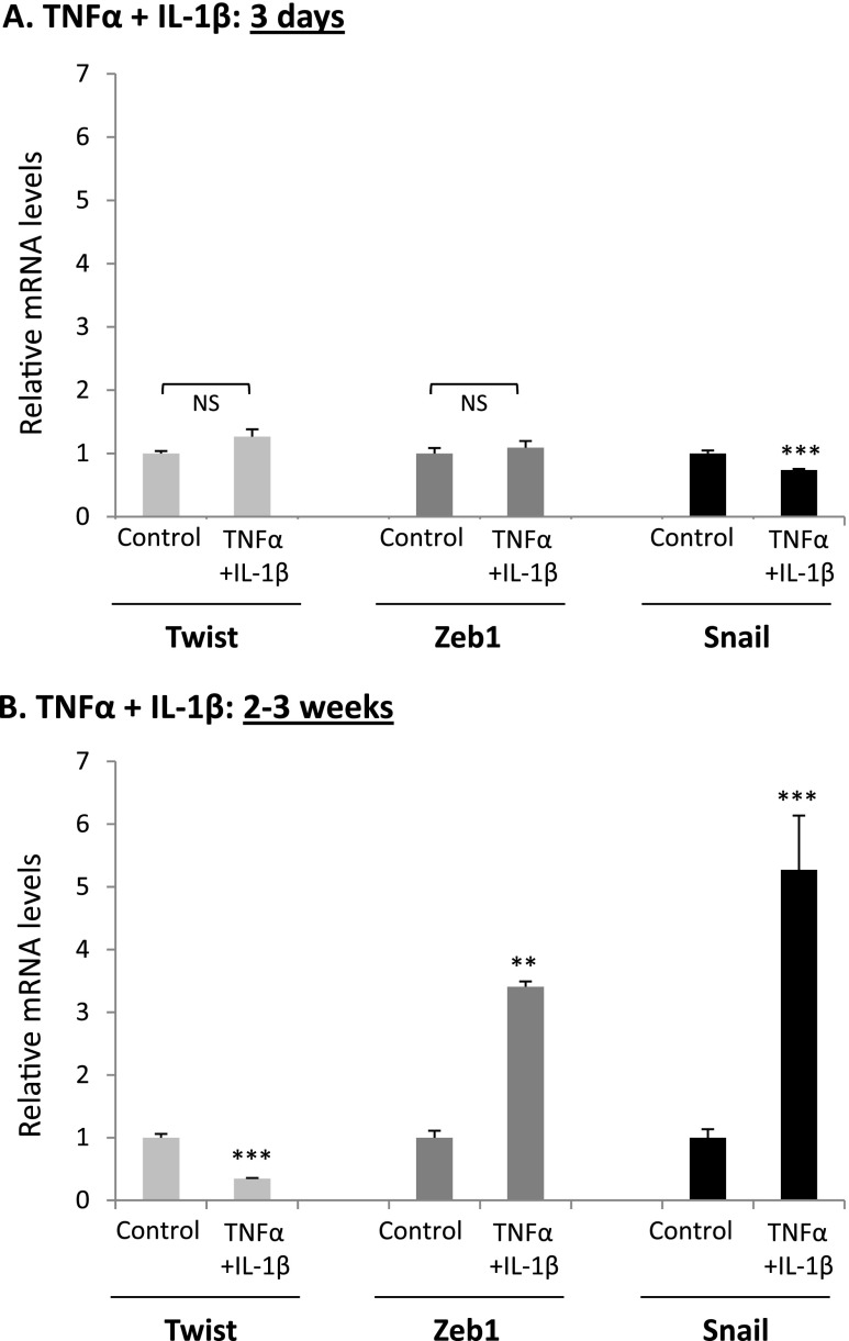
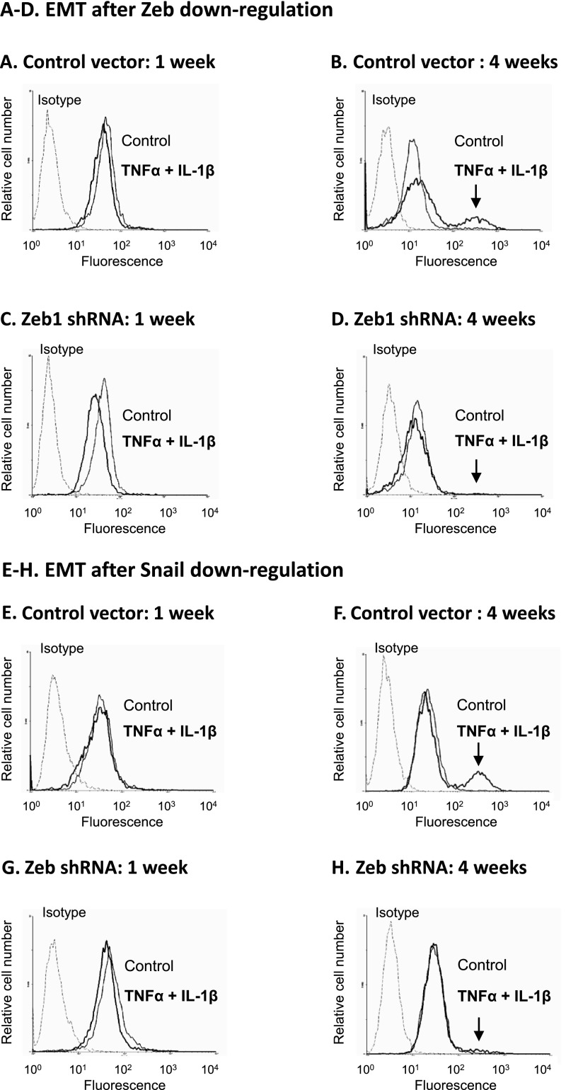
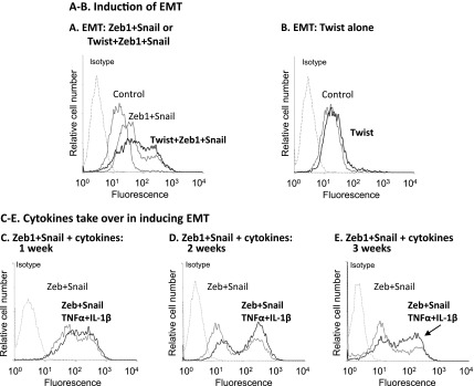
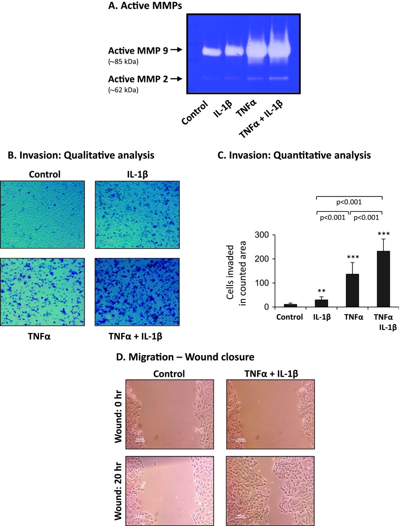
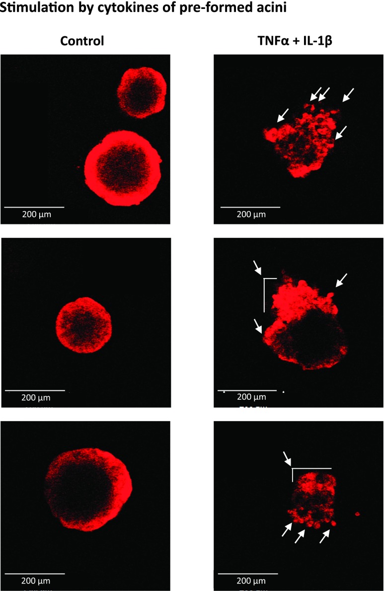
Nontransformed breast epithelial cells that are adjacent to tumor cells are constantly exposed to tumor necrosis factor-α (TNFα) and interleukin-1β (IL-1β), two inflammatory cytokines identified as having pro-tumoral causative roles. We show that continuous stimulation of nontransformed breast epithelial cells by TNFα + IL-1β for 2 to 3 weeks induced their spreading and epithelial-to-mesenchymal transition (EMT). The mechanistic bases for this slow induction of EMT by TNFα + IL-1β are: 1) it took 2 to 3 weeks for the cytokines to induce the expression of the EMT activators Zeb1 and Snail; 2) although Twist has amplified the EMT-inducing activities of Zeb1 + Snail, its expression was reduced by TNFα + IL-1β; however, the lack of Twist was compensated by prolonged stimulation with TNFα + IL-1β that has potentiated the EMT-inducing activities of Zeb1 + Snail. Stimulation by TNFα + IL-1β has induced the following dissemination-related properties in the nontransformed cells: 1) up-regulation of functional matrix metalloproteinases; 2) induction of migratory and invasive capabilities; 3) disruption of the normal phenotype of organized three-dimensional acini structures typically formed only by nontransformed breast cells and spreading of nontransformed cells out of such acini. Our findings suggest that TNFα + IL-1β induce dissemination of nontransformed breast epithelial cells and their reseeding at the primary tumor site; if, then, such detached cells are exposed to transforming events, they may form secondary malignant focus and lead to disease recurrence. Thus, our study reveals novel pathways through which the inflammatory microenvironment may contribute to relapsed disease in breast cancer.


Figures

Figure 1
Figure 1
Nontransformed breast epithelial cells respond to TNFα and IL-1β by increased release of the pro-malignancy chemokines CXCL8 and CCL2. MCF-10A cells (A, B) and HB2 cells (C, D) were stimulated by TNFα (50 ng/ml), IL-1β (500 pg/ml), or both cytokines together (TNFα + IL-1β; concentrations as above) for 24 hours, in serum-free medium. Cytokine concentrations for stimulation were selected on the basis of previous titration analyses (see Materials and Methods section for details; data not shown). Control, cells not stimulated by the cytokines (exposed to the solubilizer of the cytokines). The expression levels of CXCL8 (A, C) and CCL2 (B, D) were determined in cell supernatants by ELISA, in the linear range of absorbance. ***P < .001 for cytokine-stimulated cells compared to nonstimulated cells. In all panels, a representative experiment of n = 3 is presented.
Figure 2
Figure 2
Prolonged stimulation by TNFα and IL-1β induces morphologic changes, spreading, and formation of cellular protrusions in nontransformed breast epithelial cells. MCF-10A cells (A) and HB2 cells (B) were stimulated by TNFα, IL-1β, or TNFα + IL-1β (concentrations as in Figure 1). Control, cells not stimulated by the cytokines (exposed to the solubilizer of the cytokines). Cell images were taken immediately after cell culturing (time 0) and then after 1, 2, and 3 weeks of stimulation, at x10 to x20 magnification (x10 magnification was used only for HB2 cells, in which case photographs were adjusted to x20 magnification digitally). Arrows point to some of the cells in which cellular protrusions and spreading have been observed. In all panels, the images are representatives of many pictures taken in n = 3 experiments.
Figure 3
Figure 3
Continuous stimulation by TNFα and IL-1β leads to reduced surface expression of E-cadherin and increased expression of vimentin in nontransformed breast epithelial cells. MCF-10A cells (A, C) and HB2 cells (B, D) were stimulated by TNFα, IL-1β, or TNFα + IL-1β (concentrations as in Figure 1) for 3 weeks. Control, cells not stimulated by the cytokines (exposed to the solubilizer of the cytokines). Surface expression of E-cadherin was determined in intact cells (A, B), while intracellular expression of vimentin was determined in permeabilized cells (C, D), using flow cytometry. Isotype, nonrelevant isotype control. The arrow points out the new subpopulation of cells with high expression of vimentin that has appeared following TNFα + IL-1β stimulation in HB2 cells. In all panels, a representative experiment of at least n = 3 is presented.
Figure 4
Figure 4
Nontransformed breast epithelial cells stimulated by TNFα + IL-1β are incapable of forming organized acini structures. MCF-10A cells (A) and HB2 cells (B) were stimulated by TNFα + IL-1β (concentrations as in Figure 1) for 3 weeks. Control, cells not stimulated by the cytokines (exposed to the solubilizer of the cytokines). Then, the cells were plated onto nonadherent conditions on Matrigel to allow for acini formation. After 7 days, pictures of the structures were taken by confocal microscopy. In all panels, the images are representatives of many pictures taken in n = 3 experiments. See text (Results section) for explanation on the acini structures formed by the control nonstimulated cells.
Figure 5
Figure 5
Continuous stimulation by TNFα + IL-1β induces upregulation in Zeb1 and Snail expression and downregulates Twist expression in nontransformed breast epithelial cells. HB2 cells were stimulated by TNFα, IL-1β, or TNFα + IL-1β (concentrations as in Figure 1) for 3 days (A) or for 2 to 3 weeks (B). Control, cells not stimulated by the cytokines (exposed to the solubilizer of the cytokines). At each time point, the expression of Zeb1, Snail, and Twist was determined by qPCR. **P < .01, ***P < .001; NS, not significant for cytokine-stimulated cells compared to nonstimulated cells. Nonstimulated cells were given the value of 1. In all panels, a representative experiment of n = 3 is presented.
Figure 6
Figure 6
Zeb1 and Snail are essential for EMT induction in nontransformed breast epithelial cells stimulated by TNFα + IL-1β. HB2 cells were infected to express shRNA to Zeb1, shRNA to Snail, or control shRNA vector (down-regulation of Zeb1 and Snail expression was verified by qPCR, as shown in Figure W3). The cells were either stimulated by TNFα + IL-1β (concentrations as in Figure 1) for 1 or 4 weeks or nonstimulated by the cytokines (exposed to the solubilizer of the cytokines) for the same time course. Following downregulation of these factors and cytokine stimulation, the expression of vimentin was determined by flow cytometry in permeabilized cells, as proxy for cells undergoing EMT. Isotype, nonrelevant isotype control. (A–D) The impact of Zeb1 down-regulation on EMT. (A, C) Cells grown with or without cytokine stimulation for 1 week. (B, D) Cells grown with or without cytokine stimulation for 4 weeks. (A, B) Cells infected with control vector. (C, D) Cells infected with shRNA for Zeb1. (E–H) The impact of Snail down-regulation on EMT. (E, G) Cells grown with or without cytokine stimulation for 1 week. (F, H) Cells grown with or without cytokine stimulation for 4 weeks. (E, F) Cells infected with control vector. (G, H) Cells infected with shRNA for Snail. The arrows point out the new subpopulation of cells with high expression of vimentin that has appeared following cytokine stimulation in control HB2 cells and has disappeared upon down-regulation of Zeb1 and Snail. In all panels, a representative experiment of at least n = 3 is presented.
Figure 7
Figure 7
The EMT-inducing activities of Zeb1 and Snail on nontransformed breast epithelial cells are potentiated by Twist or alternatively by prolonged stimulation with TNFα + IL-1β. HB2 cells were infected to overexpress Zeb1+ Snail or Twist + Zeb1+ Snail (A) or Twist alone (B) (the increased expression of these EMT inducers following infection was verified by qPCR, as shown in FigureW4). Control, cells infected with control vector. Following expression of these factors, the expression of vimentin was determined by flow cytometry in permeabilized cells, as proxy for cells undergoing EMT. Isotype, nonrelevant isotype control. In all panels, a representative experiment of n = 3 is presented. (C–E) HB2 cells were infected to overexpress Zeb1 + Snail (termed “Zeb + Snail”; the expression of these EMT inducers following infection was verified by qPCR as shown in Figure W4). The cells were either not stimulated by the cytokines (exposed to the solubilizer of the cytokines) or stimulated for 1 week (C), 2 weeks (D), or 3 weeks (E) by TNFα + IL-1β (concentrations as in Figure 1; termed “Zeb + Snail, TNFα + IL-1β”). At each time point, the expression of vimentin was determined by flow cytometry in permeabilized cells, as proxy for cells undergoing EMT. Isotype, nonrelevant isotype control. In all panels, a representative experiment of n = 3 is presented.
Figure 8
Figure 8
TNFα + IL-1β induce high MMP levels and increased invasive and migratory phenotype in nontransformed breast epithelial cells. HB2 cells were stimulated by TNFα, IL-1β, or TNFα + IL-1β (concentrations as in Figure 1) for 3 weeks. Control, cells not stimulated by the cytokines (exposed to the solubilizer of the cytokines). (A) Release of functional MMPs. CM were collected, and the presence of active MMPs was determined by zymography. A representative experiment of n = 3 is presented. (B, C) Invasion through Matrigel, determined after 8 hours in response to serum-containing medium, in transwells. Of note, the constitutive level of basal migration (no serum included) was not affected by cytokine stimulation (Figure W5). In cytokine-stimulated groups, TNFα and/or IL-1β were present throughout the time of assay in the upper wells to enable constant stimulation and also in the bottom wells of the chamber to prevent cytokine gradients. (B) Representative images of cells that have invaded, in control and cytokine-stimulated groups. In all panels, the images are representatives of many pictures taken in n = 3 experiments. (C) Quantitative analysis of the number of cells that have invaded in each of the groups. **P < .01, ***P < .001 for cytokine-stimulated cells compared to nonstimulated cells. In all panels, a representative experiment of n = 3 is presented. (D) Migration in wound closure assays. Following plating, the cells reached full confluence and a scratch was performed. Images of the cells were taken at the time of scratching (wound: 0 hour) and after 20 hours (wound: 20 hours). TNFα + IL-1β were present throughout the time of assay, and in parallel analyses were not found to induce cell growth (data not shown). In all panels, the images are representatives of many pictures taken in n = 3 experiments.
Figure 9
Figure 9
TNFα + IL-1β stimulation of nontransformed breast epithelial cells ruins pre-formed acini structures and is accompanied by cell spreading out of acini. HB2 cells were plated onto nonadherent conditions on Matrigel to allow for acini formation. After 6 days, when acini have formed, the cells were stimulated for 2 weeks by TNFα + IL-1β (concentrations as in Figure 1). Control, cells not stimulated by the cytokines (exposed to the solubilizer of the cytokines). Pictures of structures were taken by confocal microscopy. The arrows point to cells that have spread out of pre-formed acini, as a result of cytokine stimulation. In all panels, the images are representatives of many pictures taken in n = 3 experiments. See text (Results section) for explanation on the acini structures formed by the control nonstimulated cells.

Similar articles

Cited by

References

    1. Colotta F, Allavena P, Sica A, Garlanda C, Mantovani A. Cancer-related inflammation, the seventh hallmark of cancer: links to genetic instability. Carcinogenesis. 2009;30:1073–1081. - PubMed
    1. Allavena P, Sica A, Solinas G, Porta C, Mantovani A. The inflammatory micro-environment in tumor progression: the role of tumor-associated macrophages. Crit Rev Oncol Hematol. 2007;66:1–9. - PubMed
    1. Jiang X, Shapiro DJ. The immune system and inflammation in breast cancer. Mol Cell Endocrinol. 2013;382:673–682. - PMC - PubMed
    1. Goldberg JE, Schwertfeger KL. Proinflammatory cytokines in breast cancer: mechanisms of action and potential targets for therapeutics. Curr Drug Targets. 2013;11:1133–1146. - PubMed
    1. Jin L, Yuan RQ, Fuchs A, Yao Y, Joseph A, Schwall R, Schnitt SJ, Guida A, Hastings HM, Andres J, et al. Expression of interleukin-1beta in human breast carcinoma. Cancer. 1997;80:421–434. - PubMed

Publication types

MeSH terms