

Magnetic Resonance Imaging versus Computed Tomography in Transient Ischemic Attack and Minor Stroke: The More Υou See the More You Know
- PMID: 24403904
- PMCID: PMC3884208
- DOI: 10.1159/000355024
Magnetic Resonance Imaging versus Computed Tomography in Transient Ischemic Attack and Minor Stroke: The More Υou See the More You Know
Abstract
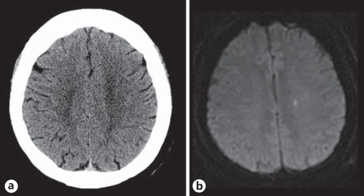
Background: Magnetic resonance imaging (MRI) is proposed as the preferred imaging modality to investigate patients with transient ischemic attack (TIA). This is mainly based on a higher yield of small acute ischemic lesions; however, direct prospective comparisons are lacking. In this study, we aimed to directly compare the yield of acute ischemic lesions on MRI and computed tomography (CT) in the emergency diagnosis of suspected TIA or minor stroke.
Methods: Consecutive patients aged 18 years or older presenting with minor stroke (NIHSS <4) or high-risk TIA and who were examined by a stroke neurologist within 24 h of symptom onset were prospectively enrolled in the CATCH study. Patients who had undergone both a baseline CT and an MRI within 24 h of symptom onset were included in this substudy. Baseline MRI and CT were interpreted independently to identify an acute ischemic lesion. The rates of acute ischemic lesions on CT and MRI were compared, and the volume of acute ischemic lesions was measured on MRI. In addition, the volume of acute ischemic lesions on MRI was compared between patients who had evidence of acute ischemia on CT and in those who did not.
Results: A total of 347 patients were included, 168 with TIAs, 147 with minor strokes and 32 with a final diagnosis of a mimic. Acute ischemic lesions were detected in 39% of TIAs by using MRI versus 8% by using CT (p < 0.0001) and in 86% of minor strokes by using MRI versus 18% by using CT (p < 0.0001). Compared to MRI, CT had a sensitivity of 20% and a specificity of 98% in identifying an acute ischemic lesion. The infarct volume on diffusion-weighted MRI was larger in cases where the CT also showed an acute ischemic lesion (median 5.07 ml, IQR 10) as compared to lesions seen only on MRI (median 0.68 ml, IQR 1.31, p < 0.0001).
Conclusion: MRI is superior to CT in detecting the small ischemic lesions occurring after TIA and minor stroke. Since these lesions are clinically relevant, MRI should be the preferred imaging modality in this setting.
Keywords: Computed tomography; Diffusion-weighted magnetic resonance imaging; Magnetic resonance imaging; Mild stroke; Transient ischemic attack.
Figures
References
-
- Merwick A, Albers GW, Amarenco P, Arsava EM, Ay H, Calvet D, Coutts SB, Cucchiara BL, Demchuk AM, Furie KL, Giles MF, Labreuche J, Lavallee PC, Mas JL, Olivot JM, Purroy F, Rothwell PM, Saver JL, Sheehan OC, Stack JP, Walsh C, Kelly PJ. Addition of brain and carotid imaging to the ABCD2 score to identify patients at early risk of stroke after transient ischaemic attack: a multicentre observational study. Lancet Neurol. 2010;9:1060–1069. - PubMed
-
- Giles MF, Albers GW, Amarenco P, Arsava EM, Asimos AW, Ay H, Calvet D, Coutts SB, Cucchiara BL, Demchuk AM, Johnston SC, Kelly PJ, Kim AS, Labreuche J, Lavallee PC, Mas JL, Merwick A, Olivot JM, Purroy F, Rosamond WD, Sciolla R, Rothwell PM. Early stroke risk and ABCD2 score performance in tissue- vs time-defined TIA: a multicenter study. Neurology. 2011;77:1222–1228. - PMC - PubMed
-
- Coutts SB, Hill MD, Simon JE, Sohn CH, Scott JN, Demchuk AM. Silent ischemia in minor stroke and TIA patients identified on MR imaging. Neurology. 2005;65:513–517. - PubMed
-
- Moreau F, Modi J, Almekhlafi M, Bal S, Goyal M, Hill MD, Coutts SB. Early magnetic resonance imaging in transient ischemic attack and minor stroke: do it or lose it. Stroke. 2013;44:671–674. - PubMed
LinkOut - more resources
Full Text Sources
Other Literature Sources
