

Optimal invasive key-hole neurosurgery with a miniaturized 3D chip on the tip: Microendoscopic device
- PMID: 24403954
- PMCID: PMC3877498
- DOI: 10.4103/1793-5482.121681
Optimal invasive key-hole neurosurgery with a miniaturized 3D chip on the tip: Microendoscopic device
Abstract
Objective: The goal of the performed study was to evaluate the possibility of a three-dimensional endoscope to become a combined microscope-endoscope device in one. We analyzed the ergonomy of the device, the implementation into the surgical workflow, the image quality, and the future perspectives such devices could have for the next generation of neurosurgeons.
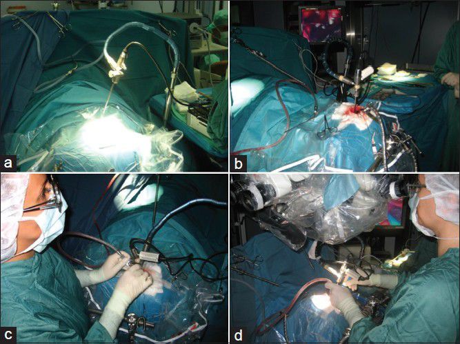
Materials and methods: Within 6 months, 22 patients (10 male, 12 female, 20-65 age) underwent surgery in neuroaxis using the new 3D-microendoscope (ME). The new 3D-ME has (a) the ability to visualize the surgical field from out- to inside with all advantages offered by a microscope, and in the same moment, (b) its design is like a small diameter endoscope that allows stereoscopic views extracorporal, intracorporal, and panoramic "para-side" of the lesion.
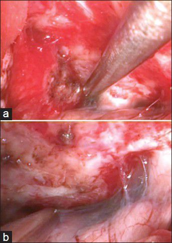
Results: In general, transcranial 3D-"microendoscopy" was performed in all patients with high-resolution 3D quality. No severe complications were observed intra- or postoperatively. With the addition of depth perception, the anatomic structures were well seen and observed.
Conclusion: The 3D-microendoscopy is a very promising surgical concept associated with new technological developments. The surgeon is able to switch to a modern visualization instrument reaching the most optimal surgical approach without compromising safety, effectiveness, and visual information.
Keywords: Endoscopy; microendoscopy; skull base surgery; three dimensionality.
Conflict of interest statement
Figures
Similar articles
-
A high-resolution, three-dimensional thin endoscope for fetal surgery.Surg Endosc. 2009 Nov;23(11):2450-3. doi: 10.1007/s00464-009-0413-7. Epub 2009 Mar 14. Surg Endosc. 2009. PMID: 19288156
-
Rigid, Variable-View Endoscope in Neurosurgery: First Intraoperative Experience.Surg Innov. 2015 Aug;22(4):390-3. doi: 10.1177/1553350614543382. Epub 2014 Jul 21. Surg Innov. 2015. PMID: 25049320
-
Multiple brain tumor nodule resections under direct visualization of a neuronavigated endoscope.Minim Invasive Neurosurg. 2007 Aug;50(4):227-32. doi: 10.1055/s-2007-985861. Minim Invasive Neurosurg. 2007. PMID: 17948182
-
Three-dimensional endoscopy in sinus surgery.Curr Opin Otolaryngol Head Neck Surg. 2013 Feb;21(1):3-10. doi: 10.1097/MOO.0b013e32835bf58c. Curr Opin Otolaryngol Head Neck Surg. 2013. PMID: 23277168 Review.
-
Endoscopy in neuro-otologic surgery.Otolaryngol Clin North Am. 2002 Apr;35(2):297-323. doi: 10.1016/s0030-6665(02)00015-4. Otolaryngol Clin North Am. 2002. PMID: 12391620 Review.
Cited by
-
Endoscopic vitreoretinal surgery: principles, applications and new directions.Int J Retina Vitreous. 2019 Jun 18;5:15. doi: 10.1186/s40942-019-0165-z. eCollection 2019. Int J Retina Vitreous. 2019. PMID: 31236288 Free PMC article. Review.
-
Trigeminal Neuralgia.Asian J Neurosurg. 2017 Oct-Dec;12(4):585-597. doi: 10.4103/ajns.AJNS_67_14. Asian J Neurosurg. 2017. PMID: 29114270 Free PMC article. Review.
-
Endoscopic endonasal trans-sphenoid management of craniopharyngiomas.Asian J Neurosurg. 2015 Jan-Mar;10(1):10-6. doi: 10.4103/1793-5482.151502. Asian J Neurosurg. 2015. PMID: 25767569 Free PMC article.
-
Intraocular endoscopy: A review.Indian J Ophthalmol. 2021 Jan;69(1):14-25. doi: 10.4103/ijo.IJO_1029_20. Indian J Ophthalmol. 2021. PMID: 33323565 Free PMC article. Review.
References
-
- Fries G, Perneczky A. Intracranial endoscopy. Adv Tech Stand Neurosurg. 1999;25:21–60. - PubMed
-
- Hopf NJ, Perneczky A. Endoscopic neurosurgery and endoscope-assisted microneurosurgery for the treatment of intracranial cysts. Neurosurgery. 1998;43:1330–6. - PubMed
-
- Perneczky A, Fries G. Endoscope-assisted brain surgery: Part 1 – evolution, basic concept, and current technique. Neurosurgery. 1998;42:219–24. - PubMed
-
- Perneczky A, Boecher-Schwarz HG. Endoscope-assisted microsurgery for cerebral aneurysms. Neurol Med Chir (Tokyo) 1998;38:33–4. - PubMed
-
- van Lindert E, Hopf N, Perneczky A. Endoscopic treatment of mesencephalic ependymal cysts: Technical case report. Neurosurgery. 1998;43:1234–41. - PubMed
LinkOut - more resources
Full Text Sources
Other Literature Sources