Review
. 2013 Dec 13:12:Doc05.
doi: 10.3205/cto000097.

Surgery of the ear and the lateral skull base: pitfalls and complications

Affiliations
Review

Surgery of the ear and the lateral skull base: pitfalls and complications

Bernhard Schick et al. GMS Curr Top Otorhinolaryngol Head Neck Surg. .

Abstract

Surgery of the ear and the lateral skull base is a fascinating, yet challenging field in otorhinolaryngology. A thorough knowledge of the associated complications and pitfalls is indispensable for the surgeon, not only to provide the best possible care to his patients, but also to further improve his surgical skills. Following a summary about general aspects in pre-, intra-and postoperative care of patients with disorders of the ear/lateral skull base, this article covers the most common pitfalls and complications in stapes surgery, cochlear implantation and surgery of vestibular schwannomas and jugulotympanal paragangliomas. Based on these exemplary procedures, basic "dos and don'ts" of skull base surgery are explained, which the reader can easily transfer to other disorders. Special emphasis is laid on functional aspects, such as hearing, balance and facial nerve function. Furthermore, the topics of infection, bleeding, skull base defects, quality of life and indication for revision surgery are discussed. An open communication about complications and pitfalls in ear/lateral skull base surgery among surgeons is a prerequisite for the further advancement of this fascinating field in ENT surgery. This article is meant to be a contribution to this process.


Figures

Table 1
Table 1. Complication rates of vestibular schwannoma surgery dependent on the selected approach [274]
Table 2
Table 2. Prevalence of preoperative cranial nerve palsies in jugulotympanal paragangliomas
Table 3
Table 3. Pre- and postoperative facial nerve function in patients with jugulotympanal paragangliomas and no evidence of facial nerve infiltration [386]
Table 4
Table 4. Complications following jugular paraganglioma resection
Figure 1
Figure 1. Recording of ocular and cervical vestibular evoked myogenic potentials (VEMPs) from the patient described in case 1. Asymmetry of n10 and p13-n23 amplitudes indicates hypofunction of the left utriculus and sacculus, which fits with the clinical diagnosis of left-sided vestibular neuritis.
Figure 2
Figure 2. Vestibular evaluation before (a, b) and after (c, d) intratympanic application of gentamicin during vestibular prehabilitation before vestibular schwannoma surgery (case 2). a.) Caloric testing (x-axis: time [min], y-axis: slow phase velocity [°/sec]): right-sided hypofunction. b.) n10 amplitudes of ocular vestibular evoked myogenic potentials (oVEMPs) indicate symmetric utricular function before gentamicin treatment. c.) Loss of right-sided caloric excitability following three applications of intratympanic gentamicin. d.) The marked asymmetry of oVEMP n10 amplitudes indicates gentamicin-induced loss of utricular function on the right side.
Figure 3
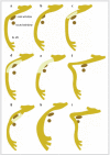
Figure 3. Anatomical variants and anomalies of the facial nerve (overview, taken from [82]). a.) normal course, b.) subtotal obstruction of the oval window by an overhanging facial nerve, c.) lack of bony cover in the tympanic segment, d.) dehiscent nerve crossing the oval window, e.) herniation of the dehiscent nerve into the middle ear and obstruction of the oval window, f.) elongation of the second genu resulting in a sharp angle between tympanic and mastoidal segment of the nerve, g.) duplication of the tympanic portion, h.) lateralization of the mastoid portion, i.) anterior displacement of the mastoid portion.
Figure 4
Figure 4. Three-dimensional reconstruction (3D) of micro-CT images from human temporal bone illustrating the spatial relationship between stapes footplate/piston, utriclus and sacculus (previously unpublished figures, with kind permission of Ian Curthoys and Payal Mukherjee, University of Sydney, Australia). a.) view of a stapes piston (insertion depth: 0.25 mm) in relation to the stapes footplate (green), utricular macula (light blue) and saccular macula (orange). The membranous structures of utriculus and sacculus are shown in dark blue and red, respectively. b.) 3D reconstruction of stapes (green), utricular (light blue) and saccular macula (orange). Both figures show that the posterosuperior utricular macula is located closest to the stapes footplate (for details see [182]).
Figure 5
Figure 5. High-resolution computed tomography of the temporal bone (coronal reconstruction) showing misplacement of a cochlear implant electrode in a hypotympanic air cell (filled arrow). The open arrow points to the basal turn of the cochlea.
Figure 6
Figure 6. Characteristic traces of intraoperative facial nerve electromyography (EMG) during vestibular schwannoma surgery (simulated data according to [279]). a.) “A train”: high-frequency signals with sudden onset / end. b.) “B train” with spikes. c.) “C train” with continuous irregular EMG activity.

Similar articles

Cited by

References

    1. Linder TE, Lin F. Felsenbeinchirurgie. Komplikationen und unerwünschte Operationsfolgen. HNO. 2011;59:974–979. doi: 10.1007/s00106-011-2359-z. Available from: http://dx.doi.org/10.1007/s00106-011-2359-z. - DOI - DOI - PubMed
    1. Dlugaiczyk J, Schick B. Klinische Differenzialdiagnose vestibulärer Symptome. CME Hals Nasen Ohrenheilkd. 2010;3:128–149.
    1. Haid CT. Vestibularisprüfung und vestibuläre Erkrankungen. Berlin, Heidelberg, New York: Springer-Verlag; 1990. Available from: http://dx.doi.org/10.1007/978-3-662-10791-1. - DOI - DOI
    1. Enticott JC, Tari S, Koh SM, Dowell RC, O'Leary SJ. Cochlear implant and vestibular function. Otol Neurotol. 2006 Sep;27(6):824–830. doi: 10.1097/01.mao.0000227903.47483.a6. Available from: http://dx.doi.org/10.1097/01.mao.0000227903.47483.a6. - DOI - DOI - PubMed
    1. Fina M, Skinner M, Goebel JA, Piccirillo JF, Neely JG, Black O. Vestibular dysfunction after cochlear implantation. Otol Neurotol. 2003 Mar;24(2):234–242. doi: 10.1097/00129492-200303000-00018. Available from: http://dx.doi.org/10.1097/00129492-200303000-00018. - DOI - DOI - PubMed