Review
. 2013 Dec 13:12:Doc06.
doi: 10.3205/cto000098.

Danger points, complications and medico-legal aspects in endoscopic sinus surgery

Affiliations
Review

Danger points, complications and medico-legal aspects in endoscopic sinus surgery

W Hosemann et al. GMS Curr Top Otorhinolaryngol Head Neck Surg. .

Abstract

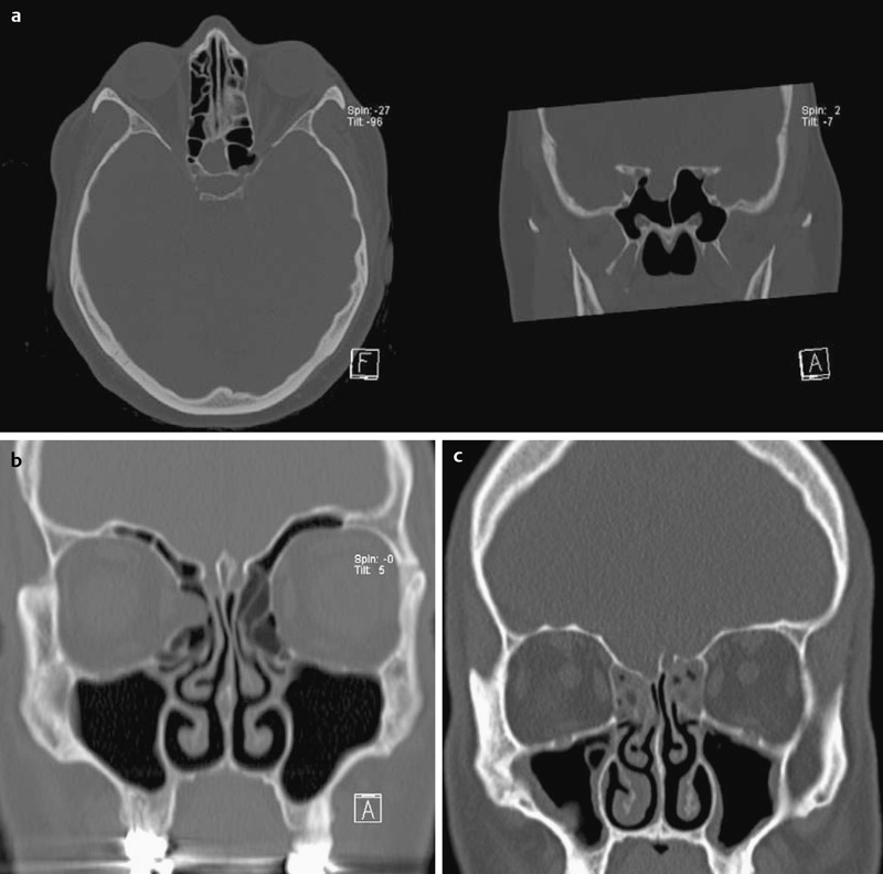
Endoscopic endonasal sinus surgery represents the overall accepted type of surgical treatment for chronic rhinosinusitis. Notwithstanding raised and still evolving quality standards, surgeons performing routine endoscopic interventions are faced with minor complications in 5% and major complications in 0.5-1%. A comprehensive review on all minor and major complications of endoscopic surgery of the paranasal sinuses and also on the anterior skull base is presented listing the actual scientific literature. The pathogenesis, signs and symptoms of each complication are reviewed and therapeutic regimens are discussed in detail relating to actual publication references. Potential medico-legal aspects are explicated and recent algorithms of avoidance are mentioned taking into account options in surgical training and education.

Keywords: FESS; complications; endoscopic sinus surgery; medico-legal aspects; review; skull base surgery.


Figures

Table 1
Table 1. Complications of endonasal sinus surgery (based on: [62]).
Table 2
Table 2. Checklist for evaluating a CT-scan before performing routine sinus surgery (based on: [106], [135], [189], [197], [363], [450], [592], [594], [704], [705]).
Table 3
Table 3. Special keywords to consider in context with medico-legal aspects of sinus surgery for the treatment of chronic rhinosinusitis (based on: [79]).
Figure 1
Figure 1. MRI of a patient with a past medical history of “uneventful sinus surgery” 7 years ago. Patient suffers from intercurrent meningitis. Image shows residual findings of a skull base laceration (loco typico) with residual signs of trauma of the neighbouring brain tissue.
Figure 2
Figure 2. CT-scan (coronal plane) of a patient having been subjected to middle meatal antrostomy. “Missed ostium sequence” on the right side leading to persistence of maxillary sinusitis.
a. Neo-ostium of the maxillary sinus in the middle meatus. b. Preserved, intact uncinate process and persistent obstruction of the natural maxillary sinus ostium due to mucosal edema of the neighbouring mucosa.
Figure 3
Figure 3. a. Intraoperative picture of a “floppy turbinate” (right side).
b. CT-scan of a patient having been subjected to anterior ethmoidectomy. Lateralization of the right sided vertical lamella of the middle turbinate causing inflammatory retentions in the ethmoidal cavity.
Figure 4
Figure 4. a. Left nasal cavity of a patient having been subjected to extensive (“radical”) sinus surgery for chronic rhinosinusitis leading to “empty nose syndrome”.
b. Right nasal cavity of a patient having been subjected to a rhino-neurosurgical intervention for craniopharyngeoma with application of a naso-septal flap. Irrespective to use of a “reverse flap” for amelioration of postoperative crusting, significant atrophic rhinitis is seen.
Figure 5
Figure 5. View of the left maxillary sinus after middle meatal anstrostomy (70° angled optical device). The inferior orbital n. revels signs of partial destruction (small nerve ending is hanging down from the orbital roof).
Figure 6
Figure 6. a. Left-sided “preseptal”, venous orbital hematoma following sinus surgery for chronic rhinosinusitis. Healing without any residual problem may be expected.
b. Patient revealing major right-sided intraorbital hematoma in the course of ethmoidectomy. Lateral canthotomy and also inferior canthoysis have been performed.
Figure 7
Figure 7. Detail of an axial CT scan outlining the contour of the right sided eye globe in red. The respective patient had been subjected to an ineffective decompression procedure for a retrobulbar orbital hematoma. The posterior pole of the globe reveals significant tenting indicating critical expansion of the hematoma [367].
Figure 8
Figure 8. Individual anatomical relation of the posterior wall of the maxillary sinus in the frontal plane (depicted in yellow) and branches of the sphenopalatine a. supplying the interior turbinate (depicted in red) (refer to: [150, 654]). The diagram illustrates that generous fenestration of the maxillary sinus in the middle nasal meatus inevitably will cause relevant bleeding in some cases.
Figure 9
Figure 9. Laceration of the left-sided internal carotid a. as a complication of routine paranasal sinus surgery.
a. Angiogram of the internal carotid artery revealing a lesion located at the anterior bending (anterior ‘genu’). b. Occlusion of the artery by coils. c. Revision surgery of the respective sphenoid sinus revealing a partly exposed coil (green arrow: coil; white arrow: suction device). d. Post-treatment axial CT scan depicting the coils.
Figure 10
Figure 10. Postoperative axial CT-scan of the left side showing destruction of the lamina papyracea (red arrow) and also complete transsection of the medial rectus muscle in the course of routine paranasal sinus surgery (blue arrow: retracted stump of the muscle).
Figure 11
Figure 11. Endoscopic aspect of the left-sided nasal cavity with a middle meatal antrostomy (70° angled optical device: some crusts are shown inside the maxillary sinus). Anterior to the neo-ostium the nasolacrimal duct is shown, having been subjected to “unintended dacryocystorhinostomy”. This event had no functional sequel.
Figure 12
Figure 12. Postoperative sagittal MRT tomography revealing signs of a recent anterior skull base perforation by a.-p. directed force.
Figure 13
Figure 13. Axial CT-scan revealing tension pneumocephalus caused by a skull base perforation during routine paranasal sinus surgery (note: “Mount Fuji sign” of the anterior cerebral poles).
Figure 14
Figure 14. Postoperative axial CT-scan following seemingly uneventful routine paranasal sinus surgery. Obviously a major skull base perforation has happened and a piece of bone (red arrow) was transferred into the remote brain tissue. The surgeon noticed an increased intraoperative blood loss only.
Figure 15
Figure 15. a. Incidental finding of a meningocele located at the roof of the sphenoid sinus.
b. incidental finding of a prolapse of orbital tissue into the aerated ethmoidal cell system. c. Minor anatomical irregularity revealing asymmetry of the ethmoidal roof of the left and right side.

References

    1. Chen Y, Dales R, Lin M. The epidemiology of chronic rhinosinusitis in Canadians. Laryngoscope. 2003 Jul;113(7):1199–1205. doi: 10.1097/00005537-200307000-00016. Available from: http://dx.doi.org/10.1097/00005537-200307000-00016. - DOI - DOI - PubMed
    1. Fokkens WJ, Lund VJ, Mullol J, Bachert C, Alobid I, Baroody F, Cohen N, Cervin A, Douglas R, Gevaert P, Georgalas C, Goossens H, Harvey R, Hellings P, Hopkins C, Jones N, Joos G, Kalogjera L, Kern B, Kowalski M, Price D, Riechelmann H, Schlosser R, Senior B, Thomas M, Toskala E, Voegels R, Wang de Y, Wormald PJ. EPOS 2012: European position paper on rhinosinusitis and nasal polyps 2012. A summary for otorhinolaryngologists. Rhinology. 2012;50(Suppl 23):1–299. Available from: http://www.rhinologyjournal.com/supplement_23.pdf. - PubMed
    1. Hedman J, Kaprio J, Poussa T, Nieminen MM. Prevalence of asthma, aspirin intolerance, nasi polyposis and chronic obstruction pulmonary disease in a population-based study. Int J Epidemiology. 1999;28:717–722. doi: 10.1093/ije/28.4.717. Available from: http://dx.doi.org/10.1093/ije/28.4.717. - DOI - DOI - PubMed
    1. Stuck BA, Bachert C, Federspil P, Hosemann W, Klimek L, Mösges R, Pfaar O, Rudack C, Sitter H, Wagenmann M, Weber R, Hörmann K German Society of Otorhinolaryngology, Head and Neck Surgery. Leitlinie "Rhinosinusitis"--Langfassung: S2-Leitlinie der Deutschen Gesellschaft für Hals-Nasen-Ohren-Heilkunde, Kopf- und Hals-Chirurgie. [Rhinosinusitis guidelines--unabridged version: S2 guidelines from the German Society of Otorhinolaryngology, Head and Neck Surgery]. HNO. 2012 Feb;60(2):141–162. doi: 10.1007/s00106-011-2396-7. (Ger). Available from: http://dx.doi.org/10.1007/s00106-011-2396-7. - DOI - DOI - PubMed
    1. Mattos JL, Woodard CR, Payne SC. Trends in common rhinologic illnesses: analysis of U.S. healthcare surveys 1995-2007. Int Forum Allergy Rhinol. 2011 Jan-Feb;1(1):3–12. doi: 10.1002/alr.20003. Available from: http://dx.doi.org/10.1002/alr.20003. - DOI - DOI - PubMed